Movie
Movie Controller

[English] 日本語
 Yorodumi
- PDB-7w85: Structural of the filamentous Escherichia coli glutamine synthetase -
+ Open data

- Basic information
| Entry | Database: PDB / ID: 7w85 | |||||||||
|---|---|---|---|---|---|---|---|---|---|---|
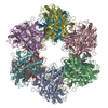
| Title | Structural of the filamentous Escherichia coli glutamine synthetase | |||||||||
 Components Components | Glutamine synthetase | |||||||||
 Keywords Keywords | BIOSYNTHETIC PROTEIN / Glutamine synthetase / filament | |||||||||
| Function / homology |  Function and homology information Function and homology informationammonia assimilation cycle / nitrogen utilization / glutamine synthetase / : / glutamine synthetase activity / response to radiation / ATP binding / membrane / metal ion binding / identical protein binding ...ammonia assimilation cycle / nitrogen utilization / glutamine synthetase / : / glutamine synthetase activity / response to radiation / ATP binding / membrane / metal ion binding / identical protein binding / cytoplasm / cytosol Similarity search - Function | |||||||||
| Biological species |  | |||||||||
| Method | ELECTRON MICROSCOPY / single particle reconstruction / cryo EM / Resolution: 2.94 Å | |||||||||
 Authors Authors | Huang, P.-C. / Chen, S.-K. / Wu, K.-P. | |||||||||
| Funding support |  Taiwan, 2items Taiwan, 2items
| |||||||||
 Citation Citation | Journal: Protein Sci / Year: 2022 Title: Structural basis for the helical filament formation of Escherichia coli glutamine synthetase. Authors: Pei-Chi Huang / Shao-Kang Chen / Wei-Hung Chiang / Meng-Ru Ho / Kuen-Phon Wu / Abstract: Escherichia coli glutamine synthetase (EcGS) spontaneously forms a dodecamer that catalytically converts glutamate to glutamine. EcGS stacks with other dodecamers to create a filament-like polymer ...Escherichia coli glutamine synthetase (EcGS) spontaneously forms a dodecamer that catalytically converts glutamate to glutamine. EcGS stacks with other dodecamers to create a filament-like polymer visible under transmission electron microscopy. Filamentous EcGS is induced by environmental metal ions. We used cryo-electron microscopy (cryo-EM) to decipher the structure of metal ion (nickel)-induced EcGS helical filament at a sub-3Å resolution. EcGS filament formation involves stacking of native dodecamers by chelating nickel ions to residues His5 and His13 in the first N-terminal helix (H1). His5 and His13 from paired parallel H1 helices provide salt bridges and hydrogen bonds to tightly stack two dodecamers. One subunit of the EcGS filament hosts two nickel ions, whereas the dodecameric interface and the ATP/Mg-binding site both host a nickel ion each. We reveal that upon adding glutamate or ATP for catalytic reactions, nickel-induced EcGS filament reverts to individual dodecamers. Such tunable filament formation is often associated with stress responses. Our results provide detailed structural information on the mechanism underlying reversible and tunable EcGS filament formation. | |||||||||
| History |
|
- Structure visualization
| Structure viewer | Molecule: MolmilJmol/JSmol |
|---|
- Downloads & links
-Download
| PDBx/mmCIF format | 7w85.cif.gz | 1.7 MB | Display | PDBx/mmCIF format |
|---|---|---|---|---|
| PDB format | pdb7w85.ent.gz | Display | PDB format | |
| PDBx/mmJSON format | 7w85.json.gz | Tree view | PDBx/mmJSON format | |
| Others |  Other downloads Other downloads |
-Validation report
| Arichive directory | https://data.pdbj.org/pub/pdb/validation_reports/w8/7w85ftp://data.pdbj.org/pub/pdb/validation_reports/w8/7w85 | HTTPS FTP |
|---|
-Related structure data
| Related structure data |  32352MC M: map data used to model this data C: citing same article ( |
|---|---|
| Similar structure data |
-Links
 PDBj
PDBj
- Assembly
| Deposited unit | 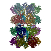
|
|---|---|
| 1 |
|
-Components
| #1: Protein | Mass: 51966.609 Da / Num. of mol.: 24 Source method: isolated from a genetically manipulated source Source: (gene. exp.) #2: Chemical | ChemComp-NI /   Mass: 58.693 Da / Num. of mol.: 42 / Source method: obtained synthetically / Formula: Ni / Feature type: SUBJECT OF INVESTIGATION Mass: 58.693 Da / Num. of mol.: 42 / Source method: obtained synthetically / Formula: Ni / Feature type: SUBJECT OF INVESTIGATIONHas ligand of interest | Y | |
|---|
-Experimental details
-Experiment
| Experiment | Method: ELECTRON MICROSCOPY |
|---|---|
| EM experiment | Aggregation state: PARTICLE / 3D reconstruction method: single particle reconstruction |
- Sample preparation
| Component | Name: Filamentous form of Ecoli glutamine synthetase / Type: COMPLEX / Entity ID: #1 / Source: RECOMBINANT |
|---|---|
| Source (natural) | Organism: |
| Source (recombinant) | Organism: |
| Buffer solution | pH: 7.4 |
| Specimen | Conc.: 0.1 mg/ml / Embedding applied: NO / Shadowing applied: NO / Staining applied: NO / Vitrification applied: YES / Details: Titan Krios |
| Specimen support | Grid material: COPPER / Grid mesh size: 200 divisions/in. / Grid type: Quantifoil R2/1 |
| Vitrification | Instrument: FEI VITROBOT MARK IV / Cryogen name: ETHANE |
- Electron microscopy imaging
| Experimental equipment |  Model: Titan Krios / Image courtesy: FEI Company |
|---|---|
| Microscopy | Model: TFS KRIOS |
| Electron gun | Electron source:  FIELD EMISSION GUN / Accelerating voltage: 300 kV / Illumination mode: FLOOD BEAM FIELD EMISSION GUN / Accelerating voltage: 300 kV / Illumination mode: FLOOD BEAM |
| Electron lens | Mode: BRIGHT FIELD / Nominal defocus max: 2000 nm / Nominal defocus min: 1000 nm |
| Image recording | Electron dose: 59.6 e/Å2 / Detector mode: COUNTING / Film or detector model: GATAN K2 SUMMIT (4k x 4k) / Num. of grids imaged: 1 / Num. of real images: 1182 |
- Processing
| EM software |
| ||||||||||||||||||||||||||||||||||||||||||||
|---|---|---|---|---|---|---|---|---|---|---|---|---|---|---|---|---|---|---|---|---|---|---|---|---|---|---|---|---|---|---|---|---|---|---|---|---|---|---|---|---|---|---|---|---|---|
| CTF correction | Type: PHASE FLIPPING AND AMPLITUDE CORRECTION | ||||||||||||||||||||||||||||||||||||||||||||
| Symmetry | Point symmetry: C6 (6 fold cyclic) | ||||||||||||||||||||||||||||||||||||||||||||
| 3D reconstruction | Resolution: 2.94 Å / Resolution method: FSC 0.143 CUT-OFF / Num. of particles: 117242 / Symmetry type: POINT | ||||||||||||||||||||||||||||||||||||||||||||
| Atomic model building | Protocol: RIGID BODY FIT / Space: REAL | ||||||||||||||||||||||||||||||||||||||||||||
| Atomic model building | PDB-ID: 2LGS Pdb chain-ID: A / Accession code: 2LGS / Source name: PDB / Type: experimental model |