[English] 日本語
 Yorodumi
- EMDB-3234: Representative tomogram as used in: Structure of bacterial chemot... -
+ Open data
- Basic information
| Entry | Database: EMDB / ID: EMD-3234 | |||||||||
|---|---|---|---|---|---|---|---|---|---|---|
| Title | Representative tomogram as used in: Structure of bacterial chemotaxis signaling CheA2-trimer core complex by cryo-electron tomography and subvolume averaging | |||||||||
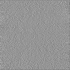
 Map data Map data | The tomogram is is binned by 4 while those used in the corresponding subTomogram work were used at full sampling. The protein array is best viewed by rotating around the X-axis 3.3 degrees, and the Y-axis 0.3 degrees as Imod Slicer angles. | |||||||||
 Sample Sample |
| |||||||||
 Keywords Keywords | Bacterial chemotaxis / Signal transduction / cryo-Electron Tomography / Molecular dynamics simulation / all-atom | |||||||||
| Function / homology |  Function and homology information Function and homology informationnegative regulation of protein modification process / detection of chemical stimulus / methyl accepting chemotaxis protein complex / positive regulation of post-translational protein modification / protein histidine kinase binding / protein histidine kinase activity / bacterial-type flagellum-dependent swimming motility / regulation of bacterial-type flagellum-dependent cell motility / aerotaxis / cell tip ...negative regulation of protein modification process / detection of chemical stimulus / methyl accepting chemotaxis protein complex / positive regulation of post-translational protein modification / protein histidine kinase binding / protein histidine kinase activity / bacterial-type flagellum-dependent swimming motility / regulation of bacterial-type flagellum-dependent cell motility / aerotaxis / cell tip / regulation of chemotaxis / thermotaxis / signal complex assembly / histidine kinase / phosphorelay signal transduction system / phosphorelay sensor kinase activity / establishment of localization in cell / cellular response to amino acid stimulus / protein homooligomerization / chemotaxis / transmembrane signaling receptor activity / protein domain specific binding / signal transduction / protein homodimerization activity / ATP binding / identical protein binding / plasma membrane / cytosol / cytoplasm Similarity search - Function | |||||||||
| Biological species |  | |||||||||
| Method | electron tomography / cryo EM | |||||||||
 Authors Authors | Cassidy CK / Himes BA / Alvarez FJ / Ma J / Zhou G / Perilla JR / Schulten K / Zhang P | |||||||||
 Citation Citation |  Journal: Elife / Year: 2015 Journal: Elife / Year: 2015Title: CryoEM and computer simulations reveal a novel kinase conformational switch in bacterial chemotaxis signaling. Authors: C Keith Cassidy / Benjamin A Himes / Frances J Alvarez / Jun Ma / Gongpu Zhao / Juan R Perilla / Klaus Schulten / Peijun Zhang /  Abstract: Chemotactic responses in bacteria require large, highly ordered arrays of sensory proteins to mediate the signal transduction that ultimately controls cell motility. A mechanistic understanding of ...Chemotactic responses in bacteria require large, highly ordered arrays of sensory proteins to mediate the signal transduction that ultimately controls cell motility. A mechanistic understanding of the molecular events underlying signaling, however, has been hampered by the lack of a high-resolution structural description of the extended array. Here, we report a novel reconstitution of the array, involving the receptor signaling domain, histidine kinase CheA, and adaptor protein CheW, as well as a density map of the core-signaling unit at 11.3 Å resolution, obtained by cryo-electron tomography and sub-tomogram averaging. Extracting key structural constraints from our density map, we computationally construct and refine an atomic model of the core array structure, exposing novel interfaces between the component proteins. Using all-atom molecular dynamics simulations, we further reveal a distinctive conformational change in CheA. Mutagenesis and chemical cross-linking experiments confirm the importance of the conformational dynamics of CheA for chemotactic function. | |||||||||
| History |
|
- Structure visualization
| Movie |
 Movie viewer Movie viewer |
|---|---|
| Structure viewer | EM map:  SurfView SurfView Molmil Molmil Jmol/JSmol Jmol/JSmol |
| Supplemental images |
- Downloads & links
-EMDB archive
| Map data |  emd_3234.map.gz emd_3234.map.gz | 71.7 MB |  EMDB map data format EMDB map data format | |
|---|---|---|---|---|
| Header (meta data) |  emd-3234-v30.xml emd-3234-v30.xml emd-3234.xml emd-3234.xml | 12.6 KB 12.6 KB | Display Display |  EMDB header EMDB header |
| Images |  EMD-3234.tif EMD-3234.tif emd_3234.tif emd_3234.tif | 200.5 KB 200.5 KB | ||
| Archive directory |  http://ftp.pdbj.org/pub/emdb/structures/EMD-3234 http://ftp.pdbj.org/pub/emdb/structures/EMD-3234 ftp://ftp.pdbj.org/pub/emdb/structures/EMD-3234 ftp://ftp.pdbj.org/pub/emdb/structures/EMD-3234 | HTTPS FTP |
-Related structure data
| Related structure data |  6319C  6320C  3ja6C C: citing same article ( |
|---|---|
| Similar structure data | Similarity search - Function & homology  F&H Search F&H Search |
- Links
Links
| EMDB pages |  EMDB (EBI/PDBe) / EMDB (EBI/PDBe) /  EMDataResource EMDataResource |
|---|---|
| Related items in Molecule of the Month |
- Map
Map
| File |  Download / File: emd_3234.map.gz / Format: CCP4 / Size: 75.4 MB / Type: IMAGE STORED AS FLOATING POINT NUMBER (4 BYTES) Download / File: emd_3234.map.gz / Format: CCP4 / Size: 75.4 MB / Type: IMAGE STORED AS FLOATING POINT NUMBER (4 BYTES) | ||||||||||||||||||||||||||||||||||||||||||||||||||||||||||||||||||||
|---|---|---|---|---|---|---|---|---|---|---|---|---|---|---|---|---|---|---|---|---|---|---|---|---|---|---|---|---|---|---|---|---|---|---|---|---|---|---|---|---|---|---|---|---|---|---|---|---|---|---|---|---|---|---|---|---|---|---|---|---|---|---|---|---|---|---|---|---|---|
| Annotation | The tomogram is is binned by 4 while those used in the corresponding subTomogram work were used at full sampling. The protein array is best viewed by rotating around the X-axis 3.3 degrees, and the Y-axis 0.3 degrees as Imod Slicer angles. | ||||||||||||||||||||||||||||||||||||||||||||||||||||||||||||||||||||
| Projections & slices | Image control
Images are generated by Spider. generated in cubic-lattice coordinate | ||||||||||||||||||||||||||||||||||||||||||||||||||||||||||||||||||||
| Voxel size | X=Y=Z: 12.04 Å | ||||||||||||||||||||||||||||||||||||||||||||||||||||||||||||||||||||
| Density |
| ||||||||||||||||||||||||||||||||||||||||||||||||||||||||||||||||||||
| Symmetry | Space group: 1 | ||||||||||||||||||||||||||||||||||||||||||||||||||||||||||||||||||||
| Details | EMDB XML:
CCP4 map header:
| ||||||||||||||||||||||||||||||||||||||||||||||||||||||||||||||||||||
-Supplemental data
- Sample components
-Entire : tarCF CheA CheW
| Entire | Name: tarCF CheA CheW |
|---|---|
| Components |
|
-Supramolecule #1000: tarCF CheA CheW
| Supramolecule | Name: tarCF CheA CheW / type: sample / ID: 1000 Oligomeric state: Trimer of (CheA dimer, dimer of tarCF trimers of dimers, 2 CheW subunits) Number unique components: 3 |
|---|---|
| Molecular weight | Theoretical: 1.66 MDa |
-Macromolecule #1: Chemotaxis protein CheA
| Macromolecule | Name: Chemotaxis protein CheA / type: protein_or_peptide / ID: 1 / Name.synonym: Bacterial Chemotaxis Histidine Kinase CheA / Number of copies: 6 / Oligomeric state: Dimer / Recombinant expression: Yes |
|---|---|
| Source (natural) | Organism:  |
| Molecular weight | Theoretical: 71.384 KDa |
| Recombinant expression | Organism:  |
| Sequence | UniProtKB: Chemotaxis protein CheA |
-Macromolecule #2: Chemotaxis protein CheW
| Macromolecule | Name: Chemotaxis protein CheW / type: protein_or_peptide / ID: 2 / Number of copies: 2 / Oligomeric state: monomeric / Recombinant expression: Yes |
|---|---|
| Source (natural) | Organism:  |
| Molecular weight | Theoretical: 18.083 KDa |
| Recombinant expression | Organism:  |
| Sequence | UniProtKB: Chemotaxis protein CheW |
-Macromolecule #3: Methyl-accepting chemotaxis protein II
| Macromolecule | Name: Methyl-accepting chemotaxis protein II / type: protein_or_peptide / ID: 3 / Name.synonym: tarCF Details: Cytoplasmic fragment of wild-type aspartate receptor Number of copies: 6 / Oligomeric state: trimer of dimers / Recombinant expression: Yes |
|---|---|
| Source (natural) | Organism:  |
| Molecular weight | Theoretical: 31.209 KDa |
| Recombinant expression | Organism:  |
| Sequence | UniProtKB: Methyl-accepting chemotaxis protein II |
-Experimental details
-Structure determination
| Method | cryo EM |
|---|---|
 Processing Processing | electron tomography |
| Aggregation state | 2D array |
- Sample preparation
| Buffer | pH: 7.4 / Details: 75 mM Tris-HCl, 100 mM KCl, 5 mM MgCl2 |
|---|---|
| Grid | Details: Perforated R2/2 Quantifoil grids precoated with 10 nm fiducial gold beads on the backside of the grid |
| Vitrification | Cryogen name: ETHANE / Instrument: HOMEMADE PLUNGER Method: Single-sided blotting to avoid disruption of the monolayer |
| Details | Pseudo-crystalline 2D monolayer reconstituted on a lipid monolayer |
- Electron microscopy
| Microscope | FEI POLARA 300 |
|---|---|
| Date | Jan 7, 2009 |
| Image recording | Category: CCD / Film or detector model: GATAN ULTRASCAN 4000 (4k x 4k) / Number real images: 60 / Average electron dose: 60 e/Å2 / Bits/pixel: 16 |
| Electron beam | Acceleration voltage: 200 kV / Electron source:  FIELD EMISSION GUN FIELD EMISSION GUN |
| Electron optics | Calibrated magnification: 49834 / Illumination mode: SPOT SCAN / Imaging mode: BRIGHT FIELD / Cs: 2.0 mm / Nominal defocus max: 8.0 µm / Nominal defocus min: 4.0 µm / Nominal magnification: 39000 |
| Sample stage | Specimen holder model: OTHER / Tilt series - Axis1 - Min angle: -70 ° / Tilt series - Axis1 - Max angle: 70 ° / Tilt series - Axis1 - Angle increment: 3 ° |
| Experimental equipment |  Model: Tecnai Polara / Image courtesy: FEI Company |
- Image processing
| Details | CTF correction (phase only) performed on projections rotated to have tilt axis coincide with Y-axis. Projections aligned using area matching with geometry refinement in Protomo. Reconstruction with SIRT in IMOD. |
|---|---|
| Final reconstruction | Algorithm: OTHER / Resolution method: OTHER / Software - Name:  IMOD / Number images used: 60 IMOD / Number images used: 60 |
| CTF correction | Details: TomoCTF (strip-based periodogram) |
| Crystal parameters | Plane group: P 1 |
 Movie
Movie Controller


 Z (Sec.)
 Y (Row.)
 X (Col.)


