 Movie
Movie Controller

[English] 日本語
 Yorodumi
- EMDB-31478: Reconstruction of the HerA-NurA complex from Deinococcus radiodurans -
+ Open data

- Basic information
| Entry |  | |||||||||
|---|---|---|---|---|---|---|---|---|---|---|
| Title | Reconstruction of the HerA-NurA complex from Deinococcus radiodurans | |||||||||
 Map data Map data | ||||||||||
 Sample Sample |
| |||||||||
 Keywords Keywords | nuclease / helicase / end resection / DNA repair / HYDROLASE | |||||||||
| Function / homology |  Function and homology information Function and homology information | |||||||||
| Biological species |  Deinococcus radiodurans R1 (radioresistant) Deinococcus radiodurans R1 (radioresistant) | |||||||||
| Method | single particle reconstruction / cryo EM / Resolution: 3.85 Å | |||||||||
 Authors Authors | Xu Y / Xu L | |||||||||
| Funding support |  China, 1 items China, 1 items
| |||||||||
 Citation Citation | Journal: Structure / Year: 2022 Title: Mechanisms of helicase activated DNA end resection in bacteria. Authors: Ying Xu / Lingyi Xu / Chen Qin / Liangyan Wang / Jiangtao Guo / Yuejin Hua / Ye Zhao / Abstract: DNA end resection mediated by the coordinated action of nuclease and helicase is a crucial step in initiating homologous recombination. The end-resection apparatus NurA nuclease and HerA helicase are ...DNA end resection mediated by the coordinated action of nuclease and helicase is a crucial step in initiating homologous recombination. The end-resection apparatus NurA nuclease and HerA helicase are present in both archaea and bacteria. Here, we report the cryo-electron microscopy structure of a bacterial HerA-NurA complex from Deinococcus radiodurans. The structure reveals a barrel-like hexameric HerA and a distinctive NurA dimer subcomplex, which has a unique extended N-terminal region (ENR) involved in bacterial NurA dimerization and activation. In addition to the long protruding linking loop and the C-terminal α helix of NurA, the flexible ENR is close to the HerA-NurA interface and divides the central channel of the DrNurA dimer into two halves, suggesting a possible mechanism of DNA end processing. In summary, this work provides new insights into the structure, assembly, and activation mechanisms of bacterial DNA end resection mediated by a minimal end-resection apparatus. | |||||||||
| History |
|
- Structure visualization
| Supplemental images |
|---|

- Downloads & links
-EMDB archive
| Map data | emd_31478.map.gz | 8.7 MB | EMDB map data format | |
|---|---|---|---|---|
| Header (meta data) | emd-31478-v30.xmlemd-31478.xml | 10.9 KB 10.9 KB | Display Display | EMDB header |
| Images | 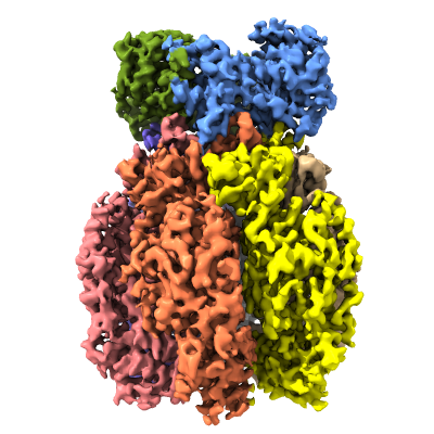 emd_31478.png emd_31478.png | 154.3 KB | ||
| Filedesc metadata | emd-31478.cif.gz | 5.4 KB | ||
| Archive directory |  http://ftp.pdbj.org/pub/emdb/structures/EMD-31478ftp://ftp.pdbj.org/pub/emdb/structures/EMD-31478 http://ftp.pdbj.org/pub/emdb/structures/EMD-31478ftp://ftp.pdbj.org/pub/emdb/structures/EMD-31478 | HTTPS FTP |
-Related structure data
| Related structure data |  7f6dMC M: atomic model generated by this map C: citing same article ( |
|---|---|
| Similar structure data |
-Links
| EMDB pages | EMDB (EBI/PDBe) / EMDataResource |
|---|
-Map
| File | Download / File: emd_31478.map.gz / Format: CCP4 / Size: 75.1 MB / Type: IMAGE STORED AS FLOATING POINT NUMBER (4 BYTES) | ||||||||||||||||||||||||||||||||||||
|---|---|---|---|---|---|---|---|---|---|---|---|---|---|---|---|---|---|---|---|---|---|---|---|---|---|---|---|---|---|---|---|---|---|---|---|---|---|
| Projections & slices | Image control
Images are generated by Spider. | ||||||||||||||||||||||||||||||||||||
| Voxel size | X=Y=Z: 1.014 Å | ||||||||||||||||||||||||||||||||||||
| Density |
| ||||||||||||||||||||||||||||||||||||
| Symmetry | Space group: 1 | ||||||||||||||||||||||||||||||||||||
| Details | EMDB XML:
|
 Z (Sec.)
 Y (Row.)
 X (Col.)


-Supplemental data
- Sample components
-Entire : Helicase-nuclease complex composed of HerA and NurA
| Entire | Name: Helicase-nuclease complex composed of HerA and NurA |
|---|---|
| Components |
|
-Supramolecule #1: Helicase-nuclease complex composed of HerA and NurA
| Supramolecule | Name: Helicase-nuclease complex composed of HerA and NurA / type: complex / ID: 1 / Parent: 0 / Macromolecule list: all |
|---|---|
| Source (natural) | Organism: Deinococcus radiodurans R1 (radioresistant) |
| Molecular weight | Theoretical: 480 KDa |
-Macromolecule #1: NurA
| Macromolecule | Name: NurA / type: protein_or_peptide / ID: 1 / Number of copies: 2 / Enantiomer: LEVO |
|---|---|
| Source (natural) | Organism: Deinococcus radiodurans R1 (radioresistant) / Strain: R1 |
| Molecular weight | Theoretical: 40.482066 KDa |
| Recombinant expression | Organism: |
| Sequence | String: MGSSHHHHHH SSGLVPRGSH MRIRLDPWPI DTFEGQLTLK PFAGLVFDVE TDRWEAIPTL GIPESVREVL VVDGKPRMEA RLLMDDDSG ELHLAAFGAY VVGAVSLCPH GTRQAELLDV RARRVLAYSS DAPLEPARLS PRNPHTGVLD YEPYAFSGRQ V EGPRAAVQ ...String: MGSSHHHHHH SSGLVPRGSH MRIRLDPWPI DTFEGQLTLK PFAGLVFDVE TDRWEAIPTL GIPESVREVL VVDGKPRMEA RLLMDDDSG ELHLAAFGAY VVGAVSLCPH GTRQAELLDV RARRVLAYSS DAPLEPARLS PRNPHTGVLD YEPYAFSGRQ V EGPRAAVQ KLMLQDEQKL SRQLASPIAL EEGEADALPE SLVLQDGPVR LGGGGSAVVG YVKTLHTDYL GADRIGLLSS LK CGERTPI LRFRVGDRGG TFSEAEGREQ RFTWYVRLCD APFYQHPLAG IMRLEMHAPE DSSFVPAAVQ QIADLSGALL SKL GSKLHK DSRAPQNLIP TAALEQAMNR SMGNLELVTR RIRTHLVTQG VVA UniProtKB: NurA domain-containing protein |
-Macromolecule #2: HerA
| Macromolecule | Name: HerA / type: protein_or_peptide / ID: 2 / Number of copies: 6 / Enantiomer: LEVO |
|---|---|
| Source (natural) | Organism: Deinococcus radiodurans R1 (radioresistant) / Strain: R1 |
| Molecular weight | Theoretical: 67.452461 KDa |
| Recombinant expression | Organism: |
| Sequence | String: MTGNDVQGAE KADAIGMVLG TEDVTPTVFW FAVSHGASVG LDDLVVVETR KPDGTPVRFY GLVDNVRKRH EGVTFESDVE DVVAGLLPA SVSYAARVLV TRVDPENFIP PQPGDHVRHA AGRELAMALS ADKMEEAAFP GGLLADGQPL PLNFRFINGE S GGHINISG ...String: MTGNDVQGAE KADAIGMVLG TEDVTPTVFW FAVSHGASVG LDDLVVVETR KPDGTPVRFY GLVDNVRKRH EGVTFESDVE DVVAGLLPA SVSYAARVLV TRVDPENFIP PQPGDHVRHA AGRELAMALS ADKMEEAAFP GGLLADGQPL PLNFRFINGE S GGHINISG ISGVATKTSY ALFLLHSIFR SGVMDRTAQG SGGRQSGTAG GRALIFNVKG EDLLFLDKPN ARMVEKEDKV VR AKGLSAD RYALLGLPAE PFRDVQLLAP PRAGAAGTAI VPQTDQRSEG VTPFVFTIRE FCARRMLPYV FSDASASLNL GFV IGNIEE KLFRLAAAQT GKGTGLIVHD WQFEDSETPP ENLDFSELGG VNLQTFEQLI SYLEYKLLEE REGEGDPKWV LKQS PGTLR AFTRRLRGVQ KYLSPLIRGD LTPEQAEGYR PDPLRRGIQL TVVDIHALSA HAQMFVVGVL LREVFEYKER VGRQD TVFV VLDELNKYAP REGDSPIKDV LLDIAERGRS LGIILIGAQQ TASEVERRIV SNAAIRVVGR LDLAEAERPE YRFLPQ SFR GRAGILQPGT MLVSQPDVPN PVLVNYPFPA WATRRDEVDD LGGKAAAEVG AGLLR UniProtKB: Helicase HerA central domain-containing protein |
-Experimental details
-Structure determination
| Method | cryo EM |
|---|---|
 Processing Processing | single particle reconstruction |
| Aggregation state | particle |
-Sample preparation
| Buffer | pH: 8 |
|---|---|
| Vitrification | Cryogen name: ETHANE |
- Electron microscopy
| Microscope | FEI TITAN KRIOS |
|---|---|
| Image recording | Film or detector model: GATAN K2 SUMMIT (4k x 4k) / Average electron dose: 64.0 e/Å2 |
| Electron beam | Acceleration voltage: 300 kV / Electron source:  FIELD EMISSION GUN FIELD EMISSION GUN |
| Electron optics | Illumination mode: OTHER / Imaging mode: BRIGHT FIELD |
| Experimental equipment |  Model: Titan Krios / Image courtesy: FEI Company |
-Image processing
| Startup model | Type of model: PDB ENTRY |
|---|---|
| Final reconstruction | Resolution.type: BY AUTHOR / Resolution: 3.85 Å / Resolution method: FSC 0.143 CUT-OFF / Number images used: 1659937 |
| Initial angle assignment | Type: OTHER |
| Final angle assignment | Type: OTHER |
