 Movie
Movie Controller

[English] 日本語
 Yorodumi
- PDB-7f6d: Reconstruction of the HerA-NurA complex from Deinococcus radiodurans -
+ Open data

- Basic information
| Entry | Database: PDB / ID: 7f6d | ||||||
|---|---|---|---|---|---|---|---|
| Title | Reconstruction of the HerA-NurA complex from Deinococcus radiodurans | ||||||
 Components Components |
| ||||||
 Keywords Keywords | HYDROLASE / nuclease / helicase / end resection / DNA repair | ||||||
| Function / homology |  Function and homology information Function and homology information | ||||||
| Biological species |  Deinococcus radiodurans R1 (radioresistant) Deinococcus radiodurans R1 (radioresistant) | ||||||
| Method | ELECTRON MICROSCOPY / single particle reconstruction / cryo EM / Resolution: 3.85 Å | ||||||
 Authors Authors | Xu, Y. / Xu, L. / Guo, J. / Hua, Y. / Zhao, Y. | ||||||
| Funding support |  China, 1items China, 1items
| ||||||
 Citation Citation | Journal: Structure / Year: 2022 Title: Mechanisms of helicase activated DNA end resection in bacteria. Authors: Ying Xu / Lingyi Xu / Chen Qin / Liangyan Wang / Jiangtao Guo / Yuejin Hua / Ye Zhao / Abstract: DNA end resection mediated by the coordinated action of nuclease and helicase is a crucial step in initiating homologous recombination. The end-resection apparatus NurA nuclease and HerA helicase are ...DNA end resection mediated by the coordinated action of nuclease and helicase is a crucial step in initiating homologous recombination. The end-resection apparatus NurA nuclease and HerA helicase are present in both archaea and bacteria. Here, we report the cryo-electron microscopy structure of a bacterial HerA-NurA complex from Deinococcus radiodurans. The structure reveals a barrel-like hexameric HerA and a distinctive NurA dimer subcomplex, which has a unique extended N-terminal region (ENR) involved in bacterial NurA dimerization and activation. In addition to the long protruding linking loop and the C-terminal α helix of NurA, the flexible ENR is close to the HerA-NurA interface and divides the central channel of the DrNurA dimer into two halves, suggesting a possible mechanism of DNA end processing. In summary, this work provides new insights into the structure, assembly, and activation mechanisms of bacterial DNA end resection mediated by a minimal end-resection apparatus. | ||||||
| History |
|
- Structure visualization
| Structure viewer | Molecule: MolmilJmol/JSmol |
|---|
- Downloads & links
-Download
| PDBx/mmCIF format | 7f6d.cif.gz | 630.1 KB | Display | PDBx/mmCIF format |
|---|---|---|---|---|
| PDB format | pdb7f6d.ent.gz | 520.2 KB | Display | PDB format |
| PDBx/mmJSON format | 7f6d.json.gz | Tree view | PDBx/mmJSON format | |
| Others |  Other downloads Other downloads |
-Validation report
| Summary document | 7f6d_validation.pdf.gz | 842.2 KB | Display | wwPDB validaton report |
|---|---|---|---|---|
| Full document | 7f6d_full_validation.pdf.gz | 883.1 KB | Display | |
| Data in XML | 7f6d_validation.xml.gz | 96 KB | Display | |
| Data in CIF | 7f6d_validation.cif.gz | 146.1 KB | Display | |
| Arichive directory | https://data.pdbj.org/pub/pdb/validation_reports/f6/7f6dftp://data.pdbj.org/pub/pdb/validation_reports/f6/7f6d | HTTPS FTP |
-Related structure data
| Related structure data | 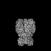 31478MC M: map data used to model this data C: citing same article ( |
|---|---|
| Similar structure data |
-Links
 PDBj
PDBj- Assembly
| Deposited unit | 
|
|---|---|
| 1 |
|
-Components
| #1: Protein | Mass: 40482.066 Da / Num. of mol.: 2 Source method: isolated from a genetically manipulated source Source: (gene. exp.) Deinococcus radiodurans R1 (radioresistant)Strain: R1 / Gene: DR_0836 Production host: References: UniProt: Q9RW33 #2: Protein | Mass: 67452.461 Da / Num. of mol.: 6 Source method: isolated from a genetically manipulated source Source: (gene. exp.) Deinococcus radiodurans R1 (radioresistant)Strain: R1 / Gene: DR_0837 Production host: References: UniProt: Q9RW32 |
|---|
-Experimental details
-Experiment
| Experiment | Method: ELECTRON MICROSCOPY |
|---|---|
| EM experiment | Aggregation state: PARTICLE / 3D reconstruction method: single particle reconstruction |
- Sample preparation
| Component | Name: Helicase-nuclease complex composed of HerA and NurA / Type: COMPLEX / Entity ID: all / Source: RECOMBINANT |
|---|---|
| Molecular weight | Value: 0.48 MDa / Experimental value: NO |
| Source (natural) | Organism: Deinococcus radiodurans R1 (radioresistant) |
| Source (recombinant) | Organism: |
| Buffer solution | pH: 8 |
| Specimen | Embedding applied: NO / Shadowing applied: NO / Staining applied: NO / Vitrification applied: YES |
| Vitrification | Cryogen name: ETHANE |
- Electron microscopy imaging
| Experimental equipment |  Model: Titan Krios / Image courtesy: FEI Company |
|---|---|
| Microscopy | Model: FEI TITAN KRIOS |
| Electron gun | Electron source:  FIELD EMISSION GUN / Accelerating voltage: 300 kV / Illumination mode: OTHER FIELD EMISSION GUN / Accelerating voltage: 300 kV / Illumination mode: OTHER |
| Electron lens | Mode: BRIGHT FIELD |
| Image recording | Electron dose: 64 e/Å2 / Film or detector model: GATAN K2 SUMMIT (4k x 4k) |
- Processing
| Software | Name: PHENIX / Version: 1.19.2_4158: / Classification: refinement | ||||||||||||||||||||||||
|---|---|---|---|---|---|---|---|---|---|---|---|---|---|---|---|---|---|---|---|---|---|---|---|---|---|
| CTF correction | Type: PHASE FLIPPING AND AMPLITUDE CORRECTION | ||||||||||||||||||||||||
| 3D reconstruction | Resolution: 3.85 Å / Resolution method: FSC 0.143 CUT-OFF / Num. of particles: 1659937 / Symmetry type: POINT | ||||||||||||||||||||||||
| Refinement | Cross valid method: NONE Stereochemistry target values: GeoStd + Monomer Library + CDL v1.2 | ||||||||||||||||||||||||
| Displacement parameters | Biso mean: 62.42 Å2 | ||||||||||||||||||||||||
| Refine LS restraints |
|
