+ Open data
- Basic information
| Entry | Database: EMDB / ID: EMD-2914 | |||||||||
|---|---|---|---|---|---|---|---|---|---|---|
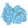
| Title | Cryo-EM reconstruction of the mammalian 55S mitoribosome | |||||||||
 Map data Map data | Reconstruction of the 55S mammalian mitoribosome | |||||||||
 Sample Sample |
| |||||||||
 Keywords Keywords | mammalian mitochondrial ribosome / 55S mitoribosome / translation / ribosomal proteins / rRNA / tRNA / mRNA / decoding / exit tunnel | |||||||||
| Function / homology |  Function and homology information Function and homology informationMitochondrial translation elongation / Mitochondrial translation termination / translation release factor activity / rRNA import into mitochondrion / mitochondrial translational elongation / mitochondrial ribosome assembly / ribonuclease III activity / microprocessor complex / Mitochondrial protein degradation / mitochondrial large ribosomal subunit ...Mitochondrial translation elongation / Mitochondrial translation termination / translation release factor activity / rRNA import into mitochondrion / mitochondrial translational elongation / mitochondrial ribosome assembly / ribonuclease III activity / microprocessor complex / Mitochondrial protein degradation / mitochondrial large ribosomal subunit / peptidyl-tRNA hydrolase / mitochondrial ribosome / mitochondrial small ribosomal subunit / mitochondrial translation / organelle membrane / ribosomal small subunit binding / RNA processing / double-stranded RNA binding / large ribosomal subunit / cell junction / regulation of translation / small ribosomal subunit / 5S rRNA binding / nuclear body / rRNA binding / ribosome / structural constituent of ribosome / ribonucleoprotein complex / translation / protein domain specific binding / mitochondrion / RNA binding / nucleoplasm / nucleus / plasma membrane / cytosol / cytoplasm Similarity search - Function | |||||||||
| Biological species |  | |||||||||
| Method | single particle reconstruction / cryo EM / Resolution: 3.8 Å | |||||||||
 Authors Authors | Greber BJ / Bieri P / Leibundgut M / Leitner A / Aebersold R / Boehringer D / Ban N | |||||||||
 Citation Citation |  Journal: Science / Year: 2015 Journal: Science / Year: 2015Title: Ribosome. The complete structure of the 55S mammalian mitochondrial ribosome. Authors: Basil J Greber / Philipp Bieri / Marc Leibundgut / Alexander Leitner / Ruedi Aebersold / Daniel Boehringer / Nenad Ban /  Abstract: Mammalian mitochondrial ribosomes (mitoribosomes) synthesize mitochondrially encoded membrane proteins that are critical for mitochondrial function. Here we present the complete atomic structure of ...Mammalian mitochondrial ribosomes (mitoribosomes) synthesize mitochondrially encoded membrane proteins that are critical for mitochondrial function. Here we present the complete atomic structure of the porcine 55S mitoribosome at 3.8 angstrom resolution by cryo-electron microscopy and chemical cross-linking/mass spectrometry. The structure of the 28S subunit in the complex was resolved at 3.6 angstrom resolution by focused alignment, which allowed building of a detailed atomic structure including all of its 15 mitoribosomal-specific proteins. The structure reveals the intersubunit contacts in the 55S mitoribosome, the molecular architecture of the mitoribosomal messenger RNA (mRNA) binding channel and its interaction with transfer RNAs, and provides insight into the highly specialized mechanism of mRNA recruitment to the 28S subunit. Furthermore, the structure contributes to a mechanistic understanding of aminoglycoside ototoxicity. | |||||||||
| History |
|
- Structure visualization
| Movie |
 Movie viewer Movie viewer |
|---|---|
| Structure viewer | EM map:  SurfView SurfView Molmil Molmil Jmol/JSmol Jmol/JSmol |
| Supplemental images |
- Downloads & links
-EMDB archive
| Map data |  emd_2914.map.gz emd_2914.map.gz | 14.2 MB |  EMDB map data format EMDB map data format | |
|---|---|---|---|---|
| Header (meta data) |  emd-2914-v30.xml emd-2914-v30.xml emd-2914.xml emd-2914.xml | 12.7 KB 12.7 KB | Display Display |  EMDB header EMDB header |
| Images |  EMDB_2914_55S_500px.png EMDB_2914_55S_500px.png | 360.2 KB | ||
| Archive directory |  http://ftp.pdbj.org/pub/emdb/structures/EMD-2914 http://ftp.pdbj.org/pub/emdb/structures/EMD-2914 ftp://ftp.pdbj.org/pub/emdb/structures/EMD-2914 ftp://ftp.pdbj.org/pub/emdb/structures/EMD-2914 | HTTPS FTP |
-Validation report
| Summary document |  emd_2914_validation.pdf.gz emd_2914_validation.pdf.gz | 258.6 KB | Display |  EMDB validaton report EMDB validaton report |
|---|---|---|---|---|
| Full document |  emd_2914_full_validation.pdf.gz emd_2914_full_validation.pdf.gz | 257.8 KB | Display | |
| Data in XML |  emd_2914_validation.xml.gz emd_2914_validation.xml.gz | 6.2 KB | Display | |
| Arichive directory |  https://ftp.pdbj.org/pub/emdb/validation_reports/EMD-2914 https://ftp.pdbj.org/pub/emdb/validation_reports/EMD-2914 ftp://ftp.pdbj.org/pub/emdb/validation_reports/EMD-2914 ftp://ftp.pdbj.org/pub/emdb/validation_reports/EMD-2914 | HTTPS FTP |
-Related structure data
| Related structure data |  5aj4MC  2913C  5aj3C M: atomic model generated by this map C: citing same article ( |
|---|---|
| Similar structure data |
- Links
Links
| EMDB pages |  EMDB (EBI/PDBe) / EMDB (EBI/PDBe) /  EMDataResource EMDataResource |
|---|---|
| Related items in Molecule of the Month |
- Map
Map
| File |  Download / File: emd_2914.map.gz / Format: CCP4 / Size: 62.5 MB / Type: IMAGE STORED AS FLOATING POINT NUMBER (4 BYTES) Download / File: emd_2914.map.gz / Format: CCP4 / Size: 62.5 MB / Type: IMAGE STORED AS FLOATING POINT NUMBER (4 BYTES) | ||||||||||||||||||||||||||||||||||||||||||||||||||||||||||||||||||||
|---|---|---|---|---|---|---|---|---|---|---|---|---|---|---|---|---|---|---|---|---|---|---|---|---|---|---|---|---|---|---|---|---|---|---|---|---|---|---|---|---|---|---|---|---|---|---|---|---|---|---|---|---|---|---|---|---|---|---|---|---|---|---|---|---|---|---|---|---|---|
| Annotation | Reconstruction of the 55S mammalian mitoribosome | ||||||||||||||||||||||||||||||||||||||||||||||||||||||||||||||||||||
| Projections & slices | Image control
Images are generated by Spider. | ||||||||||||||||||||||||||||||||||||||||||||||||||||||||||||||||||||
| Voxel size | X=Y=Z: 1.39 Å | ||||||||||||||||||||||||||||||||||||||||||||||||||||||||||||||||||||
| Density |
| ||||||||||||||||||||||||||||||||||||||||||||||||||||||||||||||||||||
| Symmetry | Space group: 1 | ||||||||||||||||||||||||||||||||||||||||||||||||||||||||||||||||||||
| Details | EMDB XML:
CCP4 map header:
| ||||||||||||||||||||||||||||||||||||||||||||||||||||||||||||||||||||
-Supplemental data
- Sample components
-Entire : 55S mammalian mitochondrial ribosome
| Entire | Name: 55S mammalian mitochondrial ribosome |
|---|---|
| Components |
|
-Supramolecule #1000: 55S mammalian mitochondrial ribosome
| Supramolecule | Name: 55S mammalian mitochondrial ribosome / type: sample / ID: 1000 / Oligomeric state: monomer / Number unique components: 2 |
|---|---|
| Molecular weight | Theoretical: 2.7 MDa |
-Supramolecule #1: 28S small subunit of the mitochondrial ribosome
| Supramolecule | Name: 28S small subunit of the mitochondrial ribosome / type: complex / ID: 1 / Recombinant expression: No Ribosome-details: ribosome-eukaryote: SSU mitochondrial 28S, SSU mitochondrial RNA 12S |
|---|---|
| Ref GO | 0: GO:0005761 |
| Source (natural) | Organism:  |
| Molecular weight | Theoretical: 1.1 MDa |
-Supramolecule #2: 39S large subunit of the mitochondrial ribosome
| Supramolecule | Name: 39S large subunit of the mitochondrial ribosome / type: complex / ID: 2 / Recombinant expression: No Ribosome-details: ribosome-eukaryote: LSU mitochondrial 39S, LSU mitochondrial RNA 16S |
|---|---|
| Ref GO | 0: GO:0005761 |
| Source (natural) | Organism:  |
| Molecular weight | Theoretical: 1.6 MDa |
-Experimental details
-Structure determination
| Method | cryo EM |
|---|---|
 Processing Processing | single particle reconstruction |
| Aggregation state | particle |
- Sample preparation
| Buffer | pH: 7.4 / Details: 20 mM HEPES-KOH, 50 mM KCl, 1 mM DTT, 40 mM MgCl2 |
|---|---|
| Grid | Details: 200 mesh Quantifoil R 2/2 holey carbon grids with a thin continuous carbon support film applied |
| Vitrification | Cryogen name: ETHANE-PROPANE MIXTURE / Instrument: HOMEMADE PLUNGER |
- Electron microscopy #1
| Microscopy ID | 1 |
|---|---|
| Microscope | FEI TITAN KRIOS |
| Details | movie mode readout in FEI EPU: 7 frames per exposure |
| Date | May 3, 2014 |
| Image recording | Category: CCD / Film or detector model: FEI FALCON II (4k x 4k) / Digitization - Sampling interval: 14 µm / Average electron dose: 20 e/Å2 Details: movie mode readout in FEI EPU: 7 frames per exposure |
| Electron beam | Acceleration voltage: 300 kV / Electron source:  FIELD EMISSION GUN FIELD EMISSION GUN |
| Electron optics | Calibrated magnification: 100720 / Illumination mode: FLOOD BEAM / Imaging mode: BRIGHT FIELD / Cs: 2.7 mm / Nominal defocus max: 3.0 µm / Nominal defocus min: 0.8 µm / Nominal magnification: 59000 |
| Sample stage | Specimen holder model: FEI TITAN KRIOS AUTOGRID HOLDER |
| Experimental equipment |  Model: Titan Krios / Image courtesy: FEI Company |
- Electron microscopy #2
| Microscopy ID | 2 |
|---|---|
| Microscope | FEI TITAN KRIOS |
| Details | movie mode readout in FEI EPU: 7 frames per exposure |
| Date | May 30, 2014 |
| Image recording | Category: CCD / Film or detector model: FEI FALCON II (4k x 4k) / Digitization - Sampling interval: 14 µm / Average electron dose: 20 e/Å2 Details: movie mode readout in FEI EPU: 7 frames per exposure |
| Electron beam | Acceleration voltage: 300 kV / Electron source:  FIELD EMISSION GUN FIELD EMISSION GUN |
| Electron optics | Calibrated magnification: 100720 / Illumination mode: FLOOD BEAM / Imaging mode: BRIGHT FIELD / Cs: 2.7 mm / Nominal defocus max: 3.0 µm / Nominal defocus min: 0.8 µm / Nominal magnification: 59000 |
| Sample stage | Specimen holder model: FEI TITAN KRIOS AUTOGRID HOLDER |
| Experimental equipment |  Model: Titan Krios / Image courtesy: FEI Company |
- Image processing
| Details | Particles were selected in batchboxer (EMAN 1.9) and extracted using RELION 1.2. |
|---|---|
| CTF correction | Details: per detector frame |
| Final reconstruction | Applied symmetry - Point group: C1 (asymmetric) / Algorithm: OTHER / Resolution.type: BY AUTHOR / Resolution: 3.8 Å / Resolution method: OTHER / Software - Name: CTFFIND3, RELION, 1.2, 1.3 / Number images used: 60872 |
-Atomic model buiding 1
| Initial model | PDB ID: |
|---|---|
| Software | Name:  Chimera Chimera |
| Details | The coordinate model of the 39S subunit was fitted into the cryo-EM density using Chimera. The model was then adjusted using COOT (RNA) and O (proteins). |
| Refinement | Space: REAL / Protocol: RIGID BODY FIT |
| Output model |  PDB-5aj4: |
-Atomic model buiding 2
| Initial model | PDB ID: |
|---|---|
| Software | Name:  Chimera Chimera |
| Details | The coordinate model of the 39S subunit was fitted into the cryo-EM density using Chimera. The model was then adjusted using COOT (RNA) and O (proteins). |
| Refinement | Space: REAL / Protocol: RIGID BODY FIT |
| Output model |  PDB-5aj4: |
 Movie
Movie Controller


 Z (Sec.)
 Y (Row.)
 X (Col.)


