 Movie
Movie Controller

[English] 日本語
 Yorodumi
- EMDB-28616: MicroED structure of an Aeropyrum pernix protoglobin metallo-carb... -
+ Open data

- Basic information
| Entry |  | |||||||||
|---|---|---|---|---|---|---|---|---|---|---|
| Title | MicroED structure of an Aeropyrum pernix protoglobin metallo-carbene complex | |||||||||
 Map data Map data | ||||||||||
 Sample Sample |
| |||||||||
 Keywords Keywords | MicroED / directed evolution / METAL BINDING PROTEIN | |||||||||
| Function / homology | Protoglobin / Globin-sensor domain / Protoglobin / Globin/Protoglobin / Globin-like superfamily / oxygen binding / heme binding / Protogloblin ApPgb Function and homology information Function and homology information | |||||||||
| Biological species |   Aeropyrum pernix (archaea) Aeropyrum pernix (archaea) | |||||||||
| Method | electron crystallography / cryo EM | |||||||||
 Authors Authors | Danelius E / Gonen T / Unge JT | |||||||||
| Funding support |  United States, 2 items United States, 2 items
| |||||||||
 Citation Citation | Journal: J Am Chem Soc / Year: 2023 Title: MicroED Structure of a Protoglobin Reactive Carbene Intermediate. Authors: Emma Danelius / Nicholas J Porter / Johan Unge / Frances H Arnold / Tamir Gonen / Abstract: Microcrystal electron diffraction (MicroED) is an emerging technique that has shown great potential for describing new chemical and biological molecular structures. Several important structures of ...Microcrystal electron diffraction (MicroED) is an emerging technique that has shown great potential for describing new chemical and biological molecular structures. Several important structures of small molecules, natural products, and peptides have been determined using methods. However, only a couple of novel protein structures have thus far been derived by MicroED. Taking advantage of recent technological advances, including higher acceleration voltage and using a low-noise detector in counting mode, we have determined the first structure of an protoglobin (Pgb) variant by MicroED using an AlphaFold2 model for phasing. The structure revealed that mutations introduced during directed evolution enhance carbene transfer activity by reorienting an α helix of Pgb into a dynamic loop, making the catalytic active site more readily accessible. After exposing the tiny crystals to the substrate, we also trapped the reactive iron-carbenoid intermediate involved in this engineered Pgb's new-to-nature activity, a challenging carbene transfer from a diazirine via a putative metallo-carbene. The bound structure discloses how an enlarged active site pocket stabilizes the carbene bound to the heme iron and, presumably, the transition state for the formation of this key intermediate. This work demonstrates that improved MicroED technology and the advancement in protein structure prediction now enable investigation of structures that was previously beyond reach. | |||||||||
| History |
|
- Structure visualization
| Supplemental images |
|---|

- Downloads & links
-EMDB archive
| Map data | emd_28616.map.gz | 6 MB | EMDB map data format | |
|---|---|---|---|---|
| Header (meta data) | emd-28616-v30.xmlemd-28616.xml | 13.6 KB 13.6 KB | Display Display | EMDB header |
| Images | 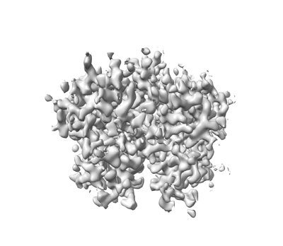 emd_28616.png emd_28616.png | 65.4 KB | ||
| Filedesc metadata | emd-28616.cif.gz | 5.7 KB | ||
| Archive directory |  http://ftp.pdbj.org/pub/emdb/structures/EMD-28616ftp://ftp.pdbj.org/pub/emdb/structures/EMD-28616 http://ftp.pdbj.org/pub/emdb/structures/EMD-28616ftp://ftp.pdbj.org/pub/emdb/structures/EMD-28616 | HTTPS FTP |
-Related structure data
| Related structure data | 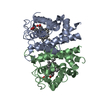 8eunMC 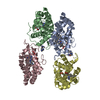 8eumC C: citing same article ( M: atomic model generated by this map |
|---|---|
| Similar structure data |

-Links
| EMDB pages | EMDB (EBI/PDBe) / EMDataResource |
|---|---|
| Related items in Molecule of the Month |


-Map
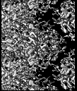
| File | Download / File: emd_28616.map.gz / Format: CCP4 / Size: 6.4 MB / Type: IMAGE STORED AS FLOATING POINT NUMBER (4 BYTES) | ||||||||||||||||||||||||||||||||||||
|---|---|---|---|---|---|---|---|---|---|---|---|---|---|---|---|---|---|---|---|---|---|---|---|---|---|---|---|---|---|---|---|---|---|---|---|---|---|
| Projections & slices | Image control
Images are generated by Spider. generated in cubic-lattice coordinate | ||||||||||||||||||||||||||||||||||||
| Voxel size | X: 0.60539 Å / Y: 0.57201 Å / Z: 0.60526 Å | ||||||||||||||||||||||||||||||||||||
| Density |
| ||||||||||||||||||||||||||||||||||||
| Symmetry | Space group: 1 | ||||||||||||||||||||||||||||||||||||
| Details | EMDB XML:
|
 X (Sec.)
 Y (Row.)
 Z (Col.)


-Supplemental data
- Sample components
-Entire : Homodimer of Aeropyrum pernix protoglobin (ApePgb) C45G, W59L, Y6...
| Entire | Name: Homodimer of Aeropyrum pernix protoglobin (ApePgb) C45G, W59L, Y60V, V63R, C102S, F145Q, I149L mutant |
|---|---|
| Components |
|
-Supramolecule #1: Homodimer of Aeropyrum pernix protoglobin (ApePgb) C45G, W59L, Y6...
| Supramolecule | Name: Homodimer of Aeropyrum pernix protoglobin (ApePgb) C45G, W59L, Y60V, V63R, C102S, F145Q, I149L mutant type: complex / ID: 1 / Parent: 0 / Macromolecule list: #1 |
|---|---|
| Source (natural) | Organism: Aeropyrum pernix (archaea) |
-Macromolecule #1: Protogloblin ApPgb
| Macromolecule | Name: Protogloblin ApPgb / type: protein_or_peptide / ID: 1 / Number of copies: 2 / Enantiomer: LEVO |
|---|---|
| Source (natural) | Organism: Aeropyrum pernix (archaea) |
| Molecular weight | Theoretical: 22.718887 KDa |
| Recombinant expression | Organism:  |
| Sequence | String: MTPSDIPGYD YGRVEKSPIT DLEFDLLKKT VMLGEKDVMY LKKAGDVLKD QVDEILDLLV GWRASNEHLI YYFSNPDTGE PIKEYLERV RARFGAWILD TTSRDYNREW LDYQYEVGLR HHRSKKGVTD GVRTVPHIPL RYLIAQIYPL TATIKPFLAK K GGSPEDIE ...String: MTPSDIPGYD YGRVEKSPIT DLEFDLLKKT VMLGEKDVMY LKKAGDVLKD QVDEILDLLV GWRASNEHLI YYFSNPDTGE PIKEYLERV RARFGAWILD TTSRDYNREW LDYQYEVGLR HHRSKKGVTD GVRTVPHIPL RYLIAQIYPL TATIKPFLAK K GGSPEDIE GMYNAWFKSV VLQVAIWSHP YTKENDW UniProtKB: Protogloblin ApPgb |
-Macromolecule #2: benzyl[3,3'-(7,12-diethenyl-3,8,13,17-tetramethylporphyrin-2,18-d...
| Macromolecule | Name: benzyl[3,3'-(7,12-diethenyl-3,8,13,17-tetramethylporphyrin-2,18-diyl-kappa~4~N~21~,N~22~,N~23~,N~24~)di(propanoato)(2-)]iron type: ligand / ID: 2 / Number of copies: 2 / Formula: WUF |
|---|---|
| Molecular weight | Theoretical: 707.618 Da |
| Chemical component information |  ChemComp-WUF: |
-Macromolecule #3: water
| Macromolecule | Name: water / type: ligand / ID: 3 / Number of copies: 64 / Formula: HOH |
|---|---|
| Molecular weight | Theoretical: 18.015 Da |
| Chemical component information |  ChemComp-HOH: |
-Experimental details
-Structure determination
| Method | cryo EM |
|---|---|
 Processing Processing | electron crystallography |
| Aggregation state | 3D array |
-Sample preparation
| Concentration | 20 mg/mL |
|---|---|
| Buffer | pH: 8 |
| Grid | Model: Quantifoil R2/1 / Material: COPPER / Support film - Material: CARBON / Support film - topology: HOLEY / Pretreatment - Type: GLOW DISCHARGE / Pretreatment - Time: 30 sec. / Pretreatment - Atmosphere: OTHER |
| Vitrification | Cryogen name: ETHANE / Chamber humidity: 90 % / Chamber temperature: 277 K / Instrument: LEICA EM GP |
- Electron microscopy
| Microscope | FEI TITAN KRIOS |
|---|---|
| Temperature | Min: 77.0 K / Max: 90.0 K |
| Image recording | Film or detector model: FEI FALCON IV (4k x 4k) / Detector mode: COUNTING / Digitization - Dimensions - Width: 4096 pixel / Digitization - Dimensions - Height: 4096 pixel / Average electron dose: 0.00025 e/Å2 |
| Electron beam | Acceleration voltage: 300 kV / Electron source:  FIELD EMISSION GUN FIELD EMISSION GUN |
| Electron optics | C2 aperture diameter: 50.0 µm / Illumination mode: OTHER / Imaging mode: DIFFRACTION / Cs: 2.7 mm / Nominal defocus max: 0.0 µm / Nominal defocus min: 0.0 µm / Camera length: 2460 mm |
| Sample stage | Specimen holder model: FEI TITAN KRIOS AUTOGRID HOLDER / Cooling holder cryogen: NITROGEN |
| Experimental equipment |  Model: Titan Krios / Image courtesy: FEI Company |
-Image processing
| Final reconstruction | Resolution method: DIFFRACTION PATTERN/LAYERLINES / Software - Name: PHENIX (ver. 1.20.1-4487-000) |
|---|---|
| Crystallography statistics | Number intensities measured: 40105 / Number structure factors: 40105 / Fourier space coverage: 70 / R merge: 0.32 / Overall phase residual: 0 / High resolution: 2.5 Å / Shell - Shell ID: 1 / Shell - High resolution: 2.5 Å / Shell - Low resolution: 24.37 Å / Shell - Number structure factors: 40105 / Shell - Phase residual: 29.27 / Shell - Fourier space coverage: 7 / Shell - Multiplicity: 4.3 |
-Atomic model buiding 1
| Refinement | Space: RECIPROCAL / Protocol: OTHER / Overall B value: 20.59 / Target criteria: maximum likelihood |
|---|---|
| Output model | PDB-8eun: |
