 Movie
Movie Controller

[English] 日本語
 Yorodumi
- EMDB-27562: Postfusion Nipah virus fusion protein in complex with Fab 1H1, lo... -
+ Open data

- Basic information
| Entry |  | |||||||||
|---|---|---|---|---|---|---|---|---|---|---|
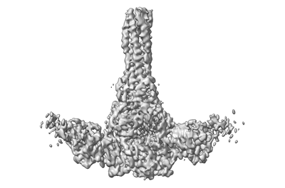
| Title | Postfusion Nipah virus fusion protein in complex with Fab 1H1, low resolution reconstruction including the six helix bundle | |||||||||
 Map data Map data | Postfusion Nipah virus fusion protein in complex with Fab 1H1, low resolution reconstruction including the six helix bundle | |||||||||
 Sample Sample |
| |||||||||
 Keywords Keywords | Nipah / Nipah virus / NiV / fusion / F / antibody / neutralizing / conserved epitope / neutralizing antibody / six helix bundle / VIRAL PROTEIN | |||||||||
| Biological species |  Nipah henipavirus Nipah henipavirus | |||||||||
| Method | single particle reconstruction / cryo EM / Resolution: 3.9 Å | |||||||||
 Authors Authors | Byrne PO / Blade EG / McLellan JS | |||||||||
| Funding support |  United States, 1 items United States, 1 items
| |||||||||
 Citation Citation | Journal: To Be Published Title: Postfusion Nipah virus fusion protein in complex with Fab 1H1 Authors: Byrne PO / Blade EG / McLellan JS | |||||||||
| History |
|
- Structure visualization
| Supplemental images |
|---|
- Downloads & links
-EMDB archive
| Map data | emd_27562.map.gz | 684 MB |  EMDB map data format EMDB map data format | |
|---|---|---|---|---|
| Header (meta data) | emd-27562-v30.xmlemd-27562.xml | 13.7 KB 13.7 KB | Display Display | EMDB header |
| FSC (resolution estimation) | emd_27562_fsc.xml | 20.8 KB | Display | FSC data file |
| Images |  emd_27562.png emd_27562.png | 51.8 KB | ||
| Masks | emd_27562_msk_1.map | 729 MB | Mask map | |
| Others | emd_27562_additional_1.map.gzemd_27562_half_map_1.map.gzemd_27562_half_map_2.map.gz | 362.8 MB 676.2 MB 676.3 MB | ||
| Archive directory |  http://ftp.pdbj.org/pub/emdb/structures/EMD-27562ftp://ftp.pdbj.org/pub/emdb/structures/EMD-27562 http://ftp.pdbj.org/pub/emdb/structures/EMD-27562ftp://ftp.pdbj.org/pub/emdb/structures/EMD-27562 | HTTPS FTP |
-Validation report
| Summary document | emd_27562_validation.pdf.gz | 1.1 MB | Display | EMDB validaton report |
|---|---|---|---|---|
| Full document | emd_27562_full_validation.pdf.gz | 1.1 MB | Display | |
| Data in XML | emd_27562_validation.xml.gz | 28.3 KB | Display | |
| Data in CIF | emd_27562_validation.cif.gz | 37.4 KB | Display | |
| Arichive directory | https://ftp.pdbj.org/pub/emdb/validation_reports/EMD-27562ftp://ftp.pdbj.org/pub/emdb/validation_reports/EMD-27562 | HTTPS FTP |
-Related structure data


-Links
| EMDB pages | EMDB (EBI/PDBe) / EMDataResource |
|---|
-Map
| File | Download / File: emd_27562.map.gz / Format: CCP4 / Size: 729 MB / Type: IMAGE STORED AS FLOATING POINT NUMBER (4 BYTES) | ||||||||||||||||||||
|---|---|---|---|---|---|---|---|---|---|---|---|---|---|---|---|---|---|---|---|---|---|
| Annotation | Postfusion Nipah virus fusion protein in complex with Fab 1H1, low resolution reconstruction including the six helix bundle | ||||||||||||||||||||
| Voxel size | X=Y=Z: 0.81 Å | ||||||||||||||||||||
| Density |
| ||||||||||||||||||||
| Symmetry | Space group: 1 | ||||||||||||||||||||
| Details | EMDB XML:
|
-Supplemental data
-Mask #1
| File | emd_27562_msk_1.map | ||||||||||||
|---|---|---|---|---|---|---|---|---|---|---|---|---|---|
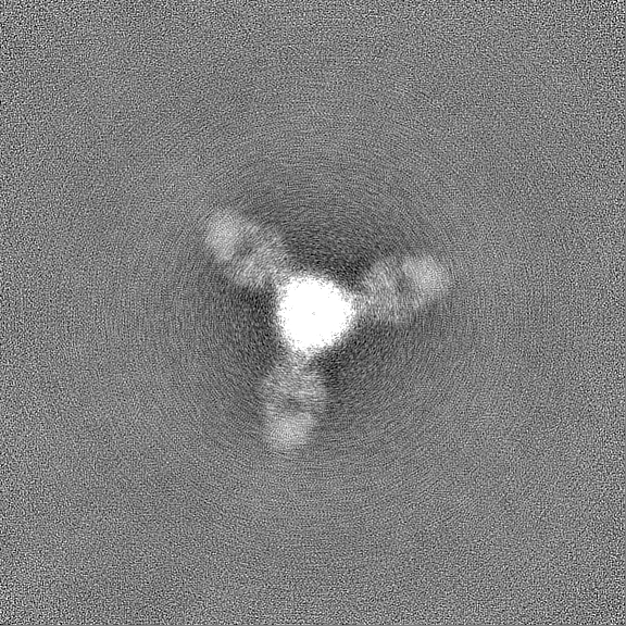
| Projections & Slices |
| ||||||||||||
| Density Histograms |
 Z
Z Y
Y X
X


-Additional map: Additional Map
| File | emd_27562_additional_1.map | ||||||||||||
|---|---|---|---|---|---|---|---|---|---|---|---|---|---|
| Annotation | Additional Map | ||||||||||||
| Projections & Slices |
| ||||||||||||
| Density Histograms |


-Half map: Half Map 1
| File | emd_27562_half_map_1.map | ||||||||||||
|---|---|---|---|---|---|---|---|---|---|---|---|---|---|
| Annotation | Half Map 1 | ||||||||||||
| Projections & Slices |
| ||||||||||||
| Density Histograms |


-Half map: Half Map 2
| File | emd_27562_half_map_2.map | ||||||||||||
|---|---|---|---|---|---|---|---|---|---|---|---|---|---|
| Annotation | Half Map 2 | ||||||||||||
| Projections & Slices |
| ||||||||||||
| Density Histograms |


- Sample components
-Entire : Postfusion Nipah virus fusion protein in complex with Fab 1H1
| Entire | Name: Postfusion Nipah virus fusion protein in complex with Fab 1H1 |
|---|---|
| Components |
|
-Supramolecule #1: Postfusion Nipah virus fusion protein in complex with Fab 1H1
| Supramolecule | Name: Postfusion Nipah virus fusion protein in complex with Fab 1H1 type: complex / ID: 1 / Parent: 0 / Macromolecule list: #1-#3 |
|---|---|
| Source (natural) | Organism: Nipah henipavirus |
-Experimental details
-Structure determination
| Method | cryo EM |
|---|---|
 Processing Processing | single particle reconstruction |
| Aggregation state | particle |
-Sample preparation
| Buffer | pH: 8 |
|---|---|
| Vitrification | Cryogen name: ETHANE |
- Electron microscopy
| Microscope | FEI TITAN KRIOS |
|---|---|
| Image recording | Film or detector model: GATAN K3 (6k x 4k) / Average electron dose: 70.0 e/Å2 |
| Electron beam | Acceleration voltage: 300 kV / Electron source:  FIELD EMISSION GUN FIELD EMISSION GUN |
| Electron optics | Illumination mode: FLOOD BEAM / Imaging mode: BRIGHT FIELD / Nominal defocus max: 2.5 µm / Nominal defocus min: 1.5 µm |
| Experimental equipment |  Model: Titan Krios / Image courtesy: FEI Company |

