National Institutes of Health/National Institute of Neurological Disorders and Stroke (NIH/NINDS)
AG 060149
United States
National Institutes of Health/National Center for Research Resources (NIH/NCRR)
1S10OD018111
United States
Citation
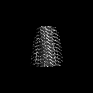
Journal: J Biol Chem / Year: 2023 Title: Cryo-EM structure of amyloid fibril formed by α-synuclein hereditary A53E mutation reveals a distinct protofilament interface. Authors: Chuanqi Sun / Kang Zhou / Peter DePaola / Woo Shik Shin / Trae Hillyer / Michael R Sawaya / Ruowei Zhu / Chao Peng / Z Hong Zhou / Lin Jiang / Abstract: Synucleinopathies like Parkinson's disease (PD), dementia with Lewy bodies (DLB), and multiple systems atrophy (MSA), have the same pathologic feature of misfolded α-synuclein protein (α-syn) ...Synucleinopathies like Parkinson's disease (PD), dementia with Lewy bodies (DLB), and multiple systems atrophy (MSA), have the same pathologic feature of misfolded α-synuclein protein (α-syn) accumulation in the brain. PD patients who carry α-syn hereditary mutations tend to have earlier onset and more severe clinical symptoms than sporadic PD patients. Therefore, revealing the effect of hereditary mutations to the α-syn fibril structure can help us understand these synucleinopathies' structural basis. Here, we present a 3.38 Å cryo-electron microscopy structure of α-synuclein fibrils containing the hereditary A53E mutation. The A53E fibril is symmetrically composed of two protofilaments, similar to other fibril structures of WT and mutant α-synuclein. The new structure is distinct from all other synuclein fibrils, not only at the interface between proto-filaments, but also between residues packed within the same proto-filament. A53E has the smallest interface with the least buried surface area among all α-syn fibrils, consisting of only two contacting residues. Within the same protofilament, A53E reveals distinct residue re-arrangement and structural variation at a cavity near its fibril core. Moreover, the A53E fibrils exhibit slower fibril formation and lower stability compared to WT and other mutants like A53T and H50Q, while also demonstrate strong cellular seeding in α-synuclein biosensor cells and primary neurons. In summary, our study aims to highlight structural differences - both within and between the protofilaments of A53E fibrils - and interpret fibril formation and cellular seeding of α-synuclein pathology in disease, which could further our understanding of the structure-activity relationship of α-synuclein mutants.
In the structure databanks used in Yorodumi, some data are registered as the other names, "COVID-19 virus" and "2019-nCoV". Here are the details of the virus and the list of structure data.
Jan 31, 2019. EMDB accession codes are about to change! (news from PDBe EMDB page)
EMDB accession codes are about to change! (news from PDBe EMDB page)
The allocation of 4 digits for EMDB accession codes will soon come to an end. Whilst these codes will remain in use, new EMDB accession codes will include an additional digit and will expand incrementally as the available range of codes is exhausted. The current 4-digit format prefixed with “EMD-” (i.e. EMD-XXXX) will advance to a 5-digit format (i.e. EMD-XXXXX), and so on. It is currently estimated that the 4-digit codes will be depleted around Spring 2019, at which point the 5-digit format will come into force.
The EM Navigator/Yorodumi systems omit the EMD- prefix.
Related info.:Q: What is EMD? / ID/Accession-code notation in Yorodumi/EM Navigator
Yorodumi is a browser for structure data from EMDB, PDB, SASBDB, etc.
This page is also the successor to EM Navigator detail page, and also detail information page/front-end page for Omokage search.
The word "yorodu" (or yorozu) is an old Japanese word meaning "ten thousand". "mi" (miru) is to see.
Related info.:EMDB / PDB / SASBDB / Comparison of 3 databanks / Yorodumi Search / Aug 31, 2016. New EM Navigator & Yorodumi / Yorodumi Papers / Jmol/JSmol / Function and homology information / Changes in new EM Navigator and Yorodumi
 Movie
Movie Controller

 Yorodumi
 Open data
 Basic information
 Map data
 Sample
 Keywords
 Function and homology information
 Homo sapiens (human)
 Authors
 United States, 2 items
  Citation
 Structure visualization
 Downloads & links
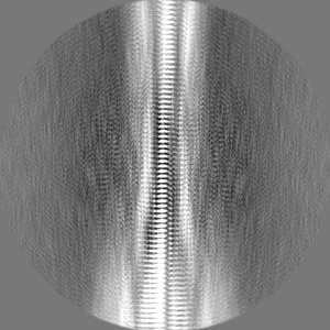
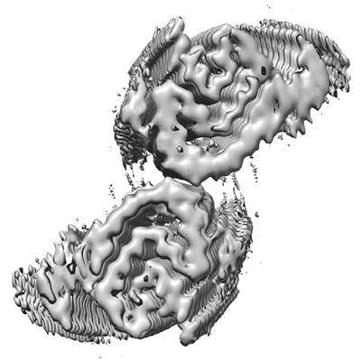 emd_26427.png
 http://ftp.pdbj.org/pub/emdb/structures/EMD-26427
 Z (Sec.)
 Y (Row.)
 X (Col.)


 Sample components
 Processing
 Electron microscopy
 FIELD EMISSION GUN