 Movie
Movie Controller
 Structure viewers
 About Yorodumi Papers


+Search query
-Structure paper
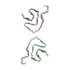
| Title | Cryo-EM structure of amyloid fibril formed by α-synuclein hereditary A53E mutation reveals a distinct protofilament interface. |
|---|---|
| Journal, issue, pages | J Biol Chem, Vol. 299, Issue 4, Page 104566, Year 2023 |
| Publish date | Mar 5, 2023 |
 Authors Authors | Chuanqi Sun / Kang Zhou / Peter DePaola / Woo Shik Shin / Trae Hillyer / Michael R Sawaya / Ruowei Zhu / Chao Peng / Z Hong Zhou / Lin Jiang /  |
| PubMed Abstract | Synucleinopathies like Parkinson's disease (PD), dementia with Lewy bodies (DLB), and multiple systems atrophy (MSA), have the same pathologic feature of misfolded α-synuclein protein (α-syn) ...Synucleinopathies like Parkinson's disease (PD), dementia with Lewy bodies (DLB), and multiple systems atrophy (MSA), have the same pathologic feature of misfolded α-synuclein protein (α-syn) accumulation in the brain. PD patients who carry α-syn hereditary mutations tend to have earlier onset and more severe clinical symptoms than sporadic PD patients. Therefore, revealing the effect of hereditary mutations to the α-syn fibril structure can help us understand these synucleinopathies' structural basis. Here, we present a 3.38 Å cryo-electron microscopy structure of α-synuclein fibrils containing the hereditary A53E mutation. The A53E fibril is symmetrically composed of two protofilaments, similar to other fibril structures of WT and mutant α-synuclein. The new structure is distinct from all other synuclein fibrils, not only at the interface between proto-filaments, but also between residues packed within the same proto-filament. A53E has the smallest interface with the least buried surface area among all α-syn fibrils, consisting of only two contacting residues. Within the same protofilament, A53E reveals distinct residue re-arrangement and structural variation at a cavity near its fibril core. Moreover, the A53E fibrils exhibit slower fibril formation and lower stability compared to WT and other mutants like A53T and H50Q, while also demonstrate strong cellular seeding in α-synuclein biosensor cells and primary neurons. In summary, our study aims to highlight structural differences - both within and between the protofilaments of A53E fibrils - and interpret fibril formation and cellular seeding of α-synuclein pathology in disease, which could further our understanding of the structure-activity relationship of α-synuclein mutants. |
 External links External links | J Biol Chem / PubMed:36871760 / PubMed Central |
| Methods | EM (helical sym.) |
| Resolution | 3.38 Å |
| Structure data | EMDB-26427, PDB-7uak: |
| Source |
|
 Keywords Keywords | PROTEIN FIBRIL / alpha-synuclein fibrils / A53E mutant / cryo-EM structure / structural protein |

 homo sapiens (human)