+ Open data
- Basic information
| Entry |  | ||||||||||||
|---|---|---|---|---|---|---|---|---|---|---|---|---|---|
| Title | The beta-tubulin folding intermediate III | ||||||||||||
 Map data Map data | sharpened map of tubulin intermediate in closed TRiC | ||||||||||||
 Sample Sample |
| ||||||||||||
 Keywords Keywords | Human chaperonin TRiC with beta-tubulin folding intermediate III / CHAPERONE | ||||||||||||
| Function / homology |  Function and homology information Function and homology informationodontoblast differentiation / zona pellucida receptor complex / scaRNA localization to Cajal body / chaperone mediated protein folding independent of cofactor / positive regulation of establishment of protein localization to telomere / positive regulation of protein localization to Cajal body / tubulin complex assembly / BBSome-mediated cargo-targeting to cilium / chaperonin-containing T-complex / cytoskeleton-dependent intracellular transport ...odontoblast differentiation / zona pellucida receptor complex / scaRNA localization to Cajal body / chaperone mediated protein folding independent of cofactor / positive regulation of establishment of protein localization to telomere / positive regulation of protein localization to Cajal body / tubulin complex assembly / BBSome-mediated cargo-targeting to cilium / chaperonin-containing T-complex / cytoskeleton-dependent intracellular transport / binding of sperm to zona pellucida / positive regulation of telomerase RNA localization to Cajal body / Folding of actin by CCT/TriC / Formation of tubulin folding intermediates by CCT/TriC / Prefoldin mediated transfer of substrate to CCT/TriC / GTPase activating protein binding / RHOBTB1 GTPase cycle / intermediate filament cytoskeleton / WD40-repeat domain binding / natural killer cell mediated cytotoxicity / intercellular bridge / regulation of synapse organization / pericentriolar material / nuclear envelope lumen / beta-tubulin binding / MHC class I protein binding / : / Association of TriC/CCT with target proteins during biosynthesis / microtubule-based process / chaperone-mediated protein complex assembly / spindle assembly / RHOBTB2 GTPase cycle / heterochromatin / chaperone-mediated protein folding / Loss of Nlp from mitotic centrosomes / Loss of proteins required for interphase microtubule organization from the centrosome / Recruitment of mitotic centrosome proteins and complexes / Recruitment of NuMA to mitotic centrosomes / protein folding chaperone / Anchoring of the basal body to the plasma membrane / positive regulation of telomere maintenance via telomerase / Gene and protein expression by JAK-STAT signaling after Interleukin-12 stimulation / AURKA Activation by TPX2 / acrosomal vesicle / cell projection / mRNA 3'-UTR binding / ATP-dependent protein folding chaperone / response to virus / cilium / structural constituent of cytoskeleton / mitotic spindle / mRNA 5'-UTR binding / microtubule cytoskeleton organization / cytoplasmic ribonucleoprotein granule / Cooperation of PDCL (PhLP1) and TRiC/CCT in G-protein beta folding / azurophil granule lumen / microtubule cytoskeleton / Regulation of PLK1 Activity at G2/M Transition / G-protein beta-subunit binding / unfolded protein binding / melanosome / protein folding / mitotic cell cycle / cell body / secretory granule lumen / ficolin-1-rich granule lumen / microtubule / Potential therapeutics for SARS / cytoskeleton / protein stabilization / cadherin binding / membrane raft / protein domain specific binding / cell division / GTPase activity / centrosome / ubiquitin protein ligase binding / Neutrophil degranulation / protein-containing complex binding / GTP binding / structural molecule activity / Golgi apparatus / ATP hydrolysis activity / protein-containing complex / RNA binding / extracellular exosome / extracellular region / nucleoplasm / ATP binding / nucleus / metal ion binding / cytosol / cytoplasm Similarity search - Function | ||||||||||||
| Biological species |  Homo sapiens (human) Homo sapiens (human) | ||||||||||||
| Method | single particle reconstruction / cryo EM / Resolution: 2.9 Å | ||||||||||||
 Authors Authors | Zhao Y / Frydman J / Chiu W | ||||||||||||
| Funding support |  United States, 3 items United States, 3 items
| ||||||||||||
 Citation Citation |  Journal: Cell / Year: 2022 Journal: Cell / Year: 2022Title: Structural visualization of the tubulin folding pathway directed by human chaperonin TRiC/CCT. Authors: Daniel Gestaut / Yanyan Zhao / Junsun Park / Boxue Ma / Alexander Leitner / Miranda Collier / Grigore Pintilie / Soung-Hun Roh / Wah Chiu / Judith Frydman /    Abstract: The ATP-dependent ring-shaped chaperonin TRiC/CCT is essential for cellular proteostasis. To uncover why some eukaryotic proteins can only fold with TRiC assistance, we reconstituted the folding of ...The ATP-dependent ring-shaped chaperonin TRiC/CCT is essential for cellular proteostasis. To uncover why some eukaryotic proteins can only fold with TRiC assistance, we reconstituted the folding of β-tubulin using human prefoldin and TRiC. We find unstructured β-tubulin is delivered by prefoldin to the open TRiC chamber followed by ATP-dependent chamber closure. Cryo-EM resolves four near-atomic-resolution structures containing progressively folded β-tubulin intermediates within the closed TRiC chamber, culminating in native tubulin. This substrate folding pathway appears closely guided by site-specific interactions with conserved regions in the TRiC chamber. Initial electrostatic interactions between the TRiC interior wall and both the folded tubulin N domain and its C-terminal E-hook tail establish the native substrate topology, thus enabling C-domain folding. Intrinsically disordered CCT C termini within the chamber promote subsequent folding of tubulin's core and middle domains and GTP-binding. Thus, TRiC's chamber provides chemical and topological directives that shape the folding landscape of its obligate substrates. | ||||||||||||
| History |
|
- Structure visualization
| Supplemental images |
|---|
- Downloads & links
-EMDB archive
| Map data |  emd_26123.map.gz emd_26123.map.gz | 112 MB |  EMDB map data format EMDB map data format | |
|---|---|---|---|---|
| Header (meta data) |  emd-26123-v30.xml emd-26123-v30.xml emd-26123.xml emd-26123.xml | 28.5 KB 28.5 KB | Display Display |  EMDB header EMDB header |
| Images |  emd_26123.png emd_26123.png | 111.6 KB | ||
| Filedesc metadata |  emd-26123.cif.gz emd-26123.cif.gz | 9.4 KB | ||
| Others |  emd_26123_additional_1.map.gz emd_26123_additional_1.map.gz | 98.5 MB | ||
| Archive directory |  http://ftp.pdbj.org/pub/emdb/structures/EMD-26123 http://ftp.pdbj.org/pub/emdb/structures/EMD-26123 ftp://ftp.pdbj.org/pub/emdb/structures/EMD-26123 ftp://ftp.pdbj.org/pub/emdb/structures/EMD-26123 | HTTPS FTP |
-Validation report
| Summary document |  emd_26123_validation.pdf.gz emd_26123_validation.pdf.gz | 652.6 KB | Display |  EMDB validaton report EMDB validaton report |
|---|---|---|---|---|
| Full document |  emd_26123_full_validation.pdf.gz emd_26123_full_validation.pdf.gz | 652.1 KB | Display | |
| Data in XML |  emd_26123_validation.xml.gz emd_26123_validation.xml.gz | 6.8 KB | Display | |
| Data in CIF |  emd_26123_validation.cif.gz emd_26123_validation.cif.gz | 7.7 KB | Display | |
| Arichive directory |  https://ftp.pdbj.org/pub/emdb/validation_reports/EMD-26123 https://ftp.pdbj.org/pub/emdb/validation_reports/EMD-26123 ftp://ftp.pdbj.org/pub/emdb/validation_reports/EMD-26123 ftp://ftp.pdbj.org/pub/emdb/validation_reports/EMD-26123 | HTTPS FTP |
-Related structure data
| Related structure data | 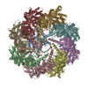 7tttMC 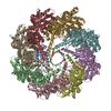 7trgC  7ttnC 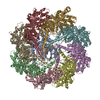 7tubC 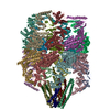 7wu7C M: atomic model generated by this map C: citing same article ( |
|---|---|
| Similar structure data | Similarity search - Function & homology  F&H Search F&H Search |
- Links
Links
| EMDB pages |  EMDB (EBI/PDBe) / EMDB (EBI/PDBe) /  EMDataResource EMDataResource |
|---|---|
| Related items in Molecule of the Month |
- Map
Map
| File |  Download / File: emd_26123.map.gz / Format: CCP4 / Size: 125 MB / Type: IMAGE STORED AS FLOATING POINT NUMBER (4 BYTES) Download / File: emd_26123.map.gz / Format: CCP4 / Size: 125 MB / Type: IMAGE STORED AS FLOATING POINT NUMBER (4 BYTES) | ||||||||||||||||||||||||||||||||||||
|---|---|---|---|---|---|---|---|---|---|---|---|---|---|---|---|---|---|---|---|---|---|---|---|---|---|---|---|---|---|---|---|---|---|---|---|---|---|
| Annotation | sharpened map of tubulin intermediate in closed TRiC | ||||||||||||||||||||||||||||||||||||
| Projections & slices | Image control
Images are generated by Spider. | ||||||||||||||||||||||||||||||||||||
| Voxel size | X=Y=Z: 1.1 Å | ||||||||||||||||||||||||||||||||||||
| Density |
| ||||||||||||||||||||||||||||||||||||
| Symmetry | Space group: 1 | ||||||||||||||||||||||||||||||||||||
| Details | EMDB XML:
|
-Supplemental data
-Additional map: unsharpened map of tubulin intermediate in closed TRiC
| File | emd_26123_additional_1.map | ||||||||||||
|---|---|---|---|---|---|---|---|---|---|---|---|---|---|
| Annotation | unsharpened map of tubulin intermediate in closed TRiC | ||||||||||||
| Projections & Slices |
| ||||||||||||
| Density Histograms |
- Sample components
+Entire : Closed form human TRiC in complex with beta-tubulin under ATP/AlF...
+Supramolecule #1: Closed form human TRiC in complex with beta-tubulin under ATP/AlF...
+Macromolecule #1: Tubulin beta chain
+Macromolecule #2: T-complex protein 1 subunit theta
+Macromolecule #3: T-complex protein 1 subunit eta
+Macromolecule #4: T-complex protein 1 subunit epsilon
+Macromolecule #5: T-complex protein 1 subunit beta
+Macromolecule #6: T-complex protein 1 subunit delta
+Macromolecule #7: T-complex protein 1 subunit alpha
+Macromolecule #8: T-complex protein 1 subunit gamma
+Macromolecule #9: T-complex protein 1 subunit zeta
+Macromolecule #10: MAGNESIUM ION
+Macromolecule #11: ADENOSINE-5'-DIPHOSPHATE
+Macromolecule #12: ALUMINUM FLUORIDE
+Macromolecule #13: water
-Experimental details
-Structure determination
| Method | cryo EM |
|---|---|
 Processing Processing | single particle reconstruction |
| Aggregation state | particle |
- Sample preparation
| Concentration | 1 mg/mL | ||||||||||||||||||||||||
|---|---|---|---|---|---|---|---|---|---|---|---|---|---|---|---|---|---|---|---|---|---|---|---|---|---|
| Buffer | pH: 7.4 Component:
| ||||||||||||||||||||||||
| Grid | Model: Quantifoil R1.2/1.3 / Support film - Material: CARBON / Support film - topology: HOLEY | ||||||||||||||||||||||||
| Vitrification | Cryogen name: ETHANE |
- Electron microscopy
| Microscope | FEI TITAN KRIOS |
|---|---|
| Image recording | Film or detector model: GATAN K2 SUMMIT (4k x 4k) / Detector mode: COUNTING / Average electron dose: 1.21 e/Å2 |
| Electron beam | Acceleration voltage: 300 kV / Electron source:  FIELD EMISSION GUN FIELD EMISSION GUN |
| Electron optics | Illumination mode: FLOOD BEAM / Imaging mode: BRIGHT FIELD / Nominal defocus max: 3.0 µm / Nominal defocus min: 0.5 µm |
| Experimental equipment |  Model: Titan Krios / Image courtesy: FEI Company |
- Image processing
| Startup model | Type of model: NONE |
|---|---|
| Final reconstruction | Resolution.type: BY AUTHOR / Resolution: 2.9 Å / Resolution method: FSC 0.143 CUT-OFF / Number images used: 94955 |
| Initial angle assignment | Type: MAXIMUM LIKELIHOOD |
| Final angle assignment | Type: MAXIMUM LIKELIHOOD |
-Atomic model buiding 1
| Refinement | Space: REAL / Protocol: RIGID BODY FIT |
|---|---|
| Output model |  PDB-7ttt: |
 Movie
Movie Controller


 X (Sec.)
 Y (Row.)
 Z (Col.)


 Trichoplusia ni (cabbage looper)
