National Institutes of Health/National Institute of General Medical Sciences (NIH/NIGMS)
R01GM127440, R01GM092917, S10OD012272
United States
Citation
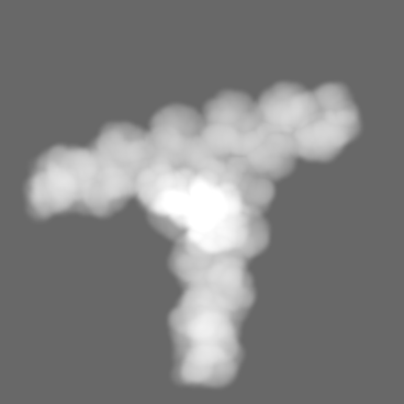
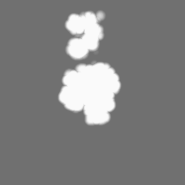
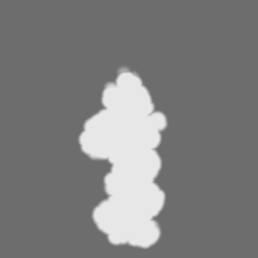
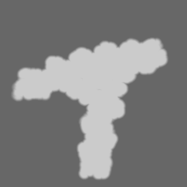
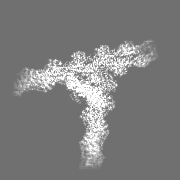
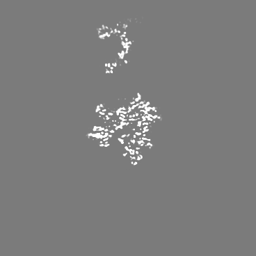
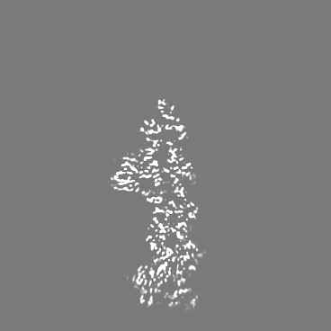
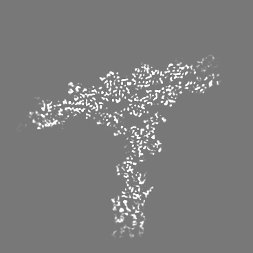
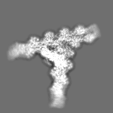
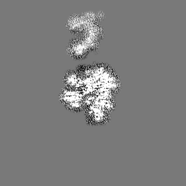
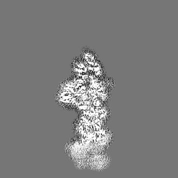
Journal: Proc Natl Acad Sci U S A / Year: 2022 Title: Structure of Arp2/3 complex at a branched actin filament junction resolved by single-particle cryo-electron microscopy. Authors: Bojian Ding / Heidy Y Narvaez-Ortiz / Yuvraj Singh / Glen M Hocky / Saikat Chowdhury / Brad J Nolen / Abstract: Arp2/3 complex nucleates branched actin filaments that provide pushing forces to drive cellular processes such as lamellipodial protrusion and endocytosis. Arp2/3 complex is intrinsically inactive, ...Arp2/3 complex nucleates branched actin filaments that provide pushing forces to drive cellular processes such as lamellipodial protrusion and endocytosis. Arp2/3 complex is intrinsically inactive, and multiple classes of nucleation promoting factors (NPFs) stimulate its nucleation activity. When activated by WASP family NPFs, the complex must bind to the side of a preexisting (mother) filament of actin to complete the nucleation process, ensuring that WASP-mediated activation creates branched rather than linear actin filaments. How actin filaments contribute to activation is currently not understood, largely due to the lack of high-resolution structures of activated Arp2/3 complex bound to the side of a filament. Here, we present the 3.9-Å cryo-electron microscopy structure of the Arp2/3 complex at a branch junction. The structure reveals contacts between Arp2/3 complex and the side of the mother actin filament that likely stimulate subunit flattening, a conformational change that allows the actin-related protein subunits in the complex (Arp2 and Arp3) to mimic filamentous actin subunits. In contrast, limited contact between the bottom half of the complex and the mother filament suggests that clamp twisting, a second major conformational change observed in the active state, is not stimulated by actin filaments, potentially explaining why actin filaments are required but insufficient to trigger nucleation during WASP-mediated activation. Along with biochemical and live-cell imaging data and molecular dynamics simulations, the structure reveals features critical for the interaction of Arp2/3 complex with actin filaments and regulated assembly of branched actin filament networks in cells.
Name: Actin-related protein 3 / type: protein_or_peptide / ID: 1 Details: Missing sequences correspond to unmodeled regions due to poor density map Number of copies: 1 / Enantiomer: LEVO
Name: Actin-related protein 2 / type: protein_or_peptide / ID: 2 Details: Missing sequences correspond to unmodelled regions due to poor density map Number of copies: 1 / Enantiomer: LEVO
Macromolecule #3: Actin-related protein 2/3 complex subunit 1B
Macromolecule
Name: Actin-related protein 2/3 complex subunit 1B / type: protein_or_peptide / ID: 3 Details: Missing sequences correspond to unmodeled regions due to poor density map Number of copies: 1 / Enantiomer: LEVO
Macromolecule #4: Actin-related protein 2/3 complex subunit 2
Macromolecule
Name: Actin-related protein 2/3 complex subunit 2 / type: protein_or_peptide / ID: 4 Details: Missing sequences correspond to unmodeled regions due to poor density map Number of copies: 1 / Enantiomer: LEVO
Macromolecule #5: Actin-related protein 2/3 complex subunit 3
Macromolecule
Name: Actin-related protein 2/3 complex subunit 3 / type: protein_or_peptide / ID: 5 Details: Missing sequences correspond to unmodeled regions due to poor density map Number of copies: 1 / Enantiomer: LEVO
Macromolecule #7: Actin-related protein 2/3 complex subunit 5
Macromolecule
Name: Actin-related protein 2/3 complex subunit 5 / type: protein_or_peptide / ID: 7 Details: Missing sequences correspond to unmodeled regions due to poor density map Number of copies: 1 / Enantiomer: LEVO
Name: Actin, alpha skeletal muscle / type: protein_or_peptide / ID: 8 Details: Missing sequences correspond to unmodeled regions due to poor density map. Actin subunits corresponding to chains J,K,L,M,T and U have been trimmed to c-beta due to lack of density for ...Details: Missing sequences correspond to unmodeled regions due to poor density map. Actin subunits corresponding to chains J,K,L,M,T and U have been trimmed to c-beta due to lack of density for modeling sidechains. These subunits were rigid body fitted into the map. Number of copies: 14 / Enantiomer: LEVO
Name: Phalloidin / type: protein_or_peptide / ID: 9 Details: Phalloidin from Amanita phalloides is a rigid bicyclic heptapeptide. This is a small molecule and does not have conventional planar peptide bonds present in proteins. Number of copies: 15 / Enantiomer: LEVO
Source (natural)
Organism: Amanita phalloides (death cap)
Molecular weight
Theoretical: 808.899 Da
Sequence
String:
W(EEP)A(DTH)C(HYP)A
+
Macromolecule #10: MAGNESIUM ION
Macromolecule
Name: MAGNESIUM ION / type: ligand / ID: 10 / Number of copies: 16 / Formula: MG
Molecular weight
Theoretical: 24.305 Da
+
Macromolecule #11: ADENOSINE-5'-DIPHOSPHATE
Macromolecule
Name: ADENOSINE-5'-DIPHOSPHATE / type: ligand / ID: 11 / Number of copies: 16 / Formula: ADP
Data were collected by shifting of stage to targeted exposure position. Stage was tilted to different angles: including 40 degree, 36 degree, 30 degree, 25 degree, 15 degree, 0 degree, -20 degree, and -33 degree during data collection.
Image recording
Film or detector model: FEI FALCON III (4k x 4k) / Detector mode: COUNTING / Digitization - Dimensions - Width: 4096 pixel / Digitization - Dimensions - Height: 4096 pixel / Digitization - Sampling interval: 14.0 µm / Number grids imaged: 3 / Number real images: 7436 / Average exposure time: 70.0 sec. / Average electron dose: 41.97 e/Å2 Details: Each micrograph was acquired as dose-fractionated movies consisting of 82 frames per movie.
Electron beam
Acceleration voltage: 200 kV / Electron source: FIELD EMISSION GUN
Model: Talos Arctica / Image courtesy: FEI Company
+
Image processing
Particle selection
Number selected: 2290562
CTF correction
Software - Name: cryoSPARC (ver. version 2) / Software - details: Patch CTF Details: Particles were CTF-corrected during projection matching and back projection
Startup model
Type of model: INSILICO MODEL In silico model: Ab-initio initial model was determined using CryoSPARC v2
Final reconstruction
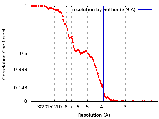
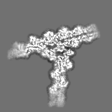
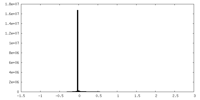
Applied symmetry - Point group: C1 (asymmetric) / Algorithm: BACK PROJECTION / Resolution.type: BY AUTHOR / Resolution: 3.9 Å / Resolution method: FSC 0.143 CUT-OFF / Software - Name: cryoSPARC (ver. version 2) / Number images used: 127093
Initial angle assignment
Type: NOT APPLICABLE
Final angle assignment
Type: MAXIMUM LIKELIHOOD / Software - Name: cryoSPARC (ver. Version 2)
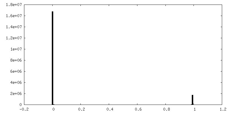
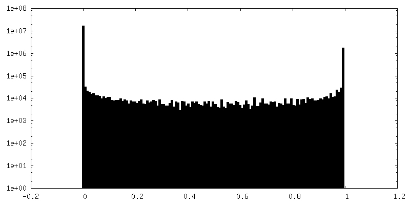
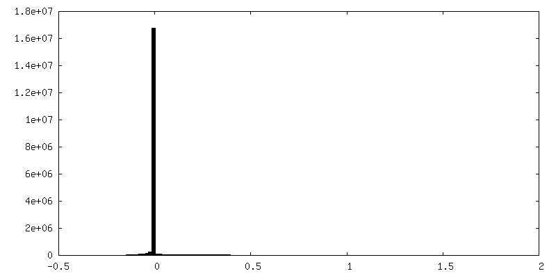
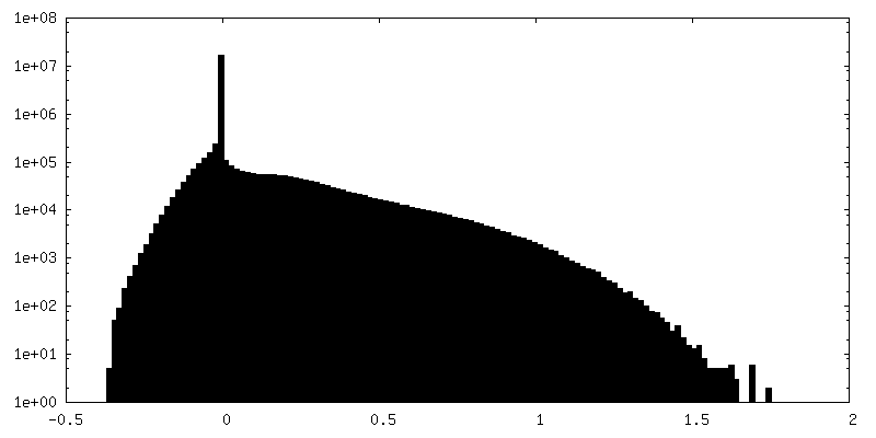
FSC plot (resolution estimation)
-
Atomic model buiding 1
Refinement
Space: REAL / Protocol: FLEXIBLE FIT
Output model
PDB-7tpt: Single-particle Cryo-EM structure of Arp2/3 complex at branched-actin junction.
+
About Yorodumi
-
News
-
Feb 9, 2022. New format data for meta-information of EMDB entries
New format data for meta-information of EMDB entries
Version 3 of the EMDB header file is now the official format.
The previous official version 1.9 will be removed from the archive.
In the structure databanks used in Yorodumi, some data are registered as the other names, "COVID-19 virus" and "2019-nCoV". Here are the details of the virus and the list of structure data.
Jan 31, 2019. EMDB accession codes are about to change! (news from PDBe EMDB page)
EMDB accession codes are about to change! (news from PDBe EMDB page)
The allocation of 4 digits for EMDB accession codes will soon come to an end. Whilst these codes will remain in use, new EMDB accession codes will include an additional digit and will expand incrementally as the available range of codes is exhausted. The current 4-digit format prefixed with “EMD-” (i.e. EMD-XXXX) will advance to a 5-digit format (i.e. EMD-XXXXX), and so on. It is currently estimated that the 4-digit codes will be depleted around Spring 2019, at which point the 5-digit format will come into force.
The EM Navigator/Yorodumi systems omit the EMD- prefix.
Related info.:Q: What is EMD? / ID/Accession-code notation in Yorodumi/EM Navigator
Yorodumi is a browser for structure data from EMDB, PDB, SASBDB, etc.
This page is also the successor to EM Navigator detail page, and also detail information page/front-end page for Omokage search.
The word "yorodu" (or yorozu) is an old Japanese word meaning "ten thousand". "mi" (miru) is to see.
Related info.:EMDB / PDB / SASBDB / Comparison of 3 databanks / Yorodumi Search / Aug 31, 2016. New EM Navigator & Yorodumi / Yorodumi Papers / Jmol/JSmol / Function and homology information / Changes in new EM Navigator and Yorodumi
 Movie
Movie Controller

 Yorodumi
 Open data
 Basic information
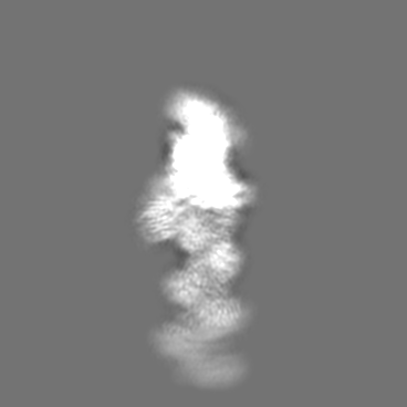
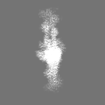
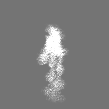
 Map data
 Sample
 Function and homology information
 Amanita phalloides (death cap)
 Authors
 United States, 1 items
  Citation
 Structure visualization
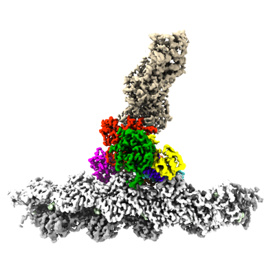
 Downloads & links
 emd_26063.png
 http://ftp.pdbj.org/pub/emdb/structures/EMD-26063


 Z
Z Y
Y X
X


 Sample components
 Processing
 Electron microscopy
 FIELD EMISSION GUN