 Movie
Movie Controller

[English] 日本語
 Yorodumi
- PDB-7tpt: Single-particle Cryo-EM structure of Arp2/3 complex at branched-a... -
+ Open data

- Basic information
| Entry | Database: PDB / ID: 7tpt | ||||||
|---|---|---|---|---|---|---|---|
| Title | Single-particle Cryo-EM structure of Arp2/3 complex at branched-actin junction. | ||||||
 Components Components |
| ||||||
 Keywords Keywords | STRUCTURAL PROTEIN / Arp2/3 / actin / cytoskeletal protein / actin regulator | ||||||
| Function / homology |  Function and homology information Function and homology informationmuscle cell projection membrane / EPHB-mediated forward signaling / Regulation of actin dynamics for phagocytic cup formation / RHO GTPases Activate WASPs and WAVEs / Arp2/3 protein complex / Arp2/3 complex-mediated actin nucleation / regulation of actin filament polymerization / Clathrin-mediated endocytosis / Neutrophil degranulation / cytoskeletal motor activator activity ...muscle cell projection membrane / EPHB-mediated forward signaling / Regulation of actin dynamics for phagocytic cup formation / RHO GTPases Activate WASPs and WAVEs / Arp2/3 protein complex / Arp2/3 complex-mediated actin nucleation / regulation of actin filament polymerization / Clathrin-mediated endocytosis / Neutrophil degranulation / cytoskeletal motor activator activity / myosin heavy chain binding / tropomyosin binding / actin filament bundle / troponin I binding / filamentous actin / mesenchyme migration / positive regulation of actin filament polymerization / cortical cytoskeleton / skeletal muscle myofibril / actin filament bundle assembly / striated muscle thin filament / skeletal muscle thin filament assembly / cilium assembly / actin monomer binding / positive regulation of double-strand break repair via homologous recombination / positive regulation of lamellipodium assembly / skeletal muscle fiber development / cell projection / stress fiber / titin binding / actin filament polymerization / positive regulation of substrate adhesion-dependent cell spreading / filopodium / actin filament / structural constituent of cytoskeleton / Hydrolases; Acting on acid anhydrides; Acting on acid anhydrides to facilitate cellular and subcellular movement / calcium-dependent protein binding / actin filament binding / cell migration / lamellipodium / synaptic vesicle membrane / site of double-strand break / actin binding / cell body / cell cortex / protein-macromolecule adaptor activity / endosome / neuron projection / postsynapse / protein domain specific binding / focal adhesion / hydrolase activity / calcium ion binding / positive regulation of gene expression / glutamatergic synapse / magnesium ion binding / positive regulation of transcription by RNA polymerase II / nucleoplasm / ATP binding / identical protein binding / nucleus / cytoplasm / cytosol Similarity search - Function | ||||||
| Biological species |   Amanita phalloides (death cap) Amanita phalloides (death cap) | ||||||
| Method | ELECTRON MICROSCOPY / single particle reconstruction / cryo EM / Resolution: 3.9 Å | ||||||
 Authors Authors | Ding, B. / Narvaez-Ortiz, H.Y. / Nolen, B.J. / Chowdhury, S. | ||||||
| Funding support |  United States, 1items United States, 1items
| ||||||
 Citation Citation | Journal: Proc Natl Acad Sci U S A / Year: 2022 Title: Structure of Arp2/3 complex at a branched actin filament junction resolved by single-particle cryo-electron microscopy. Authors: Bojian Ding / Heidy Y Narvaez-Ortiz / Yuvraj Singh / Glen M Hocky / Saikat Chowdhury / Brad J Nolen /  Abstract: Arp2/3 complex nucleates branched actin filaments that provide pushing forces to drive cellular processes such as lamellipodial protrusion and endocytosis. Arp2/3 complex is intrinsically inactive, ...Arp2/3 complex nucleates branched actin filaments that provide pushing forces to drive cellular processes such as lamellipodial protrusion and endocytosis. Arp2/3 complex is intrinsically inactive, and multiple classes of nucleation promoting factors (NPFs) stimulate its nucleation activity. When activated by WASP family NPFs, the complex must bind to the side of a preexisting (mother) filament of actin to complete the nucleation process, ensuring that WASP-mediated activation creates branched rather than linear actin filaments. How actin filaments contribute to activation is currently not understood, largely due to the lack of high-resolution structures of activated Arp2/3 complex bound to the side of a filament. Here, we present the 3.9-Å cryo-electron microscopy structure of the Arp2/3 complex at a branch junction. The structure reveals contacts between Arp2/3 complex and the side of the mother actin filament that likely stimulate subunit flattening, a conformational change that allows the actin-related protein subunits in the complex (Arp2 and Arp3) to mimic filamentous actin subunits. In contrast, limited contact between the bottom half of the complex and the mother filament suggests that clamp twisting, a second major conformational change observed in the active state, is not stimulated by actin filaments, potentially explaining why actin filaments are required but insufficient to trigger nucleation during WASP-mediated activation. Along with biochemical and live-cell imaging data and molecular dynamics simulations, the structure reveals features critical for the interaction of Arp2/3 complex with actin filaments and regulated assembly of branched actin filament networks in cells. | ||||||
| History |
|
- Structure visualization
| Structure viewer | Molecule: MolmilJmol/JSmol |
|---|
- Downloads & links
-Download
| PDBx/mmCIF format | 7tpt.cif.gz | 1.1 MB | Display | PDBx/mmCIF format |
|---|---|---|---|---|
| PDB format | pdb7tpt.ent.gz | 946.2 KB | Display | PDB format |
| PDBx/mmJSON format | 7tpt.json.gz | Tree view | PDBx/mmJSON format | |
| Others |  Other downloads Other downloads |
-Validation report
| Arichive directory | https://data.pdbj.org/pub/pdb/validation_reports/tp/7tptftp://data.pdbj.org/pub/pdb/validation_reports/tp/7tpt | HTTPS FTP |
|---|
-Related structure data
| Related structure data | 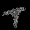 26063MC M: map data used to model this data C: citing same article ( |
|---|---|
| Similar structure data |
-Links
 PDBj
PDBj


- Assembly
| Deposited unit | 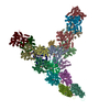
|
|---|---|
| 1 |
|
-Components
-Actin-related protein ... , 7 types, 7 molecules ABCDEFG
| #1: Protein | Mass: 47428.031 Da / Num. of mol.: 1 / Source method: isolated from a natural source Details: Missing sequences correspond to unmodeled regions due to poor density map Source: (natural) |
|---|---|
| #2: Protein | Mass: 44818.711 Da / Num. of mol.: 1 / Source method: isolated from a natural source Details: Missing sequences correspond to unmodelled regions due to poor density map Source: (natural) |
| #3: Protein | Mass: 41030.766 Da / Num. of mol.: 1 / Source method: isolated from a natural source Details: Missing sequences correspond to unmodeled regions due to poor density map Source: (natural) |
| #4: Protein | Mass: 34402.043 Da / Num. of mol.: 1 / Source method: isolated from a natural source Details: Missing sequences correspond to unmodeled regions due to poor density map Source: (natural) |
| #5: Protein | Mass: 20572.666 Da / Num. of mol.: 1 / Source method: isolated from a natural source Details: Missing sequences correspond to unmodeled regions due to poor density map Source: (natural) |
| #6: Protein | Mass: 19697.047 Da / Num. of mol.: 1 / Source method: isolated from a natural source / Source: (natural) |
| #7: Protein | Mass: 16251.308 Da / Num. of mol.: 1 / Source method: isolated from a natural source Details: Missing sequences correspond to unmodeled regions due to poor density map Source: (natural) |
-Protein / Protein/peptide , 2 types, 29 molecules HIJKLMNOPQRSTUabcdefghijklmno
| #8: Protein | Mass: 42109.973 Da / Num. of mol.: 14 / Source method: isolated from a natural source Details: Missing sequences correspond to unmodeled regions due to poor density map. Actin subunits corresponding to chains J,K,L,M,T and U have been trimmed to c-beta due to lack of density for ...Details: Missing sequences correspond to unmodeled regions due to poor density map. Actin subunits corresponding to chains J,K,L,M,T and U have been trimmed to c-beta due to lack of density for modeling sidechains. These subunits were rigid body fitted into the map. Source: (natural) #9: Protein/peptide | Type: Peptide-like / Class: Toxin / Mass: 808.899 Da / Num. of mol.: 15 / Source method: isolated from a natural source Details: Phalloidin from Amanita phalloides is a rigid bicyclic heptapeptide. This is a small molecule and does not have conventional planar peptide bonds present in proteins. Source: (natural) Amanita phalloides (death cap) / References: BIRD: PRD_002366 |
|---|
-Non-polymers , 2 types, 32 molecules 

| #10: Chemical | ChemComp-MG /  Mass: 24.305 Da / Num. of mol.: 16 / Source method: obtained synthetically / Formula: Mg Mass: 24.305 Da / Num. of mol.: 16 / Source method: obtained synthetically / Formula: Mg#11: Chemical | ChemComp-ADP /  Mass: 427.201 Da / Num. of mol.: 16 / Source method: obtained synthetically / Formula: C10H15N5O10P2 / Comment: ADP, energy-carrying molecule*YM Mass: 427.201 Da / Num. of mol.: 16 / Source method: obtained synthetically / Formula: C10H15N5O10P2 / Comment: ADP, energy-carrying molecule*YM |
|---|
-Details
| Has ligand of interest | N |
|---|
-Experimental details
-Experiment
| Experiment | Method: ELECTRON MICROSCOPY |
|---|---|
| EM experiment | Aggregation state: PARTICLE / 3D reconstruction method: single particle reconstruction |
- Sample preparation
| Component | Name: Complex consisting of Arp2/3 complex-mediated branched actin junction Type: COMPLEX / Entity ID: #1-#9 / Source: NATURAL |
|---|---|
| Molecular weight | Value: 0.834 MDa / Experimental value: NO |
| Source (natural) | Organism: |
| Buffer solution | pH: 7.5 |
| Specimen | Embedding applied: NO / Shadowing applied: NO / Staining applied: NO / Vitrification applied: YES |
| Specimen support | Details: 30 mA / Grid material: GOLD / Grid mesh size: 300 divisions/in. / Grid type: UltrAuFoil R1.2/1.3 |
| Vitrification | Instrument: HOMEMADE PLUNGER / Cryogen name: ETHANE / Humidity: 95 % / Chamber temperature: 277.15 K |
- Electron microscopy imaging
| Experimental equipment |  Model: Talos Arctica / Image courtesy: FEI Company |
|---|---|
| Microscopy | Model: FEI TALOS ARCTICA Details: Data were collected by shifting of stage to targeted exposure position. Stage was tilted to different angles: including 40 degree, 36 degree, 30 degree, 25 degree, 15 degree, 0 degree, -20 ...Details: Data were collected by shifting of stage to targeted exposure position. Stage was tilted to different angles: including 40 degree, 36 degree, 30 degree, 25 degree, 15 degree, 0 degree, -20 degree, and -33 degree during data collection. |
| Electron gun | Electron source:  FIELD EMISSION GUN / Accelerating voltage: 200 kV / Illumination mode: FLOOD BEAM FIELD EMISSION GUN / Accelerating voltage: 200 kV / Illumination mode: FLOOD BEAM |
| Electron lens | Mode: BRIGHT FIELD / Nominal magnification: 92000 X / Nominal defocus max: 1200 nm / Nominal defocus min: 800 nm / Cs: 2.7 mm / C2 aperture diameter: 70 µm / Alignment procedure: BASIC |
| Specimen holder | Cryogen: NITROGEN / Specimen holder model: FEI TITAN KRIOS AUTOGRID HOLDER |
| Image recording | Average exposure time: 70 sec. / Electron dose: 41.97 e/Å2 / Detector mode: COUNTING / Film or detector model: FEI FALCON III (4k x 4k) / Num. of grids imaged: 3 / Num. of real images: 7436 Details: Each micrograph was acquired as dose-fractionated movies consisting of 82 frames per movie. |
| Image scans | Sampling size: 14 µm / Width: 4096 / Height: 4096 |
- Processing
| EM software |
| ||||||||||||||||||||||||||||||||||||||||||||||||||
|---|---|---|---|---|---|---|---|---|---|---|---|---|---|---|---|---|---|---|---|---|---|---|---|---|---|---|---|---|---|---|---|---|---|---|---|---|---|---|---|---|---|---|---|---|---|---|---|---|---|---|---|
| CTF correction | Details: Particles were CTF-corrected during projection matching and back projection Type: PHASE FLIPPING AND AMPLITUDE CORRECTION | ||||||||||||||||||||||||||||||||||||||||||||||||||
| Particle selection | Num. of particles selected: 2290562 | ||||||||||||||||||||||||||||||||||||||||||||||||||
| Symmetry | Point symmetry: C1 (asymmetric) | ||||||||||||||||||||||||||||||||||||||||||||||||||
| 3D reconstruction | Resolution: 3.9 Å / Resolution method: FSC 0.143 CUT-OFF / Num. of particles: 127093 / Algorithm: BACK PROJECTION / Symmetry type: POINT | ||||||||||||||||||||||||||||||||||||||||||||||||||
| Atomic model building | Protocol: FLEXIBLE FIT / Space: REAL |
