[English] 日本語
 Yorodumi
- EMDB-26004: Cryo-EM map of the full-length relaxin receptor RXFP1 in complex ... -
+ Open data
- Basic information
| Entry |  | |||||||||
|---|---|---|---|---|---|---|---|---|---|---|
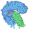
| Title | Cryo-EM map of the full-length relaxin receptor RXFP1 in complex with heterotrimeric Gs | |||||||||
 Map data Map data | Relaxin receptor RXFP1 in complex with heterotrimeric Gs | |||||||||
 Sample Sample |
| |||||||||
 Keywords Keywords | GPCR / Complex / Signaling / Relaxin / MEMBRANE PROTEIN | |||||||||
| Function / homology |  Function and homology information Function and homology informationlung connective tissue development / nipple morphogenesis / Relaxin receptors / parturition / myofibroblast differentiation / hormone binding / G protein-coupled peptide receptor activity / PKA activation in glucagon signalling / hair follicle placode formation / developmental growth ...lung connective tissue development / nipple morphogenesis / Relaxin receptors / parturition / myofibroblast differentiation / hormone binding / G protein-coupled peptide receptor activity / PKA activation in glucagon signalling / hair follicle placode formation / developmental growth / D1 dopamine receptor binding / intracellular transport / renal water homeostasis / Hedgehog 'off' state / adenylate cyclase-activating adrenergic receptor signaling pathway / activation of adenylate cyclase activity / cellular response to glucagon stimulus / adenylate cyclase activator activity / hormone-mediated signaling pathway / regulation of insulin secretion / extracellular matrix organization / trans-Golgi network membrane / G protein-coupled receptor activity / negative regulation of inflammatory response to antigenic stimulus / bone development / G-protein beta/gamma-subunit complex binding / adenylate cyclase-activating G protein-coupled receptor signaling pathway / Olfactory Signaling Pathway / Activation of the phototransduction cascade / G protein activity / G beta:gamma signalling through PLC beta / Presynaptic function of Kainate receptors / Thromboxane signalling through TP receptor / G protein-coupled acetylcholine receptor signaling pathway / G-protein activation / platelet aggregation / Activation of G protein gated Potassium channels / Inhibition of voltage gated Ca2+ channels via Gbeta/gamma subunits / Prostacyclin signalling through prostacyclin receptor / Glucagon signaling in metabolic regulation / G beta:gamma signalling through CDC42 / cognition / G beta:gamma signalling through BTK / ADP signalling through P2Y purinoceptor 12 / Sensory perception of sweet, bitter, and umami (glutamate) taste / Synthesis, secretion, and inactivation of Glucagon-like Peptide-1 (GLP-1) / photoreceptor disc membrane / Glucagon-type ligand receptors / Adrenaline,noradrenaline inhibits insulin secretion / Vasopressin regulates renal water homeostasis via Aquaporins / G alpha (z) signalling events / Glucagon-like Peptide-1 (GLP1) regulates insulin secretion / cellular response to catecholamine stimulus / ADORA2B mediated anti-inflammatory cytokines production / sensory perception of taste / ADP signalling through P2Y purinoceptor 1 / G beta:gamma signalling through PI3Kgamma / adenylate cyclase-activating dopamine receptor signaling pathway / Cooperation of PDCL (PhLP1) and TRiC/CCT in G-protein beta folding / GPER1 signaling / cellular response to prostaglandin E stimulus / Inactivation, recovery and regulation of the phototransduction cascade / G-protein beta-subunit binding / heterotrimeric G-protein complex / sensory perception of smell / G alpha (12/13) signalling events / extracellular vesicle / signaling receptor complex adaptor activity / Thrombin signalling through proteinase activated receptors (PARs) / GTPase binding / retina development in camera-type eye / positive regulation of cold-induced thermogenesis / Ca2+ pathway / phospholipase C-activating G protein-coupled receptor signaling pathway / G alpha (i) signalling events / fibroblast proliferation / Hydrolases; Acting on acid anhydrides; Acting on GTP to facilitate cellular and subcellular movement / G alpha (s) signalling events / G alpha (q) signalling events / Ras protein signal transduction / cell population proliferation / Extra-nuclear estrogen signaling / G protein-coupled receptor signaling pathway / lysosomal membrane / GTPase activity / synapse / protein-containing complex binding / GTP binding / signal transduction / extracellular exosome / membrane / metal ion binding / plasma membrane / cytosol / cytoplasm Similarity search - Function | |||||||||
| Biological species |  Homo sapiens (human) / Homo sapiens (human) /  | |||||||||
| Method | single particle reconstruction / cryo EM / Resolution: 4.2 Å | |||||||||
 Authors Authors | Erlandson SC / Rawson S / Kruse AC | |||||||||
| Funding support |  United States, 2 items United States, 2 items
| |||||||||
 Citation Citation |  Journal: Nat Chem Biol / Year: 2023 Journal: Nat Chem Biol / Year: 2023Title: The relaxin receptor RXFP1 signals through a mechanism of autoinhibition. Authors: Sarah C Erlandson / Shaun Rawson / James Osei-Owusu / Kelly P Brock / Xinyue Liu / Joao A Paulo / Julian Mintseris / Steven P Gygi / Debora S Marks / Xiaojing Cong / Andrew C Kruse /   Abstract: The relaxin family peptide receptor 1 (RXFP1) is the receptor for relaxin-2, an important regulator of reproductive and cardiovascular physiology. RXFP1 is a multi-domain G protein-coupled receptor ...The relaxin family peptide receptor 1 (RXFP1) is the receptor for relaxin-2, an important regulator of reproductive and cardiovascular physiology. RXFP1 is a multi-domain G protein-coupled receptor (GPCR) with an ectodomain consisting of a low-density lipoprotein receptor class A (LDLa) module and leucine-rich repeats. The mechanism of RXFP1 signal transduction is clearly distinct from that of other GPCRs, but remains very poorly understood. In the present study, we determine the cryo-electron microscopy structure of active-state human RXFP1, bound to a single-chain version of the endogenous agonist relaxin-2 and the heterotrimeric G protein. Evolutionary coupling analysis and structure-guided functional experiments reveal that RXFP1 signals through a mechanism of autoinhibition. Our results explain how an unusual GPCR family functions, providing a path to rational drug development targeting the relaxin receptors. | |||||||||
| History |
|
- Structure visualization
| Supplemental images |
|---|
- Downloads & links
-EMDB archive
| Map data |  emd_26004.map.gz emd_26004.map.gz | 55.9 MB |  EMDB map data format EMDB map data format | |
|---|---|---|---|---|
| Header (meta data) |  emd-26004-v30.xml emd-26004-v30.xml emd-26004.xml emd-26004.xml | 23.3 KB 23.3 KB | Display Display |  EMDB header EMDB header |
| Images |  emd_26004.png emd_26004.png | 35.3 KB | ||
| Others |  emd_26004_additional_1.map.gz emd_26004_additional_1.map.gz emd_26004_half_map_1.map.gz emd_26004_half_map_1.map.gz emd_26004_half_map_2.map.gz emd_26004_half_map_2.map.gz | 71.3 MB 71.4 MB 71.5 MB | ||
| Archive directory |  http://ftp.pdbj.org/pub/emdb/structures/EMD-26004 http://ftp.pdbj.org/pub/emdb/structures/EMD-26004 ftp://ftp.pdbj.org/pub/emdb/structures/EMD-26004 ftp://ftp.pdbj.org/pub/emdb/structures/EMD-26004 | HTTPS FTP |
-Validation report
| Summary document |  emd_26004_validation.pdf.gz emd_26004_validation.pdf.gz | 876.5 KB | Display |  EMDB validaton report EMDB validaton report |
|---|---|---|---|---|
| Full document |  emd_26004_full_validation.pdf.gz emd_26004_full_validation.pdf.gz | 876 KB | Display | |
| Data in XML |  emd_26004_validation.xml.gz emd_26004_validation.xml.gz | 12.9 KB | Display | |
| Data in CIF |  emd_26004_validation.cif.gz emd_26004_validation.cif.gz | 15.3 KB | Display | |
| Arichive directory |  https://ftp.pdbj.org/pub/emdb/validation_reports/EMD-26004 https://ftp.pdbj.org/pub/emdb/validation_reports/EMD-26004 ftp://ftp.pdbj.org/pub/emdb/validation_reports/EMD-26004 ftp://ftp.pdbj.org/pub/emdb/validation_reports/EMD-26004 | HTTPS FTP |
-Related structure data
| Related structure data |  7tmwC C: citing same article ( |
|---|---|
| Similar structure data | Similarity search - Function & homology  F&H Search F&H Search |
- Links
Links
| EMDB pages |  EMDB (EBI/PDBe) / EMDB (EBI/PDBe) /  EMDataResource EMDataResource |
|---|---|
| Related items in Molecule of the Month |
- Map
Map
| File |  Download / File: emd_26004.map.gz / Format: CCP4 / Size: 91.1 MB / Type: IMAGE STORED AS FLOATING POINT NUMBER (4 BYTES) Download / File: emd_26004.map.gz / Format: CCP4 / Size: 91.1 MB / Type: IMAGE STORED AS FLOATING POINT NUMBER (4 BYTES) | ||||||||||||||||||||||||||||||||||||
|---|---|---|---|---|---|---|---|---|---|---|---|---|---|---|---|---|---|---|---|---|---|---|---|---|---|---|---|---|---|---|---|---|---|---|---|---|---|
| Annotation | Relaxin receptor RXFP1 in complex with heterotrimeric Gs | ||||||||||||||||||||||||||||||||||||
| Projections & slices | Image control
Images are generated by Spider. | ||||||||||||||||||||||||||||||||||||
| Voxel size | X=Y=Z: 1.06 Å | ||||||||||||||||||||||||||||||||||||
| Density |
| ||||||||||||||||||||||||||||||||||||
| Symmetry | Space group: 1 | ||||||||||||||||||||||||||||||||||||
| Details | EMDB XML:
|
-Supplemental data
-Additional map: Relaxin receptor RXFP1 in complex with heterotrimeric Gs
| File | emd_26004_additional_1.map | ||||||||||||
|---|---|---|---|---|---|---|---|---|---|---|---|---|---|
| Annotation | Relaxin receptor RXFP1 in complex with heterotrimeric Gs | ||||||||||||
| Projections & Slices |
| ||||||||||||
| Density Histograms |
-Half map: Relaxin receptor RXFP1 in complex with heterotrimeric Gs
| File | emd_26004_half_map_1.map | ||||||||||||
|---|---|---|---|---|---|---|---|---|---|---|---|---|---|
| Annotation | Relaxin receptor RXFP1 in complex with heterotrimeric Gs | ||||||||||||
| Projections & Slices |
| ||||||||||||
| Density Histograms |
-Half map: Relaxin receptor RXFP1 in complex with heterotrimeric Gs
| File | emd_26004_half_map_2.map | ||||||||||||
|---|---|---|---|---|---|---|---|---|---|---|---|---|---|
| Annotation | Relaxin receptor RXFP1 in complex with heterotrimeric Gs | ||||||||||||
| Projections & Slices |
| ||||||||||||
| Density Histograms |
- Sample components
-Entire : Complex of the relaxin receptor RXFP1 and heterotrimeric Gs
| Entire | Name: Complex of the relaxin receptor RXFP1 and heterotrimeric Gs |
|---|---|
| Components |
|
-Supramolecule #1: Complex of the relaxin receptor RXFP1 and heterotrimeric Gs
| Supramolecule | Name: Complex of the relaxin receptor RXFP1 and heterotrimeric Gs type: complex / ID: 1 / Parent: 0 / Macromolecule list: all |
|---|---|
| Molecular weight | Theoretical: 180 KDa |
-Supramolecule #2: Relaxin receptor 1, Guanine nucleotide-binding protein G(s) subun...
| Supramolecule | Name: Relaxin receptor 1, Guanine nucleotide-binding protein G(s) subunit alpha isoforms short type: complex / ID: 2 / Parent: 1 / Macromolecule list: #1 |
|---|---|
| Source (natural) | Organism:  Homo sapiens (human) Homo sapiens (human) |
-Supramolecule #3: Guanine nucleotide-binding protein G(I)/G(S)/G(T) subunit beta-1,...
| Supramolecule | Name: Guanine nucleotide-binding protein G(I)/G(S)/G(T) subunit beta-1, Guanine nucleotide-binding protein G(I)/G(S)/G(O) subunit gamma-2 type: complex / ID: 3 / Parent: 1 / Macromolecule list: #2-#3 |
|---|---|
| Source (natural) | Organism:  Homo sapiens (human) Homo sapiens (human) |
-Supramolecule #4: Camelid antibody VHH fragment Nb35
| Supramolecule | Name: Camelid antibody VHH fragment Nb35 / type: complex / ID: 4 / Parent: 1 / Macromolecule list: #4 |
|---|---|
| Source (natural) | Organism:  |
-Macromolecule #1: Relaxin receptor 1, Guanine nucleotide-binding protein G(s) subun...
| Macromolecule | Name: Relaxin receptor 1, Guanine nucleotide-binding protein G(s) subunit alpha isoforms short fusion type: protein_or_peptide / ID: 1 / Enantiomer: LEVO |
|---|---|
| Source (natural) | Organism:  Homo sapiens (human) Homo sapiens (human) |
| Recombinant expression | Organism:  Homo sapiens (human) Homo sapiens (human) |
| Sequence | String: DYKDDDDGGS LEVLFQGPGG SQDVKCSLGY FPCGNITKCL PQLLHCNGVD DCGNQADEDN CGDNNGWSLQ FDKYFASYYK MTSQYPFEAE TPECLVGSVP VQCLCQGLEL DCDETNLRAV PSVSSNVTAM SLQWNLIRKL PPDCFKNYHD LQKLYLQNNK ITSISIYAFR ...String: DYKDDDDGGS LEVLFQGPGG SQDVKCSLGY FPCGNITKCL PQLLHCNGVD DCGNQADEDN CGDNNGWSLQ FDKYFASYYK MTSQYPFEAE TPECLVGSVP VQCLCQGLEL DCDETNLRAV PSVSSNVTAM SLQWNLIRKL PPDCFKNYHD LQKLYLQNNK ITSISIYAFR GLNSLTKLYL SHNRITFLKP GVFEDLHRLE WLIIEDNHLS RISPPTFYGL NSLILLVLMN NVLTRLPDKP LCQHMPRLHW LDLEGNHIHN LRNLTFISCS NLTVLVMRKN KINHLNENTF APLQKLDELD LGSNKIENLP PLIFKDLKEL SQLNLSYNPI QKIQANQFDY LVKLKSLSLE GIEISNIQQR MFRPLMNLSH IYFKKFQYCG YAPHVRSCKP NTDGISSLEN LLASIIQRVF VWVVSAVTCF GNIFVICMRP YIRSENKLYA MSIISLCCAD CLMGIYLFVI GGFDLKFRGE YNKHAQLWME STHCQLVGSL AILSTEVSVL LLTFLTLEKY ICIVYPFRCV RPGKCRTITV LILIWITGFI VAFIPLSNKE FFKNYYGTNG VCFPLHSEDT ESIGAQIYSV AIFLGINLAA FIIIVFSYGS MFYSVHQSAI TATEIRNQVK KEMILAKRFF FIVFTDALCW IPIFVVKFLS LLQVEIPGTI TSWVVIFILP INSALNPILY TLTTRPFKEM IHRFWYNYRQ RKSMDSKGQK TYAPSFIWVE MWPLQEMPPE LMKPDLNSKT EDQRNEEKAQ REANKKIEKQ LQKDKQVYRA THRLLLLGAD NSGKSTIVKQ MRIYHGGSGG SGGTSGIFET KFQVDKVNFH MFDVGGQRDE RRKWIQCFND VTAIIFVVDS SDYNRLQEAL NLFKSIWNNR WLRTISVILF LNKQDLLAEK VLAGKSKIED YFPEFARYTT PEDATPEPGE DPRVTRAKYF IRDEFLRIST ASGDGRHYCY PHFTCAVDTE NARRIFNDCR DIIQRMHLRQ YELL UniProtKB: Relaxin receptor 1, Guanine nucleotide-binding protein G(s) subunit alpha isoforms short |
-Macromolecule #2: Guanine nucleotide-binding protein G(I)/G(S)/G(T) subunit beta-1
| Macromolecule | Name: Guanine nucleotide-binding protein G(I)/G(S)/G(T) subunit beta-1 type: protein_or_peptide / ID: 2 / Enantiomer: LEVO |
|---|---|
| Source (natural) | Organism:  Homo sapiens (human) Homo sapiens (human) |
| Recombinant expression | Organism:  |
| Sequence | String: MHHHHHHGSS GSELDQLRQE AEQLKNQIRD ARKACADATL SQITNNIDPV GRIQMRTRRT LRGHLAKIYA MHWGTDSRLL VSASQDGKLI IWDSYTTNKV HAIPLRSSWV MTCAYAPSGN YVACGGLDNI CSIYNLKTRE GNVRVSRELA GHTGYLSCCR FLDDNQIVTS ...String: MHHHHHHGSS GSELDQLRQE AEQLKNQIRD ARKACADATL SQITNNIDPV GRIQMRTRRT LRGHLAKIYA MHWGTDSRLL VSASQDGKLI IWDSYTTNKV HAIPLRSSWV MTCAYAPSGN YVACGGLDNI CSIYNLKTRE GNVRVSRELA GHTGYLSCCR FLDDNQIVTS SGDTTCALWD IETGQQTTTF TGHTGDVMSL SLAPDTRLFV SGACDASAKL WDVREGMCRQ TFTGHESDIN AICFFPNGNA FATGSDDATC RLFDLRADQE LMTYSHDNII CGITSVSFSK SGRLLLAGYD DFNCNVWDAL KADRAGVLAG HDNRVSCLGV TDDGMAVATG SWDSFLKIWN UniProtKB: Guanine nucleotide-binding protein G(I)/G(S)/G(T) subunit beta-1 |
-Macromolecule #3: Guanine nucleotide-binding protein G(I)/G(S)/G(O) subunit gamma-2
| Macromolecule | Name: Guanine nucleotide-binding protein G(I)/G(S)/G(O) subunit gamma-2 type: protein_or_peptide / ID: 3 / Enantiomer: LEVO |
|---|---|
| Source (natural) | Organism:  Homo sapiens (human) Homo sapiens (human) |
| Recombinant expression | Organism:  |
| Sequence | String: MASNNTASIA QARKLVEQLK MEANIDRIKV SKAAADLMAY CEAHAKEDPL LTPVPASENP FREKKFFCAI L UniProtKB: Guanine nucleotide-binding protein G(I)/G(S)/G(O) subunit gamma-2 |
-Macromolecule #4: Camelid antibody VHH fragment Nb35
| Macromolecule | Name: Camelid antibody VHH fragment Nb35 / type: protein_or_peptide / ID: 4 / Enantiomer: LEVO |
|---|---|
| Source (natural) | Organism:  |
| Recombinant expression | Organism:  |
| Sequence | String: QVQLQESGGG LVQPGGSLRL SCAASGFTFS NYKMNWVRQA PGKGLEWVSD ISQSGASISY TGSVKGRFTI SRDNAKNTLY LQMNSLKPED TAVYYCARCP APFTRDCFDV TSTTYAYRGQ GTQVTVSSLE VLFQGPGHHH HHHHHGSEDQ VDPRLIDGK |
-Experimental details
-Structure determination
| Method | cryo EM |
|---|---|
 Processing Processing | single particle reconstruction |
| Aggregation state | particle |
- Sample preparation
| Concentration | 0.2 mg/mL |
|---|---|
| Buffer | pH: 7.5 |
| Vitrification | Cryogen name: ETHANE / Chamber humidity: 100 % / Chamber temperature: 283.15 K / Instrument: FEI VITROBOT MARK IV |
- Electron microscopy
| Microscope | FEI TITAN KRIOS |
|---|---|
| Image recording | Film or detector model: GATAN K3 BIOQUANTUM (6k x 4k) / Average electron dose: 52.0 e/Å2 |
| Electron beam | Acceleration voltage: 300 kV / Electron source:  FIELD EMISSION GUN FIELD EMISSION GUN |
| Electron optics | Illumination mode: FLOOD BEAM / Imaging mode: BRIGHT FIELD / Nominal defocus max: 2.3000000000000003 µm / Nominal defocus min: 0.8 µm / Nominal magnification: 81000 |
| Sample stage | Specimen holder model: FEI TITAN KRIOS AUTOGRID HOLDER |
| Experimental equipment |  Model: Titan Krios / Image courtesy: FEI Company |
 Movie
Movie Controller


 Z (Sec.)
 Y (Row.)
 X (Col.)


