Movie
Movie Controller

[English] 日本語
 Yorodumi
- EMDB-25875: Cryo-EM 3D map of S. cerevisiae clamp loader RFC bound to two DNA... -
+ Open data

- Basic information
| Entry |  | ||||||||||||
|---|---|---|---|---|---|---|---|---|---|---|---|---|---|
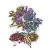
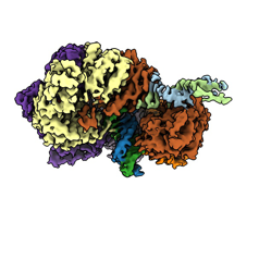
| Title | Cryo-EM 3D map of S. cerevisiae clamp loader RFC bound to two DNA molecules, one at the 5'-recessed end and the other at the 3'-recessed end | ||||||||||||
 Map data Map data | 3D cryoEM map of yeast RFC-DNA1 complex | ||||||||||||
 Sample Sample |
| ||||||||||||
 Keywords Keywords | DNA replication / DNA damage repair / RFC loader / PCNA clamp / DNA polymerase processivity factor / DNA binding protein-DNA complex | ||||||||||||
| Function / homology |  Function and homology information Function and homology informationRad17 RFC-like complex / : / Elg1 RFC-like complex / DNA replication factor C complex / Ctf18 RFC-like complex / Polymerase switching / DNA clamp loader activity / Translesion synthesis by REV1 / : / : ...Rad17 RFC-like complex / : / Elg1 RFC-like complex / DNA replication factor C complex / Ctf18 RFC-like complex / Polymerase switching / DNA clamp loader activity / Translesion synthesis by REV1 / : / : / : / DNA replication checkpoint signaling / : / Activation of ATR in response to replication stress / mitotic sister chromatid cohesion / sister chromatid cohesion / leading strand elongation / Gap-filling DNA repair synthesis and ligation in TC-NER / Dual incision in TC-NER / mismatch repair / DNA damage checkpoint signaling / DNA-templated DNA replication / mitotic cell cycle / cell division / DNA repair / ATP hydrolysis activity / DNA binding / ATP binding / nucleus / cytosol Similarity search - Function | ||||||||||||
| Biological species | synthetic construct (others) /  | ||||||||||||
| Method | single particle reconstruction / cryo EM / Resolution: 3.25 Å | ||||||||||||
 Authors Authors | Zheng F / Georgescu R | ||||||||||||
| Funding support |  United States, 3 items United States, 3 items
| ||||||||||||
 Citation Citation | Journal: Elife / Year: 2022 Title: Cryo-EM structures reveal that RFC recognizes both the 3'- and 5'-DNA ends to load PCNA onto gaps for DNA repair. Authors: Fengwei Zheng / Roxana Georgescu / Nina Y Yao / Huilin Li / Michael E O'Donnell / Abstract: RFC uses ATP to assemble PCNA onto primed sites for replicative DNA polymerases δ and ε. The RFC pentamer forms a central chamber that binds 3' ss/ds DNA junctions to load PCNA onto DNA during ...RFC uses ATP to assemble PCNA onto primed sites for replicative DNA polymerases δ and ε. The RFC pentamer forms a central chamber that binds 3' ss/ds DNA junctions to load PCNA onto DNA during replication. We show here five structures that identify a second DNA binding site in RFC that binds a 5' duplex. This 5' DNA site is located between the N-terminal BRCT domain and AAA+ module of the large Rfc1 subunit. Our structures reveal ideal binding to a 7-nt gap, which includes 2 bp unwound by the clamp loader. Biochemical studies show enhanced binding to 5 and 10 nt gaps, consistent with the structural results. Because both 3' and 5' ends are present at a ssDNA gap, we propose that the 5' site facilitates RFC's PCNA loading activity at a DNA damage-induced gap to recruit gap-filling polymerases. These findings are consistent with genetic studies showing that base excision repair of gaps greater than 1 base requires PCNA and involves the 5' DNA binding domain of Rfc1. We further observe that a 5' end facilitates PCNA loading at an RPA coated 30-nt gap, suggesting a potential role of the RFC 5'-DNA site in lagging strand DNA synthesis. | ||||||||||||
| History |
|
- Structure visualization
| Supplemental images |
|---|
- Downloads & links
-EMDB archive
| Map data | emd_25875.map.gz | 230.2 MB | EMDB map data format | |
|---|---|---|---|---|
| Header (meta data) | emd-25875-v30.xmlemd-25875.xml | 23.1 KB 23.1 KB | Display Display | EMDB header |
| Images |  emd_25875.png emd_25875.png | 45.5 KB | ||
| Filedesc metadata | emd-25875.cif.gz | 7.8 KB | ||
| Archive directory |  http://ftp.pdbj.org/pub/emdb/structures/EMD-25875ftp://ftp.pdbj.org/pub/emdb/structures/EMD-25875 http://ftp.pdbj.org/pub/emdb/structures/EMD-25875ftp://ftp.pdbj.org/pub/emdb/structures/EMD-25875 | HTTPS FTP |
-Related structure data
| Related structure data |  7tfkMC  7tfhC  7tfiC  7tfjC  7tflC C: citing same article ( M: atomic model generated by this map |
|---|---|
| Similar structure data |
-Links
| EMDB pages | EMDB (EBI/PDBe) / EMDataResource |
|---|---|
| Related items in Molecule of the Month |


-Map
| File | Download / File: emd_25875.map.gz / Format: CCP4 / Size: 244.1 MB / Type: IMAGE STORED AS FLOATING POINT NUMBER (4 BYTES) | ||||||||||||||||||||||||||||||||||||
|---|---|---|---|---|---|---|---|---|---|---|---|---|---|---|---|---|---|---|---|---|---|---|---|---|---|---|---|---|---|---|---|---|---|---|---|---|---|
| Annotation | 3D cryoEM map of yeast RFC-DNA1 complex | ||||||||||||||||||||||||||||||||||||
| Projections & slices | Image control
Images are generated by Spider. | ||||||||||||||||||||||||||||||||||||
| Voxel size | X=Y=Z: 0.828 Å | ||||||||||||||||||||||||||||||||||||
| Density |
| ||||||||||||||||||||||||||||||||||||
| Symmetry | Space group: 1 | ||||||||||||||||||||||||||||||||||||
| Details | EMDB XML:
|
 Z (Sec.)
 Y (Row.)
 X (Col.)


-Supplemental data
- Sample components
+Entire : RFC-PCNA-DNA1-DNA2
+Supramolecule #1: RFC-PCNA-DNA1-DNA2
+Supramolecule #2: dsDNA
+Supramolecule #3: Proteins
+Macromolecule #1: Replication factor C subunit 1

+Macromolecule #2: Replication factor C subunit 4
+Macromolecule #3: Replication factor C subunit 3
+Macromolecule #4: Replication factor C subunit 2
+Macromolecule #5: Replication factor C subunit 5
+Macromolecule #6: Template strand
+Macromolecule #7: Primer strand
+Macromolecule #8: PHOSPHOTHIOPHOSPHORIC ACID-ADENYLATE ESTER

+Macromolecule #9: MAGNESIUM ION
+Macromolecule #10: ADENOSINE-5'-DIPHOSPHATE

-Experimental details
-Structure determination
| Method | cryo EM |
|---|---|
 Processing Processing | single particle reconstruction |
| Aggregation state | particle |
-Sample preparation
| Buffer | pH: 7.5 |
|---|---|
| Vitrification | Cryogen name: ETHANE |
- Electron microscopy
| Microscope | FEI TITAN KRIOS |
|---|---|
| Image recording | Film or detector model: GATAN K3 (6k x 4k) / Average electron dose: 65.0 e/Å2 |
| Electron beam | Acceleration voltage: 300 kV / Electron source:  FIELD EMISSION GUN FIELD EMISSION GUN |
| Electron optics | Illumination mode: FLOOD BEAM / Imaging mode: BRIGHT FIELD / Nominal defocus max: 1.9000000000000001 µm / Nominal defocus min: 1.3 µm |
| Experimental equipment |  Model: Titan Krios / Image courtesy: FEI Company |