+ Open data
- Basic information
| Entry |  | |||||||||
|---|---|---|---|---|---|---|---|---|---|---|
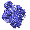
| Title | CryoEM structure of PLCg1 | |||||||||
 Map data Map data | ||||||||||
 Sample Sample |
| |||||||||
 Keywords Keywords | 1-phosphatidylinositol 4 / 5-bisphosphate phosphodiesterase gamma-1 cryo EM / HYDROLASE | |||||||||
| Function / homology |  Function and homology information Function and homology informationphosphoinositide phospholipase C / phospholipid catabolic process / phosphatidylinositol phospholipase C activity / intracellular signal transduction / calcium ion binding Similarity search - Function | |||||||||
| Biological species |  | |||||||||
| Method | single particle reconstruction / cryo EM / Resolution: 3.68 Å | |||||||||
 Authors Authors | Endo-Streeter S / Sondek J | |||||||||
| Funding support |  United States, 1 items United States, 1 items
| |||||||||
 Citation Citation |  Journal: To Be Published Journal: To Be PublishedTitle: CryoEM structure of PLCg1 Authors: Endo-Streeter S / Sondek J | |||||||||
| History |
|
- Structure visualization
| Supplemental images |
|---|
- Downloads & links
-EMDB archive
| Map data |  emd_25745.map.gz emd_25745.map.gz | 43.1 MB |  EMDB map data format EMDB map data format | |
|---|---|---|---|---|
| Header (meta data) |  emd-25745-v30.xml emd-25745-v30.xml emd-25745.xml emd-25745.xml | 13.5 KB 13.5 KB | Display Display |  EMDB header EMDB header |
| Images |  emd_25745.png emd_25745.png | 150.7 KB | ||
| Filedesc metadata |  emd-25745.cif.gz emd-25745.cif.gz | 6.4 KB | ||
| Archive directory |  http://ftp.pdbj.org/pub/emdb/structures/EMD-25745 http://ftp.pdbj.org/pub/emdb/structures/EMD-25745 ftp://ftp.pdbj.org/pub/emdb/structures/EMD-25745 ftp://ftp.pdbj.org/pub/emdb/structures/EMD-25745 | HTTPS FTP |
-Validation report
| Summary document |  emd_25745_validation.pdf.gz emd_25745_validation.pdf.gz | 472.9 KB | Display |  EMDB validaton report EMDB validaton report |
|---|---|---|---|---|
| Full document |  emd_25745_full_validation.pdf.gz emd_25745_full_validation.pdf.gz | 472.5 KB | Display | |
| Data in XML |  emd_25745_validation.xml.gz emd_25745_validation.xml.gz | 6.3 KB | Display | |
| Data in CIF |  emd_25745_validation.cif.gz emd_25745_validation.cif.gz | 7.1 KB | Display | |
| Arichive directory |  https://ftp.pdbj.org/pub/emdb/validation_reports/EMD-25745 https://ftp.pdbj.org/pub/emdb/validation_reports/EMD-25745 ftp://ftp.pdbj.org/pub/emdb/validation_reports/EMD-25745 ftp://ftp.pdbj.org/pub/emdb/validation_reports/EMD-25745 | HTTPS FTP |
-Related structure data
| Related structure data |  7t8tMC M: atomic model generated by this map C: citing same article ( |
|---|---|
| Similar structure data | Similarity search - Function & homology  F&H Search F&H Search |
- Links
Links
| EMDB pages |  EMDB (EBI/PDBe) / EMDB (EBI/PDBe) /  EMDataResource EMDataResource |
|---|---|
| Related items in Molecule of the Month |
- Map
Map
| File |  Download / File: emd_25745.map.gz / Format: CCP4 / Size: 83.7 MB / Type: IMAGE STORED AS FLOATING POINT NUMBER (4 BYTES) Download / File: emd_25745.map.gz / Format: CCP4 / Size: 83.7 MB / Type: IMAGE STORED AS FLOATING POINT NUMBER (4 BYTES) | ||||||||||||||||||||||||||||||||||||
|---|---|---|---|---|---|---|---|---|---|---|---|---|---|---|---|---|---|---|---|---|---|---|---|---|---|---|---|---|---|---|---|---|---|---|---|---|---|
| Projections & slices | Image control
Images are generated by Spider. | ||||||||||||||||||||||||||||||||||||
| Voxel size | X=Y=Z: 0.91 Å | ||||||||||||||||||||||||||||||||||||
| Density |
| ||||||||||||||||||||||||||||||||||||
| Symmetry | Space group: 1 | ||||||||||||||||||||||||||||||||||||
| Details | EMDB XML:
|
-Supplemental data
- Sample components
-Entire : 1-phosphatidylinositol 4,5-bisphosphate phosphodiesterase gamma-1
| Entire | Name: 1-phosphatidylinositol 4,5-bisphosphate phosphodiesterase gamma-1 |
|---|---|
| Components |
|
-Supramolecule #1: 1-phosphatidylinositol 4,5-bisphosphate phosphodiesterase gamma-1
| Supramolecule | Name: 1-phosphatidylinositol 4,5-bisphosphate phosphodiesterase gamma-1 type: complex / ID: 1 / Parent: 0 / Macromolecule list: #1 |
|---|---|
| Source (natural) | Organism:  |
-Macromolecule #1: 1-phosphatidylinositol 4,5-bisphosphate phosphodiesterase gamma
| Macromolecule | Name: 1-phosphatidylinositol 4,5-bisphosphate phosphodiesterase gamma type: protein_or_peptide / ID: 1 / Number of copies: 1 / Enantiomer: LEVO / EC number: phosphoinositide phospholipase C |
|---|---|
| Source (natural) | Organism:  |
| Molecular weight | Theoretical: 138.185391 KDa |
| Recombinant expression | Organism:  Trichoplusia ni (cabbage looper) Trichoplusia ni (cabbage looper) |
| Sequence | String: AEVLHLCRSL EVGTVMTLFY SKKSQRPERK TFQVKLETRQ ITWSRGADKI EGSIDIREIK EIRPGKTSRD FDRYQEDPAF RPDQSHCFV ILYGMEFRLK TLSLQATSED EVNMWIKGLT WLMEDTLQAA TPLQIERWLR KQFYSVDRNR EDRISAKDLK N MLSQVNYR ...String: AEVLHLCRSL EVGTVMTLFY SKKSQRPERK TFQVKLETRQ ITWSRGADKI EGSIDIREIK EIRPGKTSRD FDRYQEDPAF RPDQSHCFV ILYGMEFRLK TLSLQATSED EVNMWIKGLT WLMEDTLQAA TPLQIERWLR KQFYSVDRNR EDRISAKDLK N MLSQVNYR VPNMRFLRER LTDLEQRSGD ITYGQFAQLY RSLMYSAQKT MDLPFLETNT LRTGERPELC QVSLSEFQQF LL EYQGELW AVDRLQVQEF MLSFLRDPLR EIEEPYFFLD ELVTFLFSKE NSVWNSQLDA VCPETMNNPL SHYWISSSAN TYL TGDQFS SESSLEAYAR CLRMGCRCIE LDCWDGPDGM PVIYHGHTLT TKIKFSDVLH TIKEHAFVAS EYPVILSIED HCSI AQQRN MAQHFRKVLG DTLLTKPVDI AADGLPSPNQ LKRKILIKHK KLAEGSAYEE VPTSVMYSEN DISNSIKNGI LYLED PVNH EWYPHYFVLT SSKIYYSEET SSDQGNEDEE EPKEASGSTE LHSSEKWFHG KLGAGRDGRH IAERLLTEYC IETGAP DGS FLVRESETFV GDYTLSFWRN GKVQHCRIHS RQDAGTPKFF LTDNLVFDSL YDLITHYQQV PLRCNEFEMR LSEPVPQ TN AHESKEWYHA SLTRAQAEHM LMRVPRDGAF LVRKRNEPNS YAISFRAEGK IKHCRVQQEG QTVMLGNSEF DSLVDLIS Y YEKHPLYRKM KLRYPINEEA LEKIGTAEPD YGALYEGRNP GFYVEANPMP TFKCAVKALF DYKAQREDEL TFTKSAIIQ NVEKQDGGWW RGDYGGKKQL WFPSNYVEEM INPAILEPER EHLDENSPLG DLLRGVLDVP ACQIAIRPEG KNNRLFVFSI SMPSVAQWS LDVAADSQEE LQDWVKKIRE VAQTADARLT EGKMMERRKK IALELSELVV YCRPVPFDEE KIGTERACYR D MSSFPETK AEKYVNKAKG KKFLQYNRLQ LSRIYPKGQR LDSSNYDPLP MWICGSQLVA LNFQTPDKPM QMNQALFMAG GH CGYVLQP STMRDEAFDP FDKSSLRGLE PCVICIEVLG ARHLPKNGRG IVCPFVEIEV AGAEYDSTKQ KTEFVVDNGL NPV WPAKPF HFQISNPEFA FLRFVVYEED MFSDQNFLAQ ATFPVKGLKT GYRAVPLKNN YSEDLELASL LIKIDIFPAK UniProtKB: 1-phosphatidylinositol 4,5-bisphosphate phosphodiesterase gamma |
-Macromolecule #2: CALCIUM ION
| Macromolecule | Name: CALCIUM ION / type: ligand / ID: 2 / Number of copies: 1 / Formula: CA |
|---|---|
| Molecular weight | Theoretical: 40.078 Da |
-Experimental details
-Structure determination
| Method | cryo EM |
|---|---|
 Processing Processing | single particle reconstruction |
| Aggregation state | particle |
- Sample preparation
| Concentration | 10 mg/mL | ||||||||||||||||||
|---|---|---|---|---|---|---|---|---|---|---|---|---|---|---|---|---|---|---|---|
| Buffer | pH: 7.4 Component:
| ||||||||||||||||||
| Grid | Model: C-flat-1.2/1.3 / Material: COPPER / Mesh: 300 / Support film - Material: CARBON / Support film - topology: HOLEY / Support film - Film thickness: 12 / Pretreatment - Type: PLASMA CLEANING | ||||||||||||||||||
| Vitrification | Cryogen name: ETHANE-PROPANE / Chamber humidity: 95 % / Chamber temperature: 295 K / Instrument: FEI VITROBOT MARK IV |
- Electron microscopy
| Microscope | FEI TALOS ARCTICA |
|---|---|
| Image recording | Film or detector model: GATAN K3 BIOQUANTUM (6k x 4k) / Digitization - Dimensions - Width: 5760 pixel / Digitization - Dimensions - Height: 4092 pixel / Number grids imaged: 1 / Number real images: 4450 / Average electron dose: 55.0 e/Å2 |
| Electron beam | Acceleration voltage: 200 kV / Electron source:  FIELD EMISSION GUN FIELD EMISSION GUN |
| Electron optics | Illumination mode: FLOOD BEAM / Imaging mode: BRIGHT FIELD / Cs: 2.7 mm |
| Sample stage | Cooling holder cryogen: NITROGEN |
| Experimental equipment |  Model: Talos Arctica / Image courtesy: FEI Company |
- Image processing
| Startup model | Type of model: NONE |
|---|---|
| Final reconstruction | Number classes used: 13 / Applied symmetry - Point group: C1 (asymmetric) / Resolution.type: BY AUTHOR / Resolution: 3.68 Å / Resolution method: FSC 0.143 CUT-OFF / Software - Name: cryoSPARC (ver. 3.2) / Number images used: 238458 |
| Initial angle assignment | Type: MAXIMUM LIKELIHOOD |
| Final angle assignment | Type: MAXIMUM LIKELIHOOD |
-Atomic model buiding 1
| Initial model | PDB ID: Chain - Chain ID: A / Chain - Residue range: 21-1215 / Chain - Source name: PDB / Chain - Initial model type: experimental model |
|---|---|
| Refinement | Space: REAL / Protocol: FLEXIBLE FIT / Overall B value: 135.93 / Target criteria: CC fit and model metrics |
| Output model |  PDB-7t8t: |
 Movie
Movie Controller


 Z (Sec.)
 Y (Row.)
 X (Col.)


