 Movie
Movie Controller

[English] 日本語
 Yorodumi
- EMDB-2566: Electron cryo-microscopy of yeast mitochondrial large ribosomal s... -
+ Open data

- Basic information
| Entry | Database: EMDB / ID: EMD-2566 | |||||||||
|---|---|---|---|---|---|---|---|---|---|---|
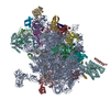
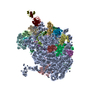
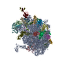
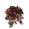
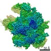
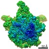
| Title | Electron cryo-microscopy of yeast mitochondrial large ribosomal subunit | |||||||||
 Map data Map data | Reconstruction of yeast mitochondrial large ribosomal subunit; this map has a mask around the large ribosomal subunit | |||||||||
 Sample Sample |
| |||||||||
 Keywords Keywords | cryo-EM / yeast mitochondrial ribosome | |||||||||
| Function / homology |  Function and homology information Function and homology informationpositive regulation of mitochondrial DNA replication / mitochondrial respiratory chain complex IV assembly / mitochondrial large ribosomal subunit assembly / Mitochondrial protein degradation / ribonuclease III activity / DNA strand exchange activity / mitochondrial large ribosomal subunit / mitochondrial translation / RNA processing / cell redox homeostasis ...positive regulation of mitochondrial DNA replication / mitochondrial respiratory chain complex IV assembly / mitochondrial large ribosomal subunit assembly / Mitochondrial protein degradation / ribonuclease III activity / DNA strand exchange activity / mitochondrial large ribosomal subunit / mitochondrial translation / RNA processing / cell redox homeostasis / double-stranded RNA binding / single-stranded DNA binding / large ribosomal subunit / transferase activity / ribosome biogenesis / cellular response to oxidative stress / large ribosomal subunit rRNA binding / DNA recombination / mitochondrial inner membrane / negative regulation of translation / rRNA binding / structural constituent of ribosome / ribosome / translation / mRNA binding / regulation of DNA-templated transcription / mitochondrion / DNA binding / RNA binding / zinc ion binding / nucleus / cytoplasm Similarity search - Function | |||||||||
| Biological species |  | |||||||||
| Method | single particle reconstruction / cryo EM / negative staining / Resolution: 3.2 Å | |||||||||
 Authors Authors | Amunts A / Brown A / Bai XC / Llacer JL / Hussain T / Emsley P / Long F / Murshudov G / Scheres SHW / Ramakrishnan V | |||||||||
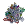
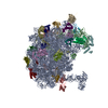
 Citation Citation | Journal: Science / Year: 2014 Title: Structure of the yeast mitochondrial large ribosomal subunit. Authors: Alexey Amunts / Alan Brown / Xiao-Chen Bai / Jose L Llácer / Tanweer Hussain / Paul Emsley / Fei Long / Garib Murshudov / Sjors H W Scheres / V Ramakrishnan /  Abstract: Mitochondria have specialized ribosomes that have diverged from their bacterial and cytoplasmic counterparts. We have solved the structure of the yeast mitoribosomal large subunit using single- ...Mitochondria have specialized ribosomes that have diverged from their bacterial and cytoplasmic counterparts. We have solved the structure of the yeast mitoribosomal large subunit using single-particle cryo-electron microscopy. The resolution of 3.2 angstroms enabled a nearly complete atomic model to be built de novo and refined, including 39 proteins, 13 of which are unique to mitochondria, as well as expansion segments of mitoribosomal RNA. The structure reveals a new exit tunnel path and architecture, unique elements of the E site, and a putative membrane docking site. | |||||||||
| History |
|
- Structure visualization
| Movie |
Movie viewer |
|---|---|
| Structure viewer | EM map: SurfViewMolmilJmol/JSmol |
| Supplemental images |

- Downloads & links
-EMDB archive
| Map data | emd_2566.map.gz | 22.8 MB | EMDB map data format | |
|---|---|---|---|---|
| Header (meta data) | emd-2566-v30.xmlemd-2566.xml | 10.2 KB 10.2 KB | Display Display | EMDB header |
| FSC (resolution estimation) | emd_2566_fsc.xml | 10.5 KB | Display | FSC data file |
| Images | 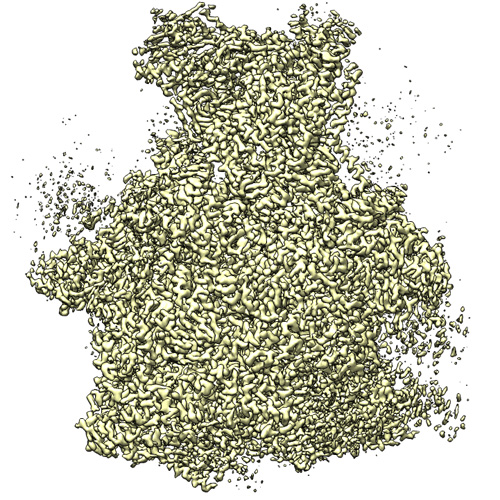 EMD_2566.jpg EMD_2566.jpg | 209.4 KB | ||
| Archive directory |  http://ftp.pdbj.org/pub/emdb/structures/EMD-2566ftp://ftp.pdbj.org/pub/emdb/structures/EMD-2566 http://ftp.pdbj.org/pub/emdb/structures/EMD-2566ftp://ftp.pdbj.org/pub/emdb/structures/EMD-2566 | HTTPS FTP |
-Related structure data
| Related structure data |  3j6bMC M: atomic model generated by this map C: citing same article ( |
|---|---|
| Similar structure data |


-Links
| EMDB pages | EMDB (EBI/PDBe) / EMDataResource |
|---|---|
| Related items in Molecule of the Month |


-Map
| File | Download / File: emd_2566.map.gz / Format: CCP4 / Size: 173.8 MB / Type: IMAGE STORED AS FLOATING POINT NUMBER (4 BYTES) | ||||||||||||||||||||||||||||||||||||||||||||||||||||||||||||||||||||
|---|---|---|---|---|---|---|---|---|---|---|---|---|---|---|---|---|---|---|---|---|---|---|---|---|---|---|---|---|---|---|---|---|---|---|---|---|---|---|---|---|---|---|---|---|---|---|---|---|---|---|---|---|---|---|---|---|---|---|---|---|---|---|---|---|---|---|---|---|---|
| Annotation | Reconstruction of yeast mitochondrial large ribosomal subunit; this map has a mask around the large ribosomal subunit | ||||||||||||||||||||||||||||||||||||||||||||||||||||||||||||||||||||
| Projections & slices | Image control
Images are generated by Spider. | ||||||||||||||||||||||||||||||||||||||||||||||||||||||||||||||||||||
| Voxel size | X=Y=Z: 1.34 Å | ||||||||||||||||||||||||||||||||||||||||||||||||||||||||||||||||||||
| Density |
| ||||||||||||||||||||||||||||||||||||||||||||||||||||||||||||||||||||
| Symmetry | Space group: 1 | ||||||||||||||||||||||||||||||||||||||||||||||||||||||||||||||||||||
| Details | EMDB XML:
CCP4 map header:
| ||||||||||||||||||||||||||||||||||||||||||||||||||||||||||||||||||||
 Z (Sec.)
 Y (Row.)
 X (Col.)


-Supplemental data
- Sample components
-Entire : Yeast Mitochondrial Large Ribosomal Subunit
| Entire | Name: Yeast Mitochondrial Large Ribosomal Subunit |
|---|---|
| Components |
|
-Supramolecule #1000: Yeast Mitochondrial Large Ribosomal Subunit
| Supramolecule | Name: Yeast Mitochondrial Large Ribosomal Subunit / type: sample / ID: 1000 / Number unique components: 1 |
|---|---|
| Molecular weight | Experimental: 1.9 MDa / Theoretical: 1.9 MDa |
-Supramolecule #1: Yeast Mitochondrial Large Ribosomal Subunit
| Supramolecule | Name: Yeast Mitochondrial Large Ribosomal Subunit / type: complex / ID: 1 / Name.synonym: 54S Mitochondrial Ribosome / Recombinant expression: No / Ribosome-details: ribosome-eukaryote: LSU 60S |
|---|---|
| Source (natural) | Organism: |
| Molecular weight | Experimental: 1.9 MDa / Theoretical: 1.9 MDa |
-Experimental details
-Structure determination
| Method | negative staining, cryo EM |
|---|---|
 Processing Processing | single particle reconstruction |
| Aggregation state | particle |
-Sample preparation
| Concentration | 0.15 mg/mL |
|---|---|
| Buffer | pH: 7.5 Details: 20 mM Hepes-KOH pH 7.5, 100 mM KCl, 20 mM MgOAc, 2 mM DTT |
| Staining | Type: NEGATIVE / Details: cryo EM |
| Grid | Details: 30 s on glow-discharged holey carbon grids (Quantifoil R2/2), onto which a home-made continuous carbon film |
| Vitrification | Cryogen name: ETHANE / Chamber humidity: 100 % / Chamber temperature: 90 K / Instrument: FEI VITROBOT MARK II / Method: Blot 2.5 seconds before plunging |
- Electron microscopy
| Microscope | FEI TITAN KRIOS |
|---|---|
| Temperature | Min: 80 K / Max: 90 K / Average: 85 K |
| Alignment procedure | Legacy - Astigmatism: Objective lens astigmatism was corrected at 59,000 times magnification |
| Date | May 25, 2013 |
| Image recording | Category: CCD / Film or detector model: FEI FALCON II (4k x 4k) / Digitization - Sampling interval: 14 µm / Number real images: 1030 / Average electron dose: 25 e/Å2 Details: An in-house built system was used to intercept the videos from the detector at a rate of 17 frames for the 1 s exposures. |
| Electron beam | Acceleration voltage: 300 kV / Electron source:  FIELD EMISSION GUN FIELD EMISSION GUN |
| Electron optics | Calibrated magnification: 104478 / Illumination mode: FLOOD BEAM / Imaging mode: BRIGHT FIELD / Cs: 2.7 mm / Nominal defocus max: 4.7 µm / Nominal defocus min: 1.2 µm / Nominal magnification: 59000 |
| Sample stage | Specimen holder model: FEI TITAN KRIOS AUTOGRID HOLDER |
| Experimental equipment |  Model: Titan Krios / Image courtesy: FEI Company |
-Image processing
| CTF correction | Details: Each particle |
|---|---|
| Final reconstruction | Applied symmetry - Point group: C1 (asymmetric) / Resolution.type: BY AUTHOR / Resolution: 3.2 Å / Resolution method: OTHER / Software - Name: CTFFIND3, RELION Details: Use a newly developed statistical movie processing approach to compensate for beam-induced movement. Number images used: 47124 |
| FSC plot (resolution estimation) | 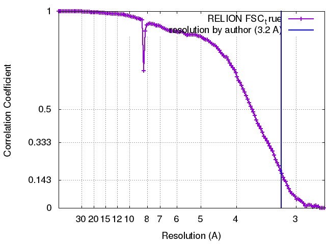 |
