[English] 日本語
Yorodumi
- EMDB-25362: Cryo-electron tomography structure of membrane-bound EHD4 complex -

+
Open data


ID or keywords:

Loading...

-
Basic information

Entry
Database: EMDB / ID: EMD-25362
TitleCryo-electron tomography structure of membrane-bound EHD4 complex
Map dataCryo-electron tomography structure of membrane-bound EHD4 complex
Sample
  • Organelle or cellular component: EHD4 with AMPPNP and liposomes
    • Protein or peptide: EH domain-containing protein 4
KeywordsEHD ATPases / dynamin family / oligomerization / membrane remodeling / membrane binding site / LIPID BINDING PROTEIN
Function / homology
Function and homology information


pinocytosis / clathrin coat of coated pit / endocytic recycling / regulation of endocytosis / endocytic vesicle / cilium assembly / protein localization to plasma membrane / protein homooligomerization / cellular response to growth factor stimulus / endocytosis ...pinocytosis / clathrin coat of coated pit / endocytic recycling / regulation of endocytosis / endocytic vesicle / cilium assembly / protein localization to plasma membrane / protein homooligomerization / cellular response to growth factor stimulus / endocytosis / recycling endosome membrane / positive regulation of peptidyl-tyrosine phosphorylation / early endosome membrane / early endosome / cadherin binding / calcium ion binding / GTP binding / perinuclear region of cytoplasm / endoplasmic reticulum / ATP binding / nucleus / plasma membrane / cytoplasm
Similarity search - Function
EH domain-containing protein, N-terminal / Domain of unknown function DUF5600 / N-terminal EH-domain containing protein / Domain of unknown function (DUF5600) / Cytoskeletal-regulatory complex EF hand / EH domain profile. / Eps15 homology domain / EH domain / Dynamin-type guanine nucleotide-binding (G) domain / Dynamin-type guanine nucleotide-binding (G) domain profile. ...EH domain-containing protein, N-terminal / Domain of unknown function DUF5600 / N-terminal EH-domain containing protein / Domain of unknown function (DUF5600) / Cytoskeletal-regulatory complex EF hand / EH domain profile. / Eps15 homology domain / EH domain / Dynamin-type guanine nucleotide-binding (G) domain / Dynamin-type guanine nucleotide-binding (G) domain profile. / Dynamin, N-terminal / Dynamin family / EF-Hand 1, calcium-binding site / EF-hand calcium-binding domain. / EF-hand calcium-binding domain profile. / EF-hand domain / EF-hand domain pair / P-loop containing nucleoside triphosphate hydrolase
Similarity search - Domain/homology
EH domain-containing protein 4
Similarity search - Component
Biological speciesMus musculus (house mouse)
Methodsubtomogram averaging / cryo EM / Resolution: 7.6 Å
AuthorsMelo AA / Noel JK
Funding support Germany, 3 items
OrganizationGrant numberCountry
German Research Foundation (DFG)SFB 958/A12 Germany
European Research Council (ERC)ERC-2013-CoG-616024 Germany
European Union (EU)iNEXT PID3536 VID5570 Germany
Citation
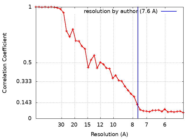
Journal: Nat Commun / Year: 2022
Title: Cryo-electron tomography reveals structural insights into the membrane remodeling mode of dynamin-like EHD filaments.
Authors: Arthur A Melo / Thiemo Sprink / Jeffrey K Noel / Elena Vázquez-Sarandeses / Chris van Hoorn / Saif Mohd / Justus Loerke / Christian M T Spahn / Oliver Daumke /
Abstract: Eps15-homology domain containing proteins (EHDs) are eukaryotic, dynamin-related ATPases involved in cellular membrane trafficking. They oligomerize on membranes into filaments that induce membrane ...Eps15-homology domain containing proteins (EHDs) are eukaryotic, dynamin-related ATPases involved in cellular membrane trafficking. They oligomerize on membranes into filaments that induce membrane tubulation. While EHD crystal structures in open and closed conformations were previously reported, little structural information is available for the membrane-bound oligomeric form. Consequently, mechanistic insights into the membrane remodeling mechanism have remained sparse. Here, by using cryo-electron tomography and subtomogram averaging, we determined structures of nucleotide-bound EHD4 filaments on membrane tubes of various diameters at an average resolution of 7.6 Å. Assembly of EHD4 is mediated via interfaces in the G-domain and the helical domain. The oligomerized EHD4 structure resembles the closed conformation, where the tips of the helical domains protrude into the membrane. The variation in filament geometry and tube radius suggests a spontaneous filament curvature of approximately 1/70 nm. Combining the available structural and functional data, we suggest a model for EHD-mediated membrane remodeling.
History
DepositionNov 1, 2021-
Header (metadata) releaseNov 9, 2022-
Map releaseNov 9, 2022-
UpdateJun 5, 2024-
Current statusJun 5, 2024Processing site: RCSB / Status: Released

-
Structure visualization

Supplemental images

Downloads & links

-
Map

FileDownload / File: emd_25362.map.gz / Format: CCP4 / Size: 3.4 MB / Type: IMAGE STORED AS FLOATING POINT NUMBER (4 BYTES)
AnnotationCryo-electron tomography structure of membrane-bound EHD4 complex
Voxel sizeX=Y=Z: 2.628 Å
Density
Contour LevelBy AUTHOR: 2.22
Minimum - Maximum-7.3744226 - 6.9472675
Average (Standard dev.)0.051608976 (±1.1095288)
SymmetrySpace group: 1
Details

EMDB XML:

Map geometry
Axis orderXYZ
Origin000
Dimensions969696
Spacing969696
CellA=B=C: 252.288 Å
α=β=γ: 90.0 °

-
Supplemental data

-
Mask #1

Fileemd_25362_msk_1.map
Projections & Slices
AxesZYX

Projections

Slices (1/2)
Density Histograms

-
Half map: Half Map 1

Fileemd_25362_half_map_1.map
AnnotationHalf Map 1
Projections & Slices
AxesZYX

Projections

Slices (1/2)
Density Histograms

-
Half map: Half Map 2

Fileemd_25362_half_map_2.map
AnnotationHalf Map 2
Projections & Slices
AxesZYX

Projections

Slices (1/2)
Density Histograms

-
Sample components

-
Entire : EHD4 with AMPPNP and liposomes

EntireName: EHD4 with AMPPNP and liposomes
Components
  • Organelle or cellular component: EHD4 with AMPPNP and liposomes
    • Protein or peptide: EH domain-containing protein 4

-
Supramolecule #1: EHD4 with AMPPNP and liposomes

SupramoleculeName: EHD4 with AMPPNP and liposomes / type: organelle_or_cellular_component / ID: 1 / Parent: 0 / Macromolecule list: all
Source (natural)Organism: Mus musculus (house mouse)
Molecular weightTheoretical: 0.59 kDa/nm

-
Macromolecule #1: EH domain-containing protein 4

MacromoleculeName: EH domain-containing protein 4 / type: protein_or_peptide / ID: 1 / Number of copies: 16 / Enantiomer: LEVO
Source (natural)Organism: Mus musculus (house mouse)
Molecular weightTheoretical: 58.674195 KDa
Recombinant expressionOrganism: Escherichia coli (E. coli)
SequenceString: QTVTGGLRSL YQRKVLPLEE AYRFHEFHSP ALEDADFENK PMILLVGQYS TGKTTFIRYL LEQDFPGMRI GPEPTTDSFI AVMYGETEG STPGNALVVD PKKPFRKLSR FGNAFLNRFM CSQLPNQVLK SISIIDSPGI LSGEKQRISR GYDFCQVLQW F AERVDRII ...String:
QTVTGGLRSL YQRKVLPLEE AYRFHEFHSP ALEDADFENK PMILLVGQYS TGKTTFIRYL LEQDFPGMRI GPEPTTDSFI AVMYGETEG STPGNALVVD PKKPFRKLSR FGNAFLNRFM CSQLPNQVLK SISIIDSPGI LSGEKQRISR GYDFCQVLQW F AERVDRII LLFDAHKLDI SDEFSEAIKA FRGQDDKIRV VLNKADQVDT QQLMRVYGAL MWSLGKVINT PEVLRVYIGS FW AQPLQNT DNRRLFEAEA QDLFRDIQSL PQKAAVRKLN DLIKRARLAK VHAYIISYLK KEMPNMFGKE NKKRELIYRL PEI YVQLQR EYQISAGDFP EVKAMQEQLE NYDFTKFHSL KPKLIEAVDN MLTNKISSLM GLISQEEMNM PTQMVQGGAF DGTT EGPFN QGYGEGAKEG ADEEEWVVAK DKPVYDELFY TLSPINGKIS GVNAKKEMVT SKLPNSVLGK IWKLADCDCD GMLDE EEFA LAKHLIKIKL DGYELPNSLP PHLVPPSHRK

UniProtKB: EH domain-containing protein 4

-
Experimental details

-
Structure determination

Methodcryo EM
Processingsubtomogram averaging
Aggregation statehelical array

-
Sample preparation

Concentration0.5 mg/mL
BufferpH: 7.5
GridModel: Quantifoil / Material: GOLD / Mesh: 200 / Support film - Material: CARBON / Support film - topology: HOLEY / Pretreatment - Type: GLOW DISCHARGE
VitrificationCryogen name: ETHANE / Chamber humidity: 100 % / Chamber temperature: 298 K / Instrument: FEI VITROBOT MARK II

-
Electron microscopy

MicroscopeFEI TITAN KRIOS
Image recordingFilm or detector model: GATAN K2 QUANTUM (4k x 4k) / Detector mode: COUNTING / Average electron dose: 2.3 e/Å2
Electron beamAcceleration voltage: 300 kV / Electron source: FIELD EMISSION GUN
Electron opticsIllumination mode: FLOOD BEAM / Imaging mode: BRIGHT FIELD
Experimental equipment
Model: Titan Krios / Image courtesy: FEI Company

+
Image processing

Final reconstructionApplied symmetry - Point group: C2 (2 fold cyclic) / Algorithm: BACK PROJECTION / Resolution.type: BY AUTHOR / Resolution: 7.6 Å / Resolution method: FSC 0.143 CUT-OFF / Software - Name: IMOD / Number subtomograms used: 23813
ExtractionNumber tomograms: 56 / Number images used: 84000 / Software - Name: Dynamo
Final 3D classificationSoftware - Name: Dynamo
Final angle assignmentType: OTHER / Software - Name: Dynamo / Details: cross correlation
FSC plot (resolution estimation)

-
Atomic model buiding 1

Initial modelPDB ID:

Chain - Source name: PDB / Chain - Initial model type: experimental model
RefinementSpace: REAL / Protocol: RIGID BODY FIT
Output model

PDB-7sox:
Cryo-electron tomography structure of membrane-bound EHD4 complex

+
About Yorodumi

-
News

-
Feb 9, 2022. New format data for meta-information of EMDB entries

New format data for meta-information of EMDB entries

  • Version 3 of the EMDB header file is now the official format.
  • The previous official version 1.9 will be removed from the archive.

Related info.:EMDB header

External links:wwPDB to switch to version 3 of the EMDB data model

-
Aug 12, 2020. Covid-19 info

Covid-19 info

URL: https://pdbjlvh1.pdbj.org/emnavi/covid19.php

New page: Covid-19 featured information page in EM Navigator.

Related info.:Covid-19 info / Mar 5, 2020. Novel coronavirus structure data

+
Mar 5, 2020. Novel coronavirus structure data

Novel coronavirus structure data

Related info.:Yorodumi Speices / Aug 12, 2020. Covid-19 info

External links:COVID-19 featured content - PDBj / Molecule of the Month (242):Coronavirus Proteases

+
Jan 31, 2019. EMDB accession codes are about to change! (news from PDBe EMDB page)

EMDB accession codes are about to change! (news from PDBe EMDB page)

  • The allocation of 4 digits for EMDB accession codes will soon come to an end. Whilst these codes will remain in use, new EMDB accession codes will include an additional digit and will expand incrementally as the available range of codes is exhausted. The current 4-digit format prefixed with “EMD-” (i.e. EMD-XXXX) will advance to a 5-digit format (i.e. EMD-XXXXX), and so on. It is currently estimated that the 4-digit codes will be depleted around Spring 2019, at which point the 5-digit format will come into force.
  • The EM Navigator/Yorodumi systems omit the EMD- prefix.

Related info.:Q: What is EMD? / ID/Accession-code notation in Yorodumi/EM Navigator

External links:EMDB Accession Codes are Changing Soon! / Contact to PDBj

+
Jul 12, 2017. Major update of PDB

Major update of PDB

  • wwPDB released updated PDB data conforming to the new PDBx/mmCIF dictionary.
  • This is a major update changing the version number from 4 to 5, and with Remediation, in which all the entries are updated.
  • In this update, many items about electron microscopy experimental information are reorganized (e.g. em_software).
  • Now, EM Navigator and Yorodumi are based on the updated data.

External links:wwPDB Remediation / Enriched Model Files Conforming to OneDep Data Standards Now Available in the PDB FTP Archive

-
Yorodumi

Thousand views of thousand structures

  • Yorodumi is a browser for structure data from EMDB, PDB, SASBDB, etc.
  • This page is also the successor to EM Navigator detail page, and also detail information page/front-end page for Omokage search.
  • The word "yorodu" (or yorozu) is an old Japanese word meaning "ten thousand". "mi" (miru) is to see.

Related info.:EMDB / PDB / SASBDB / Comparison of 3 databanks / Yorodumi Search / Aug 31, 2016. New EM Navigator & Yorodumi / Yorodumi Papers / Jmol/JSmol / Function and homology information / Changes in new EM Navigator and Yorodumi

Read more