 Movie
Movie Controller

[English] 日本語
 Yorodumi
- EMDB-24572: SP6-11 biased agonist bound to active human neurokinin 1 receptor... -
+ Open data

- Basic information
| Entry | Database: EMDB / ID: EMD-24572 | ||||||||||||||||||||||||||||||||||||
|---|---|---|---|---|---|---|---|---|---|---|---|---|---|---|---|---|---|---|---|---|---|---|---|---|---|---|---|---|---|---|---|---|---|---|---|---|---|
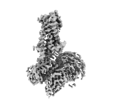
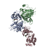
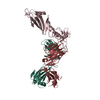
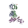
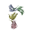
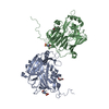
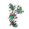
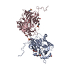
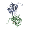
| Title | SP6-11 biased agonist bound to active human neurokinin 1 receptor in complex with miniGs/q70 | ||||||||||||||||||||||||||||||||||||
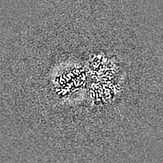
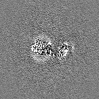
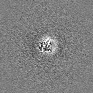
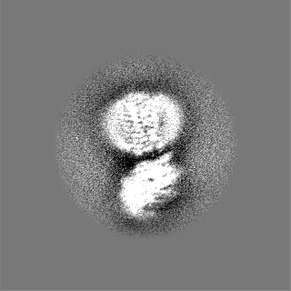
 Map data Map data | Unsharpened map | ||||||||||||||||||||||||||||||||||||
 Sample Sample |
| ||||||||||||||||||||||||||||||||||||
| Function / homology |  Function and homology information Function and homology informationsubstance P receptor activity / tachykinin receptor activity / aggressive behavior / Tachykinin receptors bind tachykinins / positive regulation of flagellated sperm motility / positive regulation of uterine smooth muscle contraction / detection of abiotic stimulus / positive regulation of synaptic transmission, cholinergic / tachykinin receptor signaling pathway / operant conditioning ...substance P receptor activity / tachykinin receptor activity / aggressive behavior / Tachykinin receptors bind tachykinins / positive regulation of flagellated sperm motility / positive regulation of uterine smooth muscle contraction / detection of abiotic stimulus / positive regulation of synaptic transmission, cholinergic / tachykinin receptor signaling pathway / operant conditioning / positive regulation of lymphocyte proliferation / sperm head / response to ozone / sperm ejaculation / positive regulation of action potential / response to auditory stimulus / smooth muscle contraction involved in micturition / regulation of smooth muscle cell proliferation / positive regulation of hormone secretion / positive regulation of blood pressure / positive regulation of vascular permeability / positive regulation of ossification / regulation of smooth muscle cell migration / positive regulation of leukocyte migration / eating behavior / positive regulation of epithelial cell migration / behavioral response to pain / associative learning / PKA activation in glucagon signalling / angiotensin-mediated drinking behavior / hair follicle placode formation / mu-type opioid receptor binding / developmental growth / corticotropin-releasing hormone receptor 1 binding / intracellular transport / D1 dopamine receptor binding / sperm flagellum / Hedgehog 'off' state / long-term memory / response to electrical stimulus / beta-2 adrenergic receptor binding / adenylate cyclase-activating adrenergic receptor signaling pathway / positive regulation of vasoconstriction / sperm midpiece / positive regulation of stress fiber assembly / activation of adenylate cyclase activity / adenylate cyclase activator activity / trans-Golgi network membrane / positive regulation of epithelial cell proliferation / response to progesterone / positive regulation of synaptic transmission, GABAergic / insulin-like growth factor receptor binding / ionotropic glutamate receptor binding / response to nicotine / G-protein beta/gamma-subunit complex binding / bone development / Olfactory Signaling Pathway / Activation of the phototransduction cascade / G beta:gamma signalling through PLC beta / Presynaptic function of Kainate receptors / Thromboxane signalling through TP receptor / adenylate cyclase-activating G protein-coupled receptor signaling pathway / G-protein activation / G protein-coupled acetylcholine receptor signaling pathway / Activation of G protein gated Potassium channels / Inhibition of voltage gated Ca2+ channels via Gbeta/gamma subunits / Prostacyclin signalling through prostacyclin receptor / Glucagon signaling in metabolic regulation / G beta:gamma signalling through CDC42 / cognition / ADP signalling through P2Y purinoceptor 12 / G beta:gamma signalling through BTK / Synthesis, secretion, and inactivation of Glucagon-like Peptide-1 (GLP-1) / Sensory perception of sweet, bitter, and umami (glutamate) taste / photoreceptor disc membrane / platelet aggregation / Adrenaline,noradrenaline inhibits insulin secretion / Glucagon-type ligand receptors / Vasopressin regulates renal water homeostasis via Aquaporins / G alpha (z) signalling events / cellular response to catecholamine stimulus / Glucagon-like Peptide-1 (GLP1) regulates insulin secretion / ADORA2B mediated anti-inflammatory cytokines production / sensory perception of taste / ADP signalling through P2Y purinoceptor 1 / adenylate cyclase-activating dopamine receptor signaling pathway / G beta:gamma signalling through PI3Kgamma / cellular response to prostaglandin E stimulus / Cooperation of PDCL (PhLP1) and TRiC/CCT in G-protein beta folding / GPER1 signaling / G-protein beta-subunit binding / Inactivation, recovery and regulation of the phototransduction cascade / heterotrimeric G-protein complex / G alpha (12/13) signalling events / extracellular vesicle / sensory perception of smell / signaling receptor complex adaptor activity / Cargo recognition for clathrin-mediated endocytosis / Thrombin signalling through proteinase activated receptors (PARs) / GTPase binding Similarity search - Function | ||||||||||||||||||||||||||||||||||||
| Biological species |  Homo sapiens (human) / synthetic construct (others) / Homo sapiens (human) / synthetic construct (others) /  | ||||||||||||||||||||||||||||||||||||
| Method | single particle reconstruction / cryo EM / Resolution: 3.2 Å | ||||||||||||||||||||||||||||||||||||
 Authors Authors | Harris JA / Faust B / Gondin AB / Daemgen MA / Suomivuori CM / Veldhuis NA / Cheng Y / Dror RO / Thal D / Manglik A | ||||||||||||||||||||||||||||||||||||
| Funding support |  United States, United States,  Australia, 11 items Australia, 11 items
| ||||||||||||||||||||||||||||||||||||
 Citation Citation | Journal: Nat Chem Biol / Year: 2022 Title: Selective G protein signaling driven by substance P-neurokinin receptor dynamics. Authors: Julian A Harris / Bryan Faust / Arisbel B Gondin / Marc André Dämgen / Carl-Mikael Suomivuori / Nicholas A Veldhuis / Yifan Cheng / Ron O Dror / David M Thal / Aashish Manglik / Abstract: The neuropeptide substance P (SP) is important in pain and inflammation. SP activates the neurokinin-1 receptor (NK1R) to signal via G and G proteins. Neurokinin A also activates NK1R, but leads to ...The neuropeptide substance P (SP) is important in pain and inflammation. SP activates the neurokinin-1 receptor (NK1R) to signal via G and G proteins. Neurokinin A also activates NK1R, but leads to selective G signaling. How two stimuli yield distinct G protein signaling at the same G protein-coupled receptor remains unclear. We determined cryogenic-electron microscopy structures of active NK1R bound to SP or the G-biased peptide SP6-11. Peptide interactions deep within NK1R are critical for receptor activation. Conversely, interactions between SP and NK1R extracellular loops are required for potent G signaling but not G signaling. Molecular dynamics simulations showed that these superficial contacts restrict SP flexibility. SP6-11, which lacks these interactions, is dynamic while bound to NK1R. Structural dynamics of NK1R agonists therefore depend on interactions with the receptor extracellular loops and regulate G protein signaling selectivity. Similar interactions between other neuropeptides and their cognate receptors may tune intracellular signaling. | ||||||||||||||||||||||||||||||||||||
| History |
|
- Structure visualization
| Movie |
Movie viewer |
|---|---|
| Structure viewer | EM map: SurfViewMolmilJmol/JSmol |
| Supplemental images |
- Downloads & links
-EMDB archive
| Map data | emd_24572.map.gz | 115.8 MB | EMDB map data format | |
|---|---|---|---|---|
| Header (meta data) | emd-24572-v30.xmlemd-24572.xml | 30.3 KB 30.3 KB | Display Display | EMDB header |
| Images |  emd_24572.png emd_24572.png | 50.2 KB | ||
| Others | emd_24572_additional_1.map.gzemd_24572_half_map_1.map.gzemd_24572_half_map_2.map.gz | 116.1 MB 18.4 MB 18.4 MB | ||
| Archive directory |  http://ftp.pdbj.org/pub/emdb/structures/EMD-24572ftp://ftp.pdbj.org/pub/emdb/structures/EMD-24572 http://ftp.pdbj.org/pub/emdb/structures/EMD-24572ftp://ftp.pdbj.org/pub/emdb/structures/EMD-24572 | HTTPS FTP |
-Validation report
| Summary document | emd_24572_validation.pdf.gz | 652 KB | Display | EMDB validaton report |
|---|---|---|---|---|
| Full document | emd_24572_full_validation.pdf.gz | 651.6 KB | Display | |
| Data in XML | emd_24572_validation.xml.gz | 14.1 KB | Display | |
| Data in CIF | emd_24572_validation.cif.gz | 16.5 KB | Display | |
| Arichive directory | https://ftp.pdbj.org/pub/emdb/validation_reports/EMD-24572ftp://ftp.pdbj.org/pub/emdb/validation_reports/EMD-24572 | HTTPS FTP |
-Related structure data
| Related structure data | 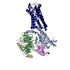 7rmiMC  7rmgC  7rmhC M: atomic model generated by this map C: citing same article ( |
|---|---|
| Similar structure data | |
| EM raw data | EMPIAR-10786 (Title: Cryo electron microscopy final particle stacks of Substance P-Neurokinin Receptor G protein complexes Data size: 170.8 Data #1: Particle stack and final .star file for SP-NK1R-miniGs399 reconstruction [picked particles - single frame - processed] Data #2: Particle stack and final .star file for SP6-11-NK1R-miniGsq70 reconstruction [picked particles - single frame - processed] Data #3: Particle stack and final .star file for SP-NK1R-miniGsq70 reconstruction [picked particles - single frame - processed]) |


-Links
| EMDB pages | EMDB (EBI/PDBe) / EMDataResource |
|---|---|
| Related items in Molecule of the Month |


-Map
| File | Download / File: emd_24572.map.gz / Format: CCP4 / Size: 125 MB / Type: IMAGE STORED AS FLOATING POINT NUMBER (4 BYTES) | ||||||||||||||||||||||||||||||||||||||||||||||||||||||||||||||||||||
|---|---|---|---|---|---|---|---|---|---|---|---|---|---|---|---|---|---|---|---|---|---|---|---|---|---|---|---|---|---|---|---|---|---|---|---|---|---|---|---|---|---|---|---|---|---|---|---|---|---|---|---|---|---|---|---|---|---|---|---|---|---|---|---|---|---|---|---|---|---|
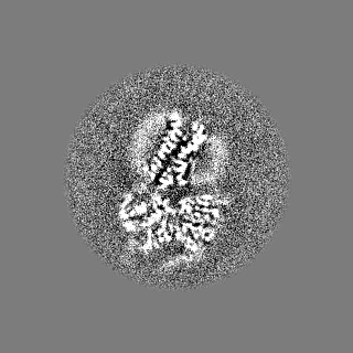
| Annotation | Unsharpened map | ||||||||||||||||||||||||||||||||||||||||||||||||||||||||||||||||||||
| Voxel size | X=Y=Z: 0.835 Å | ||||||||||||||||||||||||||||||||||||||||||||||||||||||||||||||||||||
| Density |
| ||||||||||||||||||||||||||||||||||||||||||||||||||||||||||||||||||||
| Symmetry | Space group: 1 | ||||||||||||||||||||||||||||||||||||||||||||||||||||||||||||||||||||
| Details | EMDB XML:
CCP4 map header:
| ||||||||||||||||||||||||||||||||||||||||||||||||||||||||||||||||||||
-Supplemental data
-Additional map: Sharpened map
| File | emd_24572_additional_1.map | ||||||||||||
|---|---|---|---|---|---|---|---|---|---|---|---|---|---|
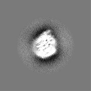
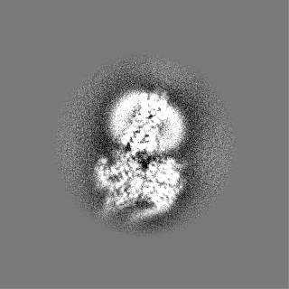
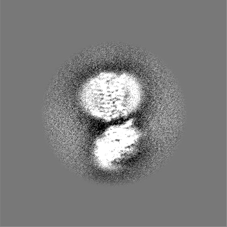
| Annotation | Sharpened map | ||||||||||||
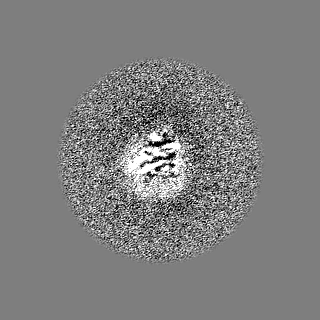
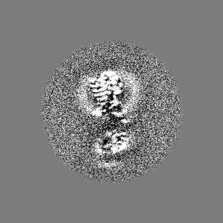
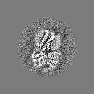
| Projections & Slices |
| ||||||||||||
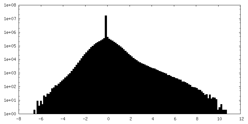
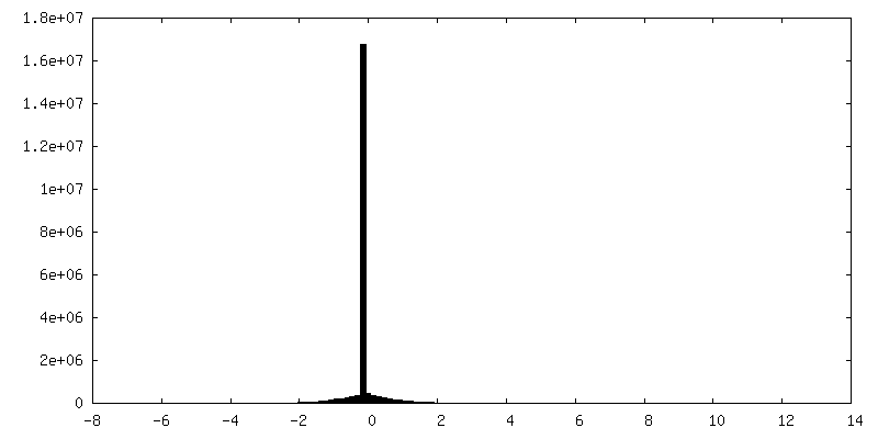
| Density Histograms |
 Z
Z Y
Y X
X


-Half map: #1
| File | emd_24572_half_map_1.map | ||||||||||||
|---|---|---|---|---|---|---|---|---|---|---|---|---|---|
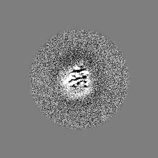
| Projections & Slices |
| ||||||||||||
| Density Histograms |

-Half map: #2
| File | emd_24572_half_map_2.map | ||||||||||||
|---|---|---|---|---|---|---|---|---|---|---|---|---|---|
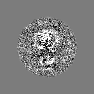
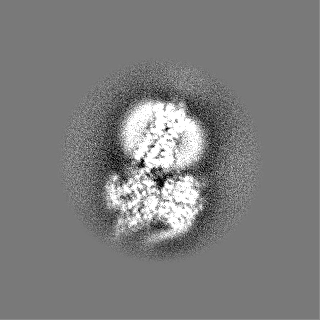
| Projections & Slices |
| ||||||||||||
| Density Histograms |

- Sample components
+Entire : Substance P bound to Neurokinin 1 receptor-miniGs/q70 complex
+Supramolecule #1: Substance P bound to Neurokinin 1 receptor-miniGs/q70 complex
+Supramolecule #2: Guanine nucleotide-binding protein G(s)/G(q) subunit alpha hybrid
+Supramolecule #3: Guanine nucleotide-binding protein G(I)/G(S)/G(T) subunit beta-1/...
 Trichoplusia ni (cabbage looper)
+Supramolecule #4: Substance-P receptor
+Supramolecule #5: Substance P 6-11
+Supramolecule #6: Nanobody 35

+Macromolecule #1: Guanine nucleotide-binding protein G(s) subunit alpha isoforms sh...
+Macromolecule #2: Substance-P receptor
+Macromolecule #3: Substance P 6-11
+Macromolecule #4: Guanine nucleotide-binding protein G(I)/G(S)/G(T) subunit beta-1
+Macromolecule #5: Guanine nucleotide-binding protein G(I)/G(S)/G(O) subunit gamma-2
+Macromolecule #6: Nanobody 35
-Experimental details
-Structure determination
| Method | cryo EM |
|---|---|
 Processing Processing | single particle reconstruction |
| Aggregation state | particle |
-Sample preparation
| Buffer | pH: 7.5 |
|---|---|
| Grid | Model: Quantifoil R1.2/1.3 / Material: GOLD / Mesh: 300 |
| Vitrification | Cryogen name: ETHANE / Chamber humidity: 100 % / Chamber temperature: 277 K / Instrument: FEI VITROBOT MARK IV |
- Electron microscopy
| Microscope | FEI TITAN KRIOS |
|---|---|
| Specialist optics | Energy filter - Name: GIF Bioquantum / Energy filter - Slit width: 20 eV |
| Image recording | Film or detector model: GATAN K3 (6k x 4k) / Average exposure time: 5.9 sec. / Average electron dose: 67.0 e/Å2 |
| Electron beam | Acceleration voltage: 300 kV / Electron source:  FIELD EMISSION GUN FIELD EMISSION GUN |
| Electron optics | Illumination mode: FLOOD BEAM / Imaging mode: BRIGHT FIELD |
| Experimental equipment |  Model: Titan Krios / Image courtesy: FEI Company |

