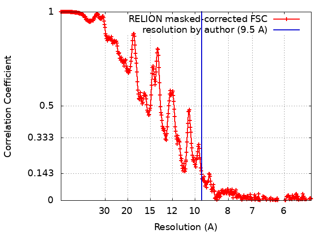
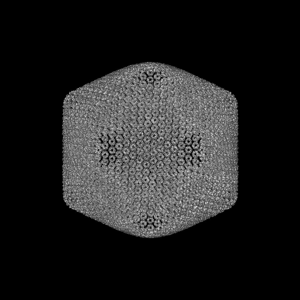
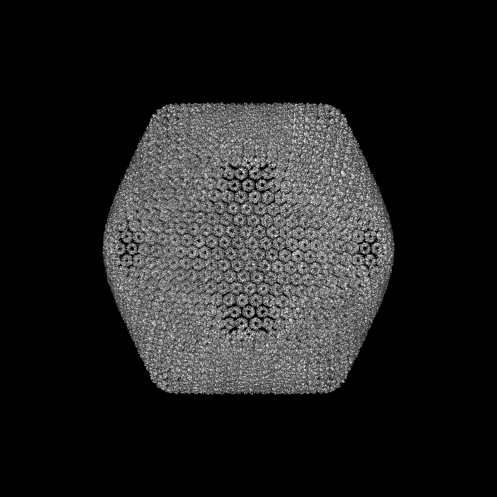
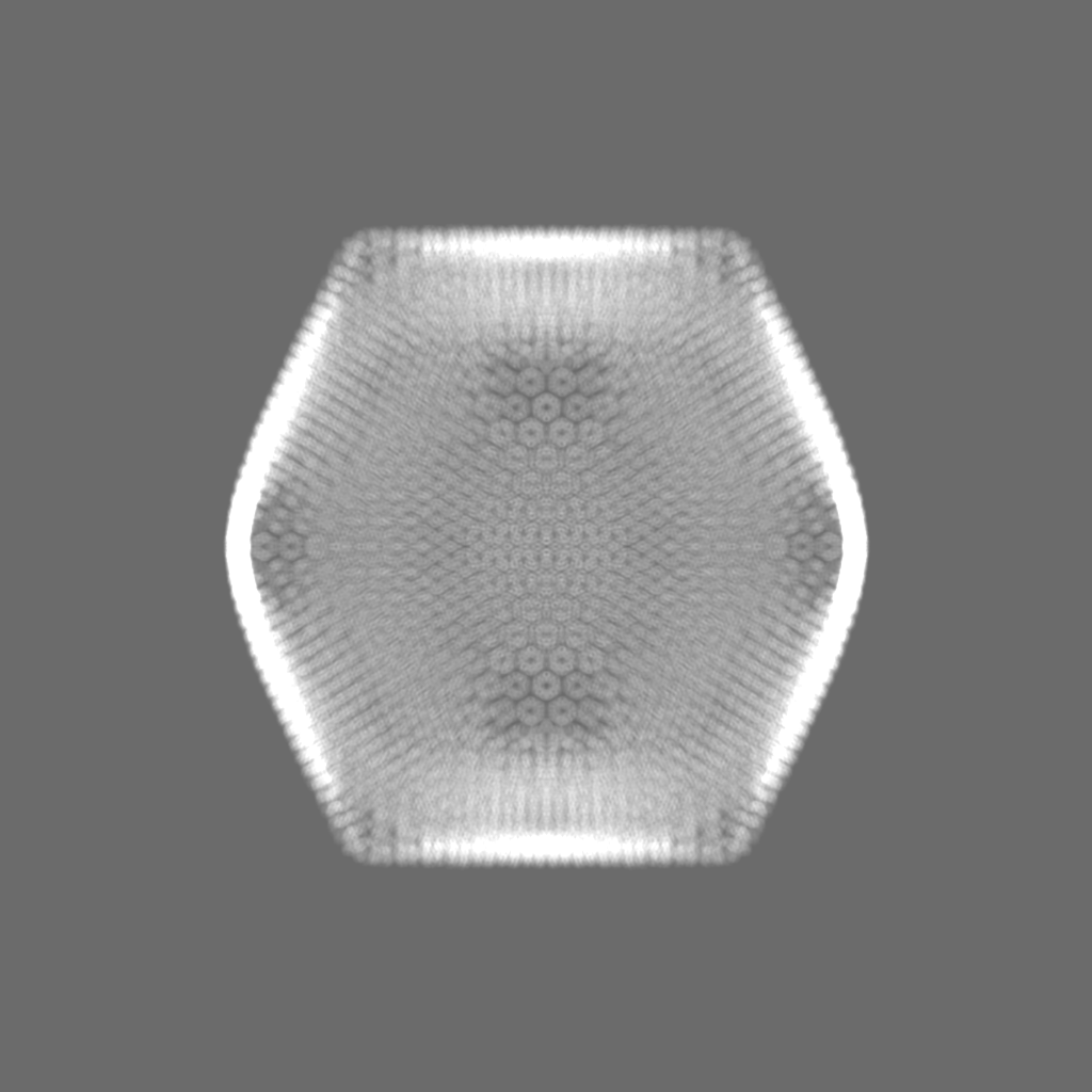
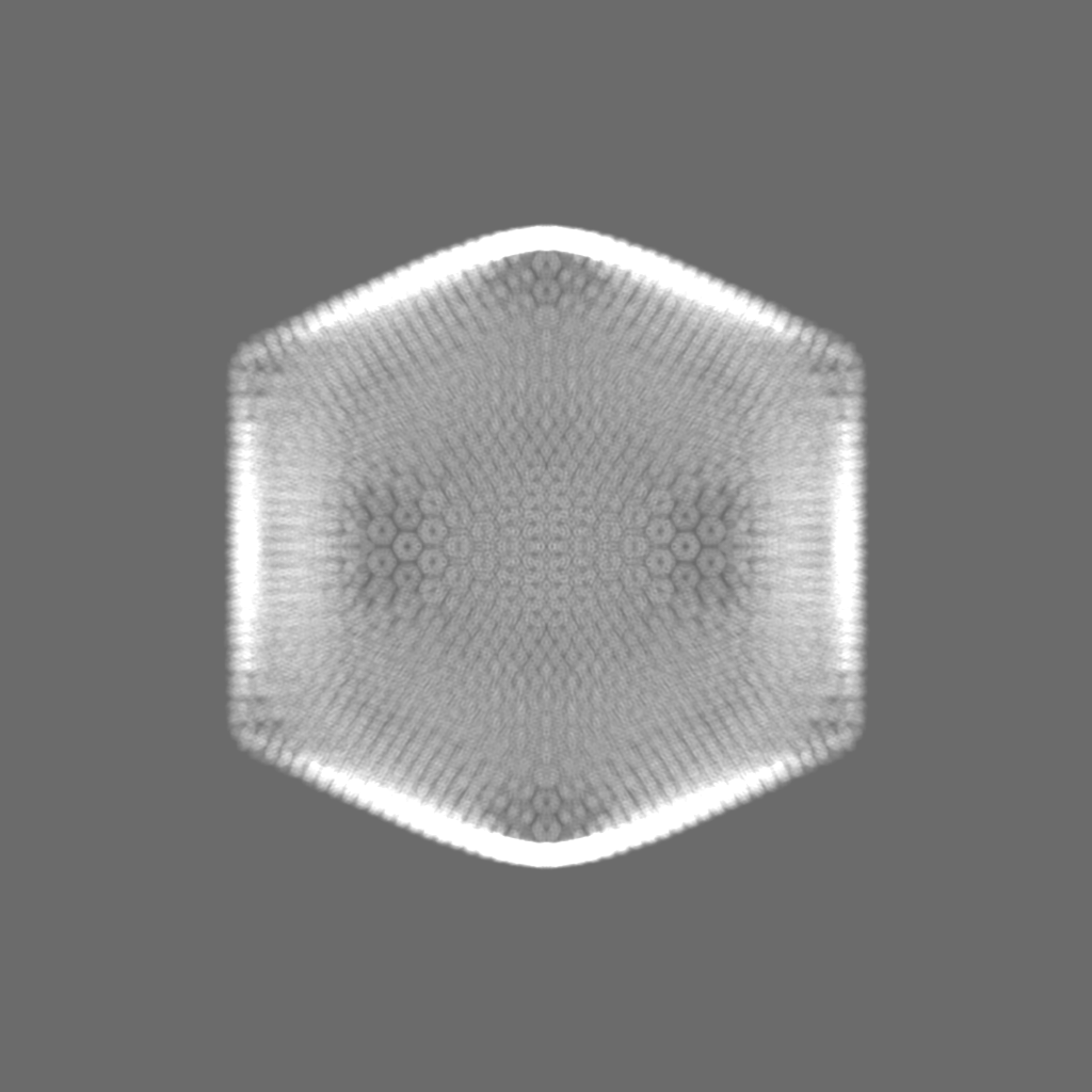
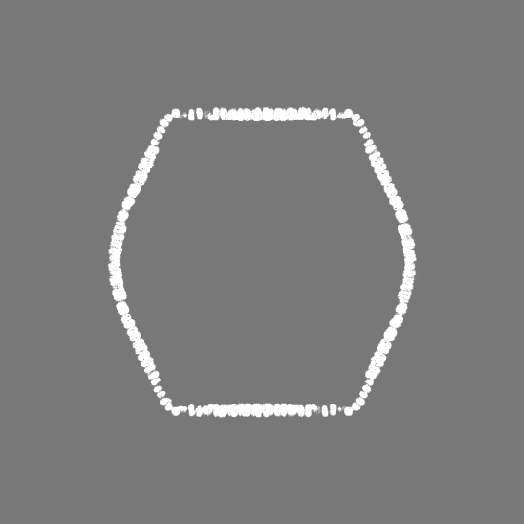
ジャーナル: Front Microbiol / 年: 2020 タイトル: Structural and Proteomic Studies of the Virus Demonstrate a Global Distribution of Virus-Encoded Carbohydrate Processing. 著者: Eric R Gann / Yuejiao Xian / Paul E Abraham / Robert L Hettich / Todd B Reynolds / Chuan Xiao / Steven W Wilhelm / 要旨: Viruses modulate the function(s) of environmentally relevant microbial populations, yet considerations of the metabolic capabilities of individual virus particles themselves are rare. We used shotgun ...Viruses modulate the function(s) of environmentally relevant microbial populations, yet considerations of the metabolic capabilities of individual virus particles themselves are rare. We used shotgun proteomics to quantitatively identify 43 virus-encoded proteins packaged within purified Virus (AaV) particles, normalizing data to the per-virion level using a 9.5-Å-resolution molecular reconstruction of the 1900-Å (AaV) particle that we generated with cryogenic electron microscopy. This packaged proteome was used to determine similarities and differences between members of different giant virus families. We noted that proteins involved in sugar degradation and binding (e.g., carbohydrate lyases) were unique to AaV among characterized giant viruses. To determine the extent to which this virally encoded metabolic capability was ecologically relevant, we examined the TARA Oceans dataset and identified genes and transcripts of viral origin. Our analyses demonstrated that putative giant virus carbohydrate lyases represented up to 17% of the marine pool for this function. In total, our observations suggest that the AaV particle has potential prepackaged metabolic capabilities and that these may be found in other giant viruses that are widespread and abundant in global oceans.
 ムービー
ムービー コントローラー


 データを開く
 基本情報
基本情報 マップデータ
 試料
試料 Aureococcus anophagefferens virus (ファージ)
 データ登録者
 引用
引用
 構造の表示
構造の表示 ムービービューア
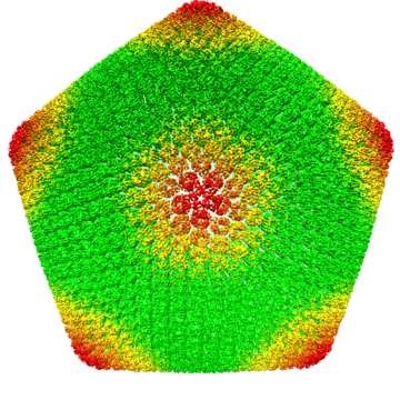
 ダウンロードとリンク
 emd_22339.png
 http://ftp.pdbj.org/pub/emdb/structures/EMD-22339


 Z (Sec.)
 Y (Row.)
 X (Col.)


 試料の構成要素
 Aureococcus anophagefferens (真核生物)
 解析
解析 電子顕微鏡法
 FIELD EMISSION GUN