+ Open data
- Basic information
| Entry | Database: EMDB / ID: EMD-2108 | |||||||||
|---|---|---|---|---|---|---|---|---|---|---|
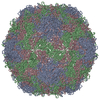
| Title | HRV2 empty 135S particle | |||||||||
 Map data Map data | Reconstruction of HRV2 135S empty particle | |||||||||
 Sample Sample |
| |||||||||
| Biological species |  Human rhinovirus 2 Human rhinovirus 2 | |||||||||
| Method | single particle reconstruction / cryo EM / Resolution: 9.9 Å | |||||||||
 Authors Authors | Pickl-Herk A / Luque D / Trus BL / Verdaguer N / Blaas D / Caston JR | |||||||||
 Citation Citation |  Journal: Proc Natl Acad Sci U S A / Year: 2013 Journal: Proc Natl Acad Sci U S A / Year: 2013Title: Uncoating of common cold virus is preceded by RNA switching as determined by X-ray and cryo-EM analyses of the subviral A-particle. Authors: Angela Pickl-Herk / Daniel Luque / Laia Vives-Adrián / Jordi Querol-Audí / Damià Garriga / Benes L Trus / Nuria Verdaguer / Dieter Blaas / José R Castón /  Abstract: During infection, viruses undergo conformational changes that lead to delivery of their genome into host cytosol. In human rhinovirus A2, this conversion is triggered by exposure to acid pH in the ...During infection, viruses undergo conformational changes that lead to delivery of their genome into host cytosol. In human rhinovirus A2, this conversion is triggered by exposure to acid pH in the endosome. The first subviral intermediate, the A-particle, is expanded and has lost the internal viral protein 4 (VP4), but retains its RNA genome. The nucleic acid is subsequently released, presumably through one of the large pores that open at the icosahedral twofold axes, and is transferred along a conduit in the endosomal membrane; the remaining empty capsids, termed B-particles, are shuttled to lysosomes for degradation. Previous structural analyses revealed important differences between the native protein shell and the empty capsid. Nonetheless, little is known of A-particle architecture or conformation of the RNA core. Using 3D cryo-electron microscopy and X-ray crystallography, we found notable changes in RNA-protein contacts during conversion of native virus into the A-particle uncoating intermediate. In the native virion, we confirmed interaction of nucleotide(s) with Trp(38) of VP2 and identified additional contacts with the VP1 N terminus. Study of A-particle structure showed that the VP2 contact is maintained, that VP1 interactions are lost after exit of the VP1 N-terminal extension, and that the RNA also interacts with residues of the VP3 N terminus at the fivefold axis. These associations lead to formation of a well-ordered RNA layer beneath the protein shell, suggesting that these interactions guide ordered RNA egress. | |||||||||
| History |
|
- Structure visualization
| Movie |
 Movie viewer Movie viewer |
|---|---|
| Structure viewer | EM map:  SurfView SurfView Molmil Molmil Jmol/JSmol Jmol/JSmol |
| Supplemental images |
- Downloads & links
-EMDB archive
| Map data |  emd_2108.map.gz emd_2108.map.gz | 51.5 MB |  EMDB map data format EMDB map data format | |
|---|---|---|---|---|
| Header (meta data) |  emd-2108-v30.xml emd-2108-v30.xml emd-2108.xml emd-2108.xml | 9.8 KB 9.8 KB | Display Display |  EMDB header EMDB header |
| Images |  emd_2108.png emd_2108.png | 356.9 KB | ||
| Archive directory |  http://ftp.pdbj.org/pub/emdb/structures/EMD-2108 http://ftp.pdbj.org/pub/emdb/structures/EMD-2108 ftp://ftp.pdbj.org/pub/emdb/structures/EMD-2108 ftp://ftp.pdbj.org/pub/emdb/structures/EMD-2108 | HTTPS FTP |
-Validation report
| Summary document |  emd_2108_validation.pdf.gz emd_2108_validation.pdf.gz | 289.3 KB | Display |  EMDB validaton report EMDB validaton report |
|---|---|---|---|---|
| Full document |  emd_2108_full_validation.pdf.gz emd_2108_full_validation.pdf.gz | 288.5 KB | Display | |
| Data in XML |  emd_2108_validation.xml.gz emd_2108_validation.xml.gz | 6.2 KB | Display | |
| Arichive directory |  https://ftp.pdbj.org/pub/emdb/validation_reports/EMD-2108 https://ftp.pdbj.org/pub/emdb/validation_reports/EMD-2108 ftp://ftp.pdbj.org/pub/emdb/validation_reports/EMD-2108 ftp://ftp.pdbj.org/pub/emdb/validation_reports/EMD-2108 | HTTPS FTP |
-Related structure data
- Links
Links
| EMDB pages |  EMDB (EBI/PDBe) / EMDB (EBI/PDBe) /  EMDataResource EMDataResource |
|---|
- Map
Map
| File |  Download / File: emd_2108.map.gz / Format: CCP4 / Size: 54.1 MB / Type: IMAGE STORED AS FLOATING POINT NUMBER (4 BYTES) Download / File: emd_2108.map.gz / Format: CCP4 / Size: 54.1 MB / Type: IMAGE STORED AS FLOATING POINT NUMBER (4 BYTES) | ||||||||||||||||||||||||||||||||||||||||||||||||||||||||||||||||||||
|---|---|---|---|---|---|---|---|---|---|---|---|---|---|---|---|---|---|---|---|---|---|---|---|---|---|---|---|---|---|---|---|---|---|---|---|---|---|---|---|---|---|---|---|---|---|---|---|---|---|---|---|---|---|---|---|---|---|---|---|---|---|---|---|---|---|---|---|---|---|
| Annotation | Reconstruction of HRV2 135S empty particle | ||||||||||||||||||||||||||||||||||||||||||||||||||||||||||||||||||||
| Projections & slices | Image control
Images are generated by Spider. | ||||||||||||||||||||||||||||||||||||||||||||||||||||||||||||||||||||
| Voxel size | X=Y=Z: 1.89 Å | ||||||||||||||||||||||||||||||||||||||||||||||||||||||||||||||||||||
| Density |
| ||||||||||||||||||||||||||||||||||||||||||||||||||||||||||||||||||||
| Symmetry | Space group: 1 | ||||||||||||||||||||||||||||||||||||||||||||||||||||||||||||||||||||
| Details | EMDB XML:
CCP4 map header:
| ||||||||||||||||||||||||||||||||||||||||||||||||||||||||||||||||||||
-Supplemental data
- Sample components
-Entire : HRV2 135S empty particle
| Entire | Name: HRV2 135S empty particle |
|---|---|
| Components |
|
-Supramolecule #1000: HRV2 135S empty particle
| Supramolecule | Name: HRV2 135S empty particle / type: sample / ID: 1000 / Oligomeric state: icosahedral / Number unique components: 1 |
|---|---|
| Molecular weight | Theoretical: 5.8 MDa |
-Supramolecule #1: Human rhinovirus 2
| Supramolecule | Name: Human rhinovirus 2 / type: virus / ID: 1 / Name.synonym: Common cold virus / NCBI-ID: 12130 / Sci species name: Human rhinovirus 2 / Virus type: VIRION / Virus isolate: SEROTYPE / Virus enveloped: No / Virus empty: Yes / Syn species name: Common cold virus |
|---|---|
| Host (natural) | Organism:  Homo sapiens (human) / synonym: VERTEBRATES Homo sapiens (human) / synonym: VERTEBRATES |
| Virus shell | Shell ID: 1 / Diameter: 316 Å / T number (triangulation number): 1 |
-Experimental details
-Structure determination
| Method | cryo EM |
|---|---|
 Processing Processing | single particle reconstruction |
| Aggregation state | particle |
- Sample preparation
| Buffer | pH: 7.4 / Details: 50 mM sodium borate |
|---|---|
| Grid | Details: R 1.2/1.3 Quantifoil grids |
| Vitrification | Cryogen name: ETHANE / Chamber humidity: 80 % / Chamber temperature: 99 K / Instrument: LEICA EM GP Method: Using a Leica EM Grid Plunger, sample was applied per grid in a humidified chamber, left for 30 s, blotted for 0.8 s and plunge-frozen |
- Electron microscopy
| Microscope | FEI TECNAI F30 |
|---|---|
| Date | Aug 1, 2010 |
| Image recording | Category: CCD / Film or detector model: GATAN ULTRASCAN 4000 (4k x 4k) / Digitization - Sampling interval: 15 µm / Number real images: 186 / Average electron dose: 20 e/Å2 / Bits/pixel: 32 |
| Electron beam | Acceleration voltage: 300 kV / Electron source:  FIELD EMISSION GUN FIELD EMISSION GUN |
| Electron optics | Calibrated magnification: 79372 / Illumination mode: FLOOD BEAM / Imaging mode: BRIGHT FIELD / Cs: 2.00 mm / Nominal defocus max: 3.5 µm / Nominal defocus min: 0.8 µm / Nominal magnification: 71949 |
| Sample stage | Specimen holder: Eucentric / Specimen holder model: PHILIPS ROTATION HOLDER |
| Experimental equipment |  Model: Tecnai F30 / Image courtesy: FEI Company |
- Image processing
| CTF correction | Details: Phase flipping & amplitude decay correction |
|---|---|
| Final reconstruction | Applied symmetry - Point group: I (icosahedral) / Algorithm: OTHER / Resolution.type: BY AUTHOR / Resolution: 9.9 Å / Resolution method: FSC 0.5 CUT-OFF / Software - Name: Xmipp / Number images used: 2976 |
-Atomic model buiding 1
| Initial model | PDB ID: Chain - #0 - Chain ID: 1 / Chain - #1 - Chain ID: 2 / Chain - #2 - Chain ID: 3 |
|---|---|
| Software | Name: URO |
| Details | Protocol: Rigid body. The asymmetric unit was initially treated as a single rigid body and then refined, treating each subunit as an independent rigid body |
| Refinement | Space: RECIPROCAL / Protocol: RIGID BODY FIT / Target criteria: CC |
 Movie
Movie Controller


 Z (Sec.)
 Y (Row.)
 X (Col.)


