 Movie
Movie Controller

[English] 日本語
 Yorodumi
- EMDB-20765: Structural basis of COMPASS eCM recognition of an unmodified nucl... -
+ Open data

- Basic information
| Entry | Database: EMDB / ID: EMD-20765 | |||||||||
|---|---|---|---|---|---|---|---|---|---|---|
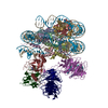
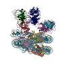
| Title | Structural basis of COMPASS eCM recognition of an unmodified nucleosome | |||||||||
 Map data Map data | COMPASS eCM/unmodified ribosome | |||||||||
 Sample Sample |
| |||||||||
 Keywords Keywords | Complex / methyltransferase / epigenetics / chromatin / nucleosome / TRANSFERASE-STRUCTURAL PROTEIN-DNA complex | |||||||||
| Function / homology |  Function and homology information Function and homology information[histone H3]-lysine4 N-trimethyltransferase / histone H3K4 trimethyltransferase activity / Set1C/COMPASS complex / : / structural constituent of chromatin / nucleosome / chromosome / methylation / histone binding / transcription cis-regulatory region binding ...[histone H3]-lysine4 N-trimethyltransferase / histone H3K4 trimethyltransferase activity / Set1C/COMPASS complex / : / structural constituent of chromatin / nucleosome / chromosome / methylation / histone binding / transcription cis-regulatory region binding / protein heterodimerization activity / positive regulation of DNA-templated transcription / DNA binding / RNA binding / nucleus / metal ion binding Similarity search - Function | |||||||||
| Biological species |  Kluyveromyces lactis (strain ATCC 8585 / CBS 2359 / DSM 70799 / NBRC 1267 / NRRL Y-1140 / WM37) (yeast) / synthetic construct (others) Kluyveromyces lactis (strain ATCC 8585 / CBS 2359 / DSM 70799 / NBRC 1267 / NRRL Y-1140 / WM37) (yeast) / synthetic construct (others) | |||||||||
| Method | single particle reconstruction / cryo EM / Resolution: 3.7 Å | |||||||||
 Authors Authors | Hsu PL / Shi H | |||||||||
| Funding support |  United States, 2 items United States, 2 items
| |||||||||
 Citation Citation | Journal: Mol Cell / Year: 2019 Title: Structural Basis of H2B Ubiquitination-Dependent H3K4 Methylation by COMPASS. Authors: Peter L Hsu / Hui Shi / Calvin Leonen / Jianming Kang / Champak Chatterjee / Ning Zheng / Abstract: The COMPASS (complex of proteins associated with Set1) complex represents the prototype of the SET1/MLL family of methyltransferases that controls gene transcription by H3K4 methylation (H3K4me). ...The COMPASS (complex of proteins associated with Set1) complex represents the prototype of the SET1/MLL family of methyltransferases that controls gene transcription by H3K4 methylation (H3K4me). Although H2B monoubiquitination (H2Bub) is well known as a prerequisite histone mark for COMPASS activity, how H2Bub activates COMPASS remains unclear. Here, we report the cryoelectron microscopy (cryo-EM) structures of an extended COMPASS catalytic module (CM) bound to the H2Bub and free nucleosome. The COMPASS CM clamps onto the nucleosome disk-face via an extensive interface to capture the flexible H3 N-terminal tail. The interface also sandwiches a critical Set1 arginine-rich motif (ARM) that autoinhibits COMPASS. Unexpectedly, without enhancing COMPASS-nucleosome interaction, H2Bub activates the enzymatic assembly by packing against Swd1 and alleviating the inhibitory effect of the Set1 ARM upon fastening it to the acidic patch. By delineating the spatial configuration of the COMPASS-H2Bub-nucleosome assembly, our studies establish the structural framework for understanding the long-studied H2Bub-H3K4me histone modification crosstalk. | |||||||||
| History |
|
- Structure visualization
| Movie |
Movie viewer |
|---|---|
| Structure viewer | EM map: SurfViewMolmilJmol/JSmol |
| Supplemental images |

- Downloads & links
-EMDB archive
| Map data | emd_20765.map.gz | 10.2 MB | EMDB map data format | |
|---|---|---|---|---|
| Header (meta data) | emd-20765-v30.xmlemd-20765.xml | 30 KB 30 KB | Display Display | EMDB header |
| Images | 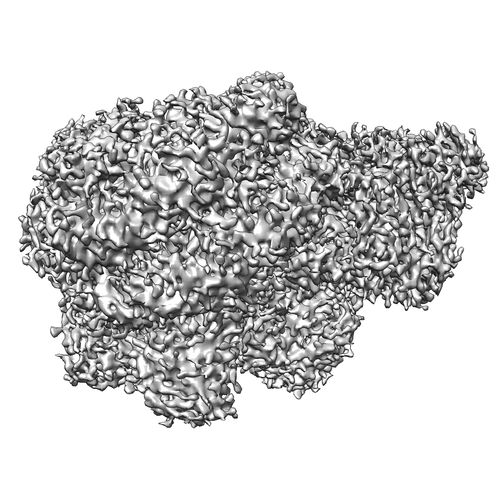 emd_20765.png emd_20765.png | 218.1 KB | ||
| Filedesc metadata | emd-20765.cif.gz | 8.1 KB | ||
| Archive directory |  http://ftp.pdbj.org/pub/emdb/structures/EMD-20765ftp://ftp.pdbj.org/pub/emdb/structures/EMD-20765 http://ftp.pdbj.org/pub/emdb/structures/EMD-20765ftp://ftp.pdbj.org/pub/emdb/structures/EMD-20765 | HTTPS FTP |
-Validation report
| Summary document | emd_20765_validation.pdf.gz | 377.4 KB | Display | EMDB validaton report |
|---|---|---|---|---|
| Full document | emd_20765_full_validation.pdf.gz | 377 KB | Display | |
| Data in XML | emd_20765_validation.xml.gz | 6.5 KB | Display | |
| Data in CIF | emd_20765_validation.cif.gz | 7.4 KB | Display | |
| Arichive directory | https://ftp.pdbj.org/pub/emdb/validation_reports/EMD-20765ftp://ftp.pdbj.org/pub/emdb/validation_reports/EMD-20765 | HTTPS FTP |
-Related structure data
| Related structure data |  6ugmMC  6uh5C C: citing same article ( M: atomic model generated by this map |
|---|---|
| Similar structure data |


-Links
| EMDB pages | EMDB (EBI/PDBe) / EMDataResource |
|---|---|
| Related items in Molecule of the Month |


-Map
| File | Download / File: emd_20765.map.gz / Format: CCP4 / Size: 129.7 MB / Type: IMAGE STORED AS FLOATING POINT NUMBER (4 BYTES) | ||||||||||||||||||||||||||||||||||||||||||||||||||||||||||||||||||||
|---|---|---|---|---|---|---|---|---|---|---|---|---|---|---|---|---|---|---|---|---|---|---|---|---|---|---|---|---|---|---|---|---|---|---|---|---|---|---|---|---|---|---|---|---|---|---|---|---|---|---|---|---|---|---|---|---|---|---|---|---|---|---|---|---|---|---|---|---|---|
| Annotation | COMPASS eCM/unmodified ribosome | ||||||||||||||||||||||||||||||||||||||||||||||||||||||||||||||||||||
| Projections & slices | Image control
Images are generated by Spider. | ||||||||||||||||||||||||||||||||||||||||||||||||||||||||||||||||||||
| Voxel size | X=Y=Z: 1.056 Å | ||||||||||||||||||||||||||||||||||||||||||||||||||||||||||||||||||||
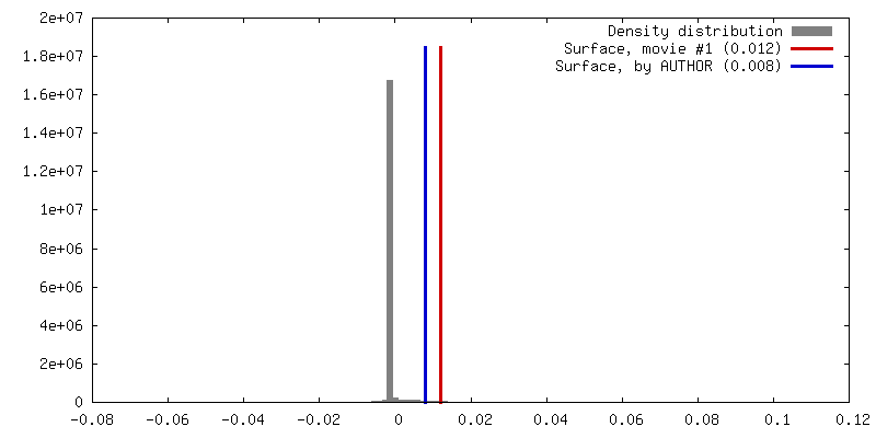
| Density |
| ||||||||||||||||||||||||||||||||||||||||||||||||||||||||||||||||||||
| Symmetry | Space group: 1 | ||||||||||||||||||||||||||||||||||||||||||||||||||||||||||||||||||||
| Details | EMDB XML:
CCP4 map header:
| ||||||||||||||||||||||||||||||||||||||||||||||||||||||||||||||||||||
 Z (Sec.)
 Y (Row.)
 X (Col.)


-Supplemental data
- Sample components
+Entire : Yeast COMPASS eCM bound to an unmodified nucleosome
+Supramolecule #1: Yeast COMPASS eCM bound to an unmodified nucleosome
+Supramolecule #2: nucleosome
+Supramolecule #3: COMPASS eCM
+Macromolecule #1: Histone H3

+Macromolecule #2: Histone H4
+Macromolecule #3: Histone H2A
+Macromolecule #4: Histone H2B
+Macromolecule #5: Histone H2B 1.1
+Macromolecule #8: Swd3
 Trichoplusia ni (cabbage looper)
+Macromolecule #9: Histone-lysine N-methyltransferase, H3 lysine-4 specific
+Macromolecule #10: Swd1
+Macromolecule #11: H3 N-terminus
+Macromolecule #12: Spp1
+Macromolecule #13: Bre2
+Macromolecule #14: Sdc1
+Macromolecule #6: DNA (146-MER)
+Macromolecule #7: DNA (146-MER)
+Macromolecule #15: S-ADENOSYLMETHIONINE

+Macromolecule #16: ZINC ION
-Experimental details
-Structure determination
| Method | cryo EM |
|---|---|
 Processing Processing | single particle reconstruction |
| Aggregation state | particle |
-Sample preparation
| Buffer | pH: 7.5 |
|---|---|
| Grid | Details: unspecified |
| Vitrification | Cryogen name: ETHANE |
- Electron microscopy
| Microscope | FEI TITAN KRIOS |
|---|---|
| Image recording | Film or detector model: GATAN K2 SUMMIT (4k x 4k) / Average electron dose: 74.0 e/Å2 |
| Electron beam | Acceleration voltage: 300 kV / Electron source:  FIELD EMISSION GUN FIELD EMISSION GUN |
| Electron optics | Illumination mode: FLOOD BEAM / Imaging mode: BRIGHT FIELD |
| Experimental equipment |  Model: Titan Krios / Image courtesy: FEI Company |
-Image processing
| Startup model | Type of model: NONE |
|---|---|
| Final reconstruction | Applied symmetry - Point group: C1 (asymmetric) / Resolution.type: BY AUTHOR / Resolution: 3.7 Å / Resolution method: FSC 0.143 CUT-OFF / Number images used: 100905 |
| Initial angle assignment | Type: MAXIMUM LIKELIHOOD |
| Final angle assignment | Type: MAXIMUM LIKELIHOOD |
