+ Open data
- Basic information
| Entry | Database: EMDB / ID: EMD-20555 | |||||||||
|---|---|---|---|---|---|---|---|---|---|---|
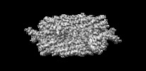
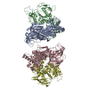
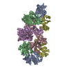
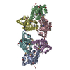
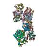
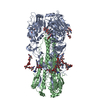
| Title | The cryo-EM structure of the SNX-BAR Mvp1 tetramer | |||||||||
 Map data Map data | Mvp1 tetramer, masked and sharpend | |||||||||
 Sample Sample |
| |||||||||
 Keywords Keywords | Mvp1 / sorting nexin / SNX / PX / BAR / SNX-BAR / LIPID BINDING PROTEIN | |||||||||
| Function / homology |  Function and homology information Function and homology informationplasma membrane tubulation / protein targeting to vacuole / phosphatidylinositol-3-phosphate binding / retrograde transport, endosome to Golgi / endosome / identical protein binding / membrane / nucleus / cytosol / cytoplasm Similarity search - Function | |||||||||
| Biological species |   | |||||||||
| Method | single particle reconstruction / cryo EM / Resolution: 4.2 Å | |||||||||
 Authors Authors | Sun D / Ford MGJ | |||||||||
| Funding support |  United States, 2 items United States, 2 items
| |||||||||
 Citation Citation |  Journal: Nat Commun / Year: 2020 Journal: Nat Commun / Year: 2020Title: The cryo-EM structure of the SNX-BAR Mvp1 tetramer. Authors: Dapeng Sun / Natalia V Varlakhanova / Bryan A Tornabene / Rajesh Ramachandran / Peijun Zhang / Marijn G J Ford /   Abstract: Sorting nexins (SNX) are a family of PX domain-containing proteins with pivotal roles in trafficking and signaling. SNX-BARs, which also have a curvature-generating Bin/Amphiphysin/Rvs (BAR) domain, ...Sorting nexins (SNX) are a family of PX domain-containing proteins with pivotal roles in trafficking and signaling. SNX-BARs, which also have a curvature-generating Bin/Amphiphysin/Rvs (BAR) domain, have membrane-remodeling functions, particularly at the endosome. The minimal PX-BAR module is a dimer mediated by BAR-BAR interactions. Many SNX-BAR proteins, however, additionally have low-complexity N-terminal regions of unknown function. Here, we present the cryo-EM structure of the full-length SNX-BAR Mvp1, which is an autoinhibited tetramer. The tetramer is a dimer of dimers, wherein the membrane-interacting BAR surfaces are sequestered and the PX lipid-binding sites are occluded. The N-terminal low-complexity region of Mvp1 is essential for tetramerization. Mvp1 lacking its N-terminus is dimeric and exhibits enhanced membrane association. Membrane binding and remodeling by Mvp1 therefore requires unmasking of the PX and BAR domain lipid-interacting surfaces. This work reveals a tetrameric configuration of a SNX-BAR protein that provides critical insight into SNX-BAR function and regulation. | |||||||||
| History |
|
- Structure visualization
| Movie |
 Movie viewer Movie viewer |
|---|---|
| Structure viewer | EM map:  SurfView SurfView Molmil Molmil Jmol/JSmol Jmol/JSmol |
| Supplemental images |
- Downloads & links
-EMDB archive
| Map data |  emd_20555.map.gz emd_20555.map.gz | 59.5 MB |  EMDB map data format EMDB map data format | |
|---|---|---|---|---|
| Header (meta data) |  emd-20555-v30.xml emd-20555-v30.xml emd-20555.xml emd-20555.xml | 11.9 KB 11.9 KB | Display Display |  EMDB header EMDB header |
| FSC (resolution estimation) |  emd_20555_fsc.xml emd_20555_fsc.xml | 9.1 KB | Display |  FSC data file FSC data file |
| Images |  emd_20555.png emd_20555.png | 177.4 KB | ||
| Filedesc metadata |  emd-20555.cif.gz emd-20555.cif.gz | 5.6 KB | ||
| Archive directory |  http://ftp.pdbj.org/pub/emdb/structures/EMD-20555 http://ftp.pdbj.org/pub/emdb/structures/EMD-20555 ftp://ftp.pdbj.org/pub/emdb/structures/EMD-20555 ftp://ftp.pdbj.org/pub/emdb/structures/EMD-20555 | HTTPS FTP |
-Validation report
| Summary document |  emd_20555_validation.pdf.gz emd_20555_validation.pdf.gz | 595.2 KB | Display |  EMDB validaton report EMDB validaton report |
|---|---|---|---|---|
| Full document |  emd_20555_full_validation.pdf.gz emd_20555_full_validation.pdf.gz | 594.8 KB | Display | |
| Data in XML |  emd_20555_validation.xml.gz emd_20555_validation.xml.gz | 11.2 KB | Display | |
| Data in CIF |  emd_20555_validation.cif.gz emd_20555_validation.cif.gz | 14.6 KB | Display | |
| Arichive directory |  https://ftp.pdbj.org/pub/emdb/validation_reports/EMD-20555 https://ftp.pdbj.org/pub/emdb/validation_reports/EMD-20555 ftp://ftp.pdbj.org/pub/emdb/validation_reports/EMD-20555 ftp://ftp.pdbj.org/pub/emdb/validation_reports/EMD-20555 | HTTPS FTP |
-Related structure data
| Related structure data |  6q0xMC M: atomic model generated by this map C: citing same article ( |
|---|---|
| Similar structure data |
- Links
Links
| EMDB pages |  EMDB (EBI/PDBe) / EMDB (EBI/PDBe) /  EMDataResource EMDataResource |
|---|
- Map
Map
| File |  Download / File: emd_20555.map.gz / Format: CCP4 / Size: 64 MB / Type: IMAGE STORED AS FLOATING POINT NUMBER (4 BYTES) Download / File: emd_20555.map.gz / Format: CCP4 / Size: 64 MB / Type: IMAGE STORED AS FLOATING POINT NUMBER (4 BYTES) | ||||||||||||||||||||||||||||||||||||||||||||||||||||||||||||||||||||
|---|---|---|---|---|---|---|---|---|---|---|---|---|---|---|---|---|---|---|---|---|---|---|---|---|---|---|---|---|---|---|---|---|---|---|---|---|---|---|---|---|---|---|---|---|---|---|---|---|---|---|---|---|---|---|---|---|---|---|---|---|---|---|---|---|---|---|---|---|---|
| Annotation | Mvp1 tetramer, masked and sharpend | ||||||||||||||||||||||||||||||||||||||||||||||||||||||||||||||||||||
| Projections & slices | Image control
Images are generated by Spider. | ||||||||||||||||||||||||||||||||||||||||||||||||||||||||||||||||||||
| Voxel size | X=Y=Z: 1.056 Å | ||||||||||||||||||||||||||||||||||||||||||||||||||||||||||||||||||||
| Density |
| ||||||||||||||||||||||||||||||||||||||||||||||||||||||||||||||||||||
| Symmetry | Space group: 1 | ||||||||||||||||||||||||||||||||||||||||||||||||||||||||||||||||||||
| Details | EMDB XML:
CCP4 map header:
| ||||||||||||||||||||||||||||||||||||||||||||||||||||||||||||||||||||
-Supplemental data
- Sample components
-Entire : Mvp1 tetramer
| Entire | Name: Mvp1 tetramer |
|---|---|
| Components |
|
-Supramolecule #1: Mvp1 tetramer
| Supramolecule | Name: Mvp1 tetramer / type: complex / ID: 1 / Parent: 0 / Macromolecule list: all / Details: recombinant purified Mvp1 |
|---|---|
| Source (natural) | Organism:  |
| Molecular weight | Theoretical: 242.362 KDa |
-Macromolecule #1: Sorting nexin MVP1
| Macromolecule | Name: Sorting nexin MVP1 / type: protein_or_peptide / ID: 1 / Number of copies: 4 / Enantiomer: LEVO |
|---|---|
| Source (natural) | Organism:  |
| Molecular weight | Theoretical: 61.902852 KDa |
| Recombinant expression | Organism:  |
| Sequence | String: MGSSHHHHHH SSGLVPRGSH MDNYEGSDPW NTSSNAWTKD DDHVVSTTNS EPSLNGISGE FNTLNFSTPL DTNEEDTGFL PTNDVLEES IWDDSRNPLG ATGMSQTPNI AANETVIDKN DARDQNIEES EADLLDWTNN VRKTYRPLDA DIIIIEEIPE R EGLLFKHA ...String: MGSSHHHHHH SSGLVPRGSH MDNYEGSDPW NTSSNAWTKD DDHVVSTTNS EPSLNGISGE FNTLNFSTPL DTNEEDTGFL PTNDVLEES IWDDSRNPLG ATGMSQTPNI AANETVIDKN DARDQNIEES EADLLDWTNN VRKTYRPLDA DIIIIEEIPE R EGLLFKHA NYLVKHLIAL PSTSPSEERT VVRRYSDFLW LREILLKRYP FRMIPELPPK RIGSQNADQL FLKKRRIGLS RF INLVMKH PKLSNDDLVL TFLTVRTDLT SWRKQATYDT SNEFADKKIS QEFMKMWKKE FAEQWNQAAS CIDTSMELWY RIT LLLERH EKRIMQMVHE RNFFETLVDN FSEVTPKLYP VQQNDTILDI NNNLSIIKKH LETTSSICKQ ETEEISGTLS PKFK IFTDI LLSLRSLFER YKIMAANNVV ELQRHVELNK EKLESMKGKP DVSGAEYDRI KKIIQKDRRS IIEQSNRAWL IRQCI LEEF TIFQETQFLI TRAFQDWAKL NSNHAGLKLN EWEKLVTSIM DMPISRE UniProtKB: Sorting nexin MVP1 |
-Experimental details
-Structure determination
| Method | cryo EM |
|---|---|
 Processing Processing | single particle reconstruction |
| Aggregation state | particle |
- Sample preparation
| Concentration | 0.32 mg/mL |
|---|---|
| Buffer | pH: 7.5 |
| Grid | Details: unspecified |
| Vitrification | Cryogen name: ETHANE |
- Electron microscopy
| Microscope | FEI TITAN KRIOS |
|---|---|
| Image recording | Film or detector model: GATAN K2 SUMMIT (4k x 4k) / Detector mode: COUNTING / Average electron dose: 58.0 e/Å2 |
| Electron beam | Acceleration voltage: 300 kV / Electron source:  FIELD EMISSION GUN FIELD EMISSION GUN |
| Electron optics | Illumination mode: FLOOD BEAM / Imaging mode: BRIGHT FIELD |
| Experimental equipment |  Model: Titan Krios / Image courtesy: FEI Company |
+ Image processing
-Atomic model buiding 1
| Initial model |
| ||||||||||
|---|---|---|---|---|---|---|---|---|---|---|---|
| Refinement | Space: REAL / Protocol: AB INITIO MODEL / Overall B value: 88.91 / Target criteria: Overall correlation coefficients | ||||||||||
| Output model |  PDB-6q0x: |
 Movie
Movie Controller





 X (Sec.)
 Y (Row.)
 Z (Col.)


