 Movie
Movie Controller


+ Open data

- Basic information
| Entry | Database: EMDB / ID: EMD-1949 | |||||||||
|---|---|---|---|---|---|---|---|---|---|---|
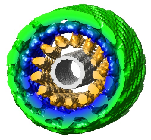
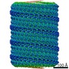
| Title | Human dynamin 1 deltaPRD polymer stabilized with GMPPCP | |||||||||
 Map data Map data | Three-dimensional volume of deltaPRD human dynamin 1 polymer | |||||||||
 Sample Sample |
| |||||||||
 Keywords Keywords | dynamin / endocytosis / GTP hydrolysis / membrane remodeling | |||||||||
| Function / homology |  Function and homology information Function and homology informationclathrin coat assembly involved in endocytosis / vesicle scission / synaptic vesicle budding from presynaptic endocytic zone membrane / interleukin-27-mediated signaling pathway / dynamin GTPase / chromaffin granule / regulation of vesicle size / Retrograde neurotrophin signalling / Toll Like Receptor 4 (TLR4) Cascade / endosome organization ...clathrin coat assembly involved in endocytosis / vesicle scission / synaptic vesicle budding from presynaptic endocytic zone membrane / interleukin-27-mediated signaling pathway / dynamin GTPase / chromaffin granule / regulation of vesicle size / Retrograde neurotrophin signalling / Toll Like Receptor 4 (TLR4) Cascade / endosome organization / Formation of annular gap junctions / Gap junction degradation / response to type I interferon / negative regulation of viral genome replication / phosphatidylinositol-3,4,5-trisphosphate binding / Recycling pathway of L1 / EPH-ephrin mediated repulsion of cells / endocytic vesicle / phosphatidylinositol-4,5-bisphosphate binding / clathrin-coated pit / MHC class II antigen presentation / receptor-mediated endocytosis / antiviral innate immune response / defense response / protein homooligomerization / ISG15 antiviral mechanism / response to virus / endocytosis / Interferon alpha/beta signaling / GDP binding / Clathrin-mediated endocytosis / presynapse / nuclear membrane / microtubule binding / defense response to virus / protein homotetramerization / microtubule / innate immune response / GTPase activity / apoptotic process / synapse / endoplasmic reticulum membrane / protein kinase binding / GTP binding / perinuclear region of cytoplasm / signal transduction / protein homodimerization activity / RNA binding / extracellular exosome / identical protein binding / nucleus / plasma membrane / cytoplasm / cytosol Similarity search - Function | |||||||||
| Biological species |  Homo sapiens (human) Homo sapiens (human) | |||||||||
| Method | helical reconstruction / cryo EM / Resolution: 12.2 Å | |||||||||
 Authors Authors | Chappie JS / Mears JA / Fang S / Leonard M / Schmid SL / Milligan RA / Hinshaw JE / Dyda F | |||||||||
 Citation Citation | Journal: Cell / Year: 2011 Title: A pseudoatomic model of the dynamin polymer identifies a hydrolysis-dependent powerstroke. Authors: Joshua S Chappie / Jason A Mears / Shunming Fang / Marilyn Leonard / Sandra L Schmid / Ronald A Milligan / Jenny E Hinshaw / Fred Dyda /  Abstract: The GTPase dynamin catalyzes membrane fission by forming a collar around the necks of clathrin-coated pits, but the specific structural interactions and conformational changes that drive this process ...The GTPase dynamin catalyzes membrane fission by forming a collar around the necks of clathrin-coated pits, but the specific structural interactions and conformational changes that drive this process remain a mystery. We present the GMPPCP-bound structures of the truncated human dynamin 1 helical polymer at 12.2 Å and a fusion protein, GG, linking human dynamin 1's catalytic G domain to its GTPase effector domain (GED) at 2.2 Å. The structures reveal the position and connectivity of dynamin fragments in the assembled structure, showing that G domain dimers only form between tetramers in sequential rungs of the dynamin helix. Using chemical crosslinking, we demonstrate that dynamin tetramers are made of two dimers, in which the G domain of one molecule interacts in trans with the GED of another. Structural comparison of GG(GMPPCP) to the GG transition-state complex identifies a hydrolysis-dependent powerstroke that may play a role in membrane-remodeling events necessary for fission. | |||||||||
| History |
|
- Structure visualization
| Movie |
Movie viewer |
|---|---|
| Structure viewer | EM map: SurfViewMolmilJmol/JSmol |
| Supplemental images |
- Downloads & links
-EMDB archive
| Map data | emd_1949.map.gz | 7.2 MB | EMDB map data format | |
|---|---|---|---|---|
| Header (meta data) | emd-1949-v30.xmlemd-1949.xml | 14.8 KB 14.8 KB | Display Display | EMDB header |
| Images | emd_1949.tif | 360.3 KB | ||
| Archive directory |  http://ftp.pdbj.org/pub/emdb/structures/EMD-1949ftp://ftp.pdbj.org/pub/emdb/structures/EMD-1949 http://ftp.pdbj.org/pub/emdb/structures/EMD-1949ftp://ftp.pdbj.org/pub/emdb/structures/EMD-1949 | HTTPS FTP |
-Related structure data
| Related structure data |  3zysMC  3zycC M: atomic model generated by this map C: citing same article ( |
|---|---|
| Similar structure data |

-Links
| EMDB pages | EMDB (EBI/PDBe) / EMDataResource |
|---|---|
| Related items in Molecule of the Month |


-Map
| File | Download / File: emd_1949.map.gz / Format: CCP4 / Size: 29.8 MB / Type: IMAGE STORED AS FLOATING POINT NUMBER (4 BYTES) | ||||||||||||||||||||||||||||||||||||||||||||||||||||||||||||||||||||
|---|---|---|---|---|---|---|---|---|---|---|---|---|---|---|---|---|---|---|---|---|---|---|---|---|---|---|---|---|---|---|---|---|---|---|---|---|---|---|---|---|---|---|---|---|---|---|---|---|---|---|---|---|---|---|---|---|---|---|---|---|---|---|---|---|---|---|---|---|---|
| Annotation | Three-dimensional volume of deltaPRD human dynamin 1 polymer | ||||||||||||||||||||||||||||||||||||||||||||||||||||||||||||||||||||
| Projections & slices | Image control
Images are generated by Spider. | ||||||||||||||||||||||||||||||||||||||||||||||||||||||||||||||||||||
| Voxel size | X=Y=Z: 2.26 Å | ||||||||||||||||||||||||||||||||||||||||||||||||||||||||||||||||||||
| Density |
| ||||||||||||||||||||||||||||||||||||||||||||||||||||||||||||||||||||
| Symmetry | Space group: 1 | ||||||||||||||||||||||||||||||||||||||||||||||||||||||||||||||||||||
| Details | EMDB XML:
CCP4 map header:
| ||||||||||||||||||||||||||||||||||||||||||||||||||||||||||||||||||||
 Z (Sec.)
 Y (Row.)
 X (Col.)


-Supplemental data
- Sample components
-Entire : GMPPCP-stablized human dynamin 1 delta PRD polymer
| Entire | Name: GMPPCP-stablized human dynamin 1 delta PRD polymer |
|---|---|
| Components |
|
-Supramolecule #1000: GMPPCP-stablized human dynamin 1 delta PRD polymer
| Supramolecule | Name: GMPPCP-stablized human dynamin 1 delta PRD polymer / type: sample / ID: 1000 / Oligomeric state: Helical assembly of dynamin tetramers / Number unique components: 3 |
|---|
-Macromolecule #1: Human dynamin 1 delta PRD
| Macromolecule | Name: Human dynamin 1 delta PRD / type: protein_or_peptide / ID: 1 / Name.synonym: Human dynamin 1 delta PRD / Details: contains bound GMPPCP / Oligomeric state: Dimer / Recombinant expression: Yes |
|---|---|
| Source (natural) | Organism: Homo sapiens (human) / synonym: Human / Location in cell: Plasma membrane and cytosol |
| Recombinant expression | Organism:  |
-Macromolecule #2: Human dynamin 1 pleckstrin homology domain
| Macromolecule | Name: Human dynamin 1 pleckstrin homology domain / type: protein_or_peptide / ID: 2 / Name.synonym: Human dynamin 1 pleckstrin homology domain / Oligomeric state: Dimer / Recombinant expression: Yes |
|---|---|
| Source (natural) | Organism: Homo sapiens (human) / synonym: Human / Location in cell: Plasma membrane and cytosol |
| Recombinant expression | Organism: |
-Macromolecule #3: Interferon-induced GTP-binding protein Mx1
| Macromolecule | Name: Interferon-induced GTP-binding protein Mx1 / type: protein_or_peptide / ID: 3 / Name.synonym: Interferon-induced GTP-binding protein Mx1 / Oligomeric state: Dimer / Recombinant expression: Yes |
|---|---|
| Source (natural) | Organism: Homo sapiens (human) / synonym: Human |
| Recombinant expression | Organism: |
-Experimental details
-Structure determination
| Method | cryo EM |
|---|---|
 Processing Processing | helical reconstruction |
| Aggregation state | filament |
-Sample preparation
| Concentration | 0.5 mg/mL |
|---|---|
| Buffer | pH: 7.5 Details: 20 mM Hepes pH 7.5, 50 mM NaCl, 2 mM EGTA, 4 mM MgCl2, 1 mM DTT, 1 mg/ml 0.4 um 1,2-dioleoyl-sn-glycero-3-phospho-L-serine (DOPS) liposomes, 2 mM GMPPCP |
| Grid | Details: 400 mesh C-flat grids |
| Vitrification | Cryogen name: ETHANE / Chamber humidity: 100 % / Chamber temperature: 93 K / Instrument: HOMEMADE PLUNGER / Details: Vitrification instrument: Manual Method: Absorbed samples to grids, blotted, washed with 20 mM Hepes pH 7.5, blotted and plunged. |
- Electron microscopy
| Microscope | FEI TECNAI 20 |
|---|---|
| Temperature | Min: 93 K / Max: 95 K |
| Alignment procedure | Legacy - Astigmatism: objective lens astigmatism was corrected at 100,000X magnification |
| Image recording | Category: CCD / Film or detector model: GATAN ULTRASCAN 4000 (4k x 4k) / Average electron dose: 10 e/Å2 / Details: Images were collected on CCD |
| Tilt angle min | 0 |
| Tilt angle max | 0 |
| Electron beam | Acceleration voltage: 120 kV / Electron source:  FIELD EMISSION GUN FIELD EMISSION GUN |
| Electron optics | Illumination mode: FLOOD BEAM / Imaging mode: BRIGHT FIELD / Cs: 2 mm / Nominal defocus max: 2.0 µm / Nominal defocus min: 0.5 µm / Nominal magnification: 50000 |
| Sample stage | Specimen holder: Gatan 626 side entry cryo-stage / Specimen holder model: GATAN LIQUID NITROGEN |
-Image processing
| Final reconstruction | Applied symmetry - Helical parameters - Δz: 7.52 Å Applied symmetry - Helical parameters - Δ&Phi: 27.3 ° Algorithm: OTHER / Resolution.type: BY AUTHOR / Resolution: 12.2 Å / Resolution method: FSC 0.5 CUT-OFF / Software - Name: Spider Details: A total of 4,814 helical segments were incorporated by the IHRSR algorithm into the final reconstruction after 50 cycles |
|---|---|
| CTF correction | Details: Each image using ACE2 |
-Atomic model buiding 1
| Initial model | PDB ID: Chain - Chain ID: A |
|---|---|
| Software | Name: YUP |
| Details | PDBEntryID_givenInChain. Protocol: Manual and Flexible fitting. Models were initially placed manually and initial positions were refined using the YUP.SCX method of the YUP software package |
| Refinement | Space: REAL / Protocol: FLEXIBLE FIT |
| Output model | PDB-3zys: |
-Atomic model buiding 2
| Initial model | PDB ID: Chain - Chain ID: A |
|---|---|
| Software | Name: YUP |
| Details | PDBEntryID_givenInChain. Protocol: Manual and Flexible fitting. Models were initially placed manually and initial positions were refined using the YUP.SCX method of the YUP software package |
| Refinement | Space: REAL / Protocol: FLEXIBLE FIT |
| Output model | PDB-3zys: |

-Atomic model buiding 3
| Initial model | PDB ID: Chain - Chain ID: A |
|---|---|
| Software | Name: YUP |
| Details | PDBEntryID_givenInChain. Protocol: Manual and Flexible fitting. Models were initially placed manually and initial positions were refined using the YUP.SCX method of the YUP software package |
| Refinement | Space: REAL / Protocol: FLEXIBLE FIT |
| Output model | PDB-3zys: |

