 Movie
Movie Controller

[English] 日本語
 Yorodumi
- EMDB-19036: Cryo-EM structure of the Emiliania huxleyi virus 201 (EhV-201) vi... -
+ Open data

- Basic information
| Entry |  | |||||||||
|---|---|---|---|---|---|---|---|---|---|---|
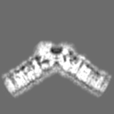
| Title | Cryo-EM structure of the Emiliania huxleyi virus 201 (EhV-201) virion vertex with a diameter of 50 nm and a mask applied on the capsid layer. | |||||||||
 Map data Map data | Result from postprocessing masked by tight mask to remove surrounding noise. | |||||||||
 Sample Sample |
| |||||||||
 Keywords Keywords | cryo-EM / subtomogram averaging / EhV-201 / enveloped virus / capsid / major capsid protein / VIRUS | |||||||||
| Function / homology |  Function and homology information Function and homology information | |||||||||
| Biological species |  Emiliania huxleyi virus 201 Emiliania huxleyi virus 201 | |||||||||
| Method | subtomogram averaging / cryo EM / Resolution: 12.0 Å | |||||||||
 Authors Authors | Homola M / Buttner CR / Fuzik T / Novacek J / Chaillet M / Forster F / Plevka P | |||||||||
| Funding support |  Czech Republic, European Union, 2 items Czech Republic, European Union, 2 items
| |||||||||
 Citation Citation | Journal: Sci Adv / Year: 2024 Title: Structure and replication cycle of a virus infecting climate-modulating alga . Authors: Miroslav Homola / Carina R Büttner / Tibor Füzik / Pavel Křepelka / Radka Holbová / Jiří Nováček / Marten L Chaillet / Jakub Žák / Danyil Grybchuk / Friedrich Förster / William H ...Authors: Miroslav Homola / Carina R Büttner / Tibor Füzik / Pavel Křepelka / Radka Holbová / Jiří Nováček / Marten L Chaillet / Jakub Žák / Danyil Grybchuk / Friedrich Förster / William H Wilson / Declan C Schroeder / Pavel Plevka /    Abstract: The globally distributed marine alga has cooling effect on the Earth's climate. The population density of is restricted by viruses, including virus 201 (EhV-201). Despite the impact of viruses ...The globally distributed marine alga has cooling effect on the Earth's climate. The population density of is restricted by viruses, including virus 201 (EhV-201). Despite the impact of viruses on the climate, there is limited information about their structure and replication. Here, we show that the dsDNA genome inside the EhV-201 virion is protected by an inner membrane, capsid, and outer membrane. EhV-201 virions infect by using fivefold vertices to bind to and fuse the virus' inner membrane with the cell plasma membrane. Progeny virions assemble in the cytoplasm at the surface of endoplasmic reticulum-derived membrane segments. Genome packaging initiates synchronously with the capsid assembly and completes through an aperture in the forming capsid. The genome-filled capsids acquire an outer membrane by budding into intracellular vesicles. EhV-201 infection induces a loss of surface protective layers from cells, which enables the continuous release of virions by exocytosis. | |||||||||
| History |
|
- Structure visualization
| Supplemental images |
|---|

- Downloads & links
-EMDB archive
| Map data | emd_19036.map.gz | 983.8 KB | EMDB map data format | |
|---|---|---|---|---|
| Header (meta data) | emd-19036-v30.xmlemd-19036.xml | 25.4 KB 25.4 KB | Display Display | EMDB header |
| FSC (resolution estimation) | emd_19036_fsc.xml | 5.9 KB | Display | FSC data file |
| Images | 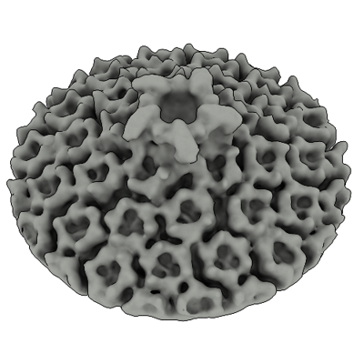 emd_19036.png emd_19036.png | 137.9 KB | ||
| Masks | emd_19036_msk_1.map | 8 MB | Mask map | |
| Filedesc metadata | emd-19036.cif.gz | 7.6 KB | ||
| Others | emd_19036_additional_1.map.gzemd_19036_half_map_1.map.gzemd_19036_half_map_2.map.gz | 1.8 MB 5.9 MB 5.9 MB | ||
| Archive directory |  http://ftp.pdbj.org/pub/emdb/structures/EMD-19036ftp://ftp.pdbj.org/pub/emdb/structures/EMD-19036 http://ftp.pdbj.org/pub/emdb/structures/EMD-19036ftp://ftp.pdbj.org/pub/emdb/structures/EMD-19036 | HTTPS FTP |
-Related structure data
| Related structure data |  8rbtMC 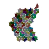 8rbsC M: atomic model generated by this map C: citing same article ( |
|---|---|
| Similar structure data |


-Links
| EMDB pages | EMDB (EBI/PDBe) / EMDataResource |
|---|---|
| Related items in Molecule of the Month |


-Map
| File | Download / File: emd_19036.map.gz / Format: CCP4 / Size: 8 MB / Type: IMAGE STORED AS FLOATING POINT NUMBER (4 BYTES) | ||||||||||||||||||||||||||||||||||||
|---|---|---|---|---|---|---|---|---|---|---|---|---|---|---|---|---|---|---|---|---|---|---|---|---|---|---|---|---|---|---|---|---|---|---|---|---|---|
| Annotation | Result from postprocessing masked by tight mask to remove surrounding noise. | ||||||||||||||||||||||||||||||||||||
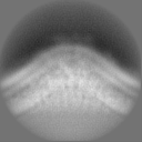
| Projections & slices | Image control
Images are generated by Spider. | ||||||||||||||||||||||||||||||||||||
| Voxel size | X=Y=Z: 4.16 Å | ||||||||||||||||||||||||||||||||||||
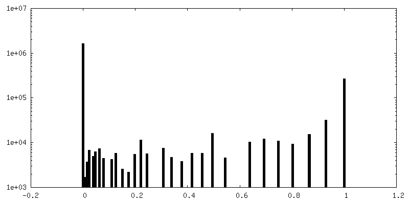
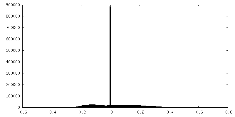
| Density |
| ||||||||||||||||||||||||||||||||||||
| Symmetry | Space group: 1 | ||||||||||||||||||||||||||||||||||||
| Details | EMDB XML:
|
 Z (Sec.)
 Y (Row.)
 X (Col.)


-Supplemental data
-Mask #1
| File | emd_19036_msk_1.map | ||||||||||||
|---|---|---|---|---|---|---|---|---|---|---|---|---|---|
| Projections & Slices |
| ||||||||||||
| Density Histograms |


-Additional map: Result from postprocessing - masked.
| File | emd_19036_additional_1.map | ||||||||||||
|---|---|---|---|---|---|---|---|---|---|---|---|---|---|
| Annotation | Result from postprocessing - masked. | ||||||||||||
| Projections & Slices |
| ||||||||||||
| Density Histograms |


-Half map: halfMap 1
| File | emd_19036_half_map_1.map | ||||||||||||
|---|---|---|---|---|---|---|---|---|---|---|---|---|---|
| Annotation | halfMap_1 | ||||||||||||
| Projections & Slices |
| ||||||||||||
| Density Histograms |


-Half map: halfMap 2
| File | emd_19036_half_map_2.map | ||||||||||||
|---|---|---|---|---|---|---|---|---|---|---|---|---|---|
| Annotation | halfMap_2 | ||||||||||||
| Projections & Slices |
| ||||||||||||
| Density Histograms |


- Sample components
-Entire : Emiliania huxleyi virus 201
| Entire | Name: Emiliania huxleyi virus 201 |
|---|---|
| Components |
|
-Supramolecule #1: Emiliania huxleyi virus 201
| Supramolecule | Name: Emiliania huxleyi virus 201 / type: virus / ID: 1 / Parent: 0 / Macromolecule list: all Details: EhV-201 was propagated on a non-calcifying Emiliania huxleyi strain (CCPM 2090). NCBI-ID: 181210 / Sci species name: Emiliania huxleyi virus 201 / Virus type: VIRION / Virus isolate: SPECIES / Virus enveloped: Yes / Virus empty: No |
|---|---|
| Host (natural) | Organism:  Emiliania huxleyi CCMP1516 (eukaryote) / Strain: CCMP 2090 Emiliania huxleyi CCMP1516 (eukaryote) / Strain: CCMP 2090 |
| Virus shell | Shell ID: 1 / Name: inner membrane |
| Virus shell | Shell ID: 2 / Name: capsid / Diameter: 1990.0 Å / T number (triangulation number): 169 |
| Virus shell | Shell ID: 3 / Name: outer membrane / Diameter: 2110.0 Å |
-Macromolecule #1: Major capsid protein
| Macromolecule | Name: Major capsid protein / type: protein_or_peptide / ID: 1 / Details: AlphaFold2 predicted structure. / Number of copies: 18 / Enantiomer: LEVO |
|---|---|
| Source (natural) | Organism: Emiliania huxleyi virus 201 |
| Molecular weight | Theoretical: 55.14577 KDa |
| Sequence | String: MSGFGGGSSG AGSLTQLLAT GSMDAALTQN ATRTFWKSSY QKHSLFALES INQPFTTQVQ FGAESHITVN RQGDLLSWMY LKIVLPGLK VQNQADTVQP TQQSFASLDN DVAAQADVSH VLPYIEGAYT EASLNTKEQL IAEAKNSYEA AKYNAAPLPV A AQMQSTEM ...String: MSGFGGGSSG AGSLTQLLAT GSMDAALTQN ATRTFWKSSY QKHSLFALES INQPFTTQVQ FGAESHITVN RQGDLLSWMY LKIVLPGLK VQNQADTVQP TQQSFASLDN DVAAQADVSH VLPYIEGAYT EASLNTKEQL IAEAKNSYEA AKYNAAPLPV A AQMQSTEM PDFDYAYWTE AIGFHLIKRA EFKVGGATID TIWSELLFAM EELMGRAGRR LTETIGRTLR RPTELMKASR QE QILYVPL PWYFTKHPSL AFPLVAATYH NIQLWVQWAQ LNSCIIKSRS NLVVLHAERN VPISDDHLRA SLECTYVHLE AAE RDALTA NAGTQLIVQH QAHLQQVSSN NVTARLNFNF PVLEFYYFLR RKANKDAGDH FNFSGIGGRD PVVSAELLFN NTAR VTQKP AVWWRAVQAL QFHSSAPLTN IYSYSFSLSP EDPITPSGSA NFSRLDSVEL ALTLQDDFGA AHDANSELFV FARSY NILK FTNGLAGLLY SN UniProtKB: Major capsid protein |
-Macromolecule #2: Penton protein
| Macromolecule | Name: Penton protein / type: protein_or_peptide / ID: 2 / Details: AlphaFold2 predicted structure. / Number of copies: 1 / Enantiomer: LEVO |
|---|---|
| Source (natural) | Organism: Emiliania huxleyi virus 201 |
| Molecular weight | Theoretical: 72.841266 KDa |
| Sequence | String: MPSIAFSGIS TKPGELEFHV PSVLSKHNRA GLLKSIDFPY SQRTIESSWN KLHYMESIRI TPESRSVSVL LTDKESGDRV EMLAMVPLT TNKIIEISTA TTDGTIILVT EEPHGFFAPG CFGKEVRNVI SSYKSIFPHG TPPFILIHGS NGSVQVDPAL F EYNDEYSV ...String: MPSIAFSGIS TKPGELEFHV PSVLSKHNRA GLLKSIDFPY SQRTIESSWN KLHYMESIRI TPESRSVSVL LTDKESGDRV EMLAMVPLT TNKIIEISTA TTDGTIILVT EEPHGFFAPG CFGKEVRNVI SSYKSIFPHG TPPFILIHGS NGSVQVDPAL F EYNDEYSV KIKYTAIRTE FKLFGKGDHG WMVTPEFPTI SMLCQVITNA MNSAIIFNHD DPAARTRMYP GTCSNTHVIE SV SGDSLAK ALLGYDGVYA GWEEITIPAG MYCYGELDLS KMIAAKMNRW HIDRESSIIF RGVSGYTWNV TLPSGNYGTP EKL AHCIQH MMNMTAKNRK NPYCVRFTLN EGSSHRGKFV FVAAEPFDLL FGDDESIDPS ILGFEPVDHI GRNSYMSEND LGAP LMKPN CNVYDVDEIP GTHQIRIGRR TRLAVDGKIR GYSGGTLRLN TVNRTTGAPK CHGMNKGDVV TLTTVMPAPD GATGT KREG FTFRAKNKIM GVVVADENEN PDASSLHVSV PSMSWTLGIG SYITIDSQAA PISVALFDPG KNQFFRDSIG ASRLGF SNG VSTGQHGVVV SQCAVNLEPR TVDVSFIEGS MTSASTEMYN QNGKSLITQV PSDRSSGPVP QGMVRFNNAL QQFKLEF TN PDGSPYHFNH ASLSLLMEFD D UniProtKB: Uncharacterized protein |
-Experimental details
-Structure determination
| Method | cryo EM |
|---|---|
 Processing Processing | subtomogram averaging |
| Aggregation state | particle |
-Sample preparation
| Buffer | pH: 8 / Component - Name: sea salt Details: Sea salts (Sigma Aldrich - S9883) were dissolved in distilled water (40 g/L w/v), filtered through a 0.22 um filter, and pH adjusted to 8. |
|---|---|
| Grid | Model: Quantifoil R2/1 / Material: COPPER / Mesh: 200 / Support film - Material: CARBON / Support film - topology: HOLEY / Support film - Film thickness: 12 / Pretreatment - Type: GLOW DISCHARGE / Pretreatment - Time: 15 sec. / Pretreatment - Atmosphere: OTHER / Pretreatment - Pressure: 5e-05 kPa / Details: top side only |
| Vitrification | Cryogen name: ETHANE / Chamber humidity: 100 % / Chamber temperature: 283.15 K / Instrument: FEI VITROBOT MARK IV Details: Sample: 3.5 ul; Wait time: 10 s; Blot time: 3 s; Blot force: -2; Drain time: 0 s. |
| Details | The viral sample was concentrated down to 1x10^10 plaque-forming units per ml (PFU ml^-1) |
- Electron microscopy
| Microscope | FEI TITAN KRIOS |
|---|---|
| Temperature | Min: 77.0 K / Max: 77.0 K |
| Specialist optics | Energy filter - Name: GIF Bioquantum / Energy filter - Slit width: 10 eV |
| Image recording | Film or detector model: GATAN K3 BIOQUANTUM (6k x 4k) / Digitization - Dimensions - Width: 5760 pixel / Digitization - Dimensions - Height: 4092 pixel / Number real images: 4323 / Average exposure time: 1.5 sec. / Average electron dose: 2.42 e/Å2 |
| Electron beam | Acceleration voltage: 300 kV / Electron source:  FIELD EMISSION GUN FIELD EMISSION GUN |
| Electron optics | C2 aperture diameter: 30.0 µm / Calibrated defocus max: 4.0 µm / Calibrated defocus min: 2.0 µm / Calibrated magnification: 42000 / Illumination mode: FLOOD BEAM / Imaging mode: BRIGHT FIELD / Cs: 2.7 mm / Nominal defocus max: 4.0 µm / Nominal defocus min: 2.0 µm / Nominal magnification: 42000 |
| Sample stage | Specimen holder model: FEI TITAN KRIOS AUTOGRID HOLDER / Cooling holder cryogen: NITROGEN |
| Experimental equipment |  Model: Titan Krios / Image courtesy: FEI Company |
+Image processing

-Atomic model buiding 1
| Initial model |
| ||||||
|---|---|---|---|---|---|---|---|
| Refinement | Space: REAL / Protocol: RIGID BODY FIT / Target criteria: Cross-correlation coefficient | ||||||
| Output model | PDB-8rbt: |


