+ Open data
- Basic information
| Entry | Database: EMDB / ID: EMD-1862 | |||||||||
|---|---|---|---|---|---|---|---|---|---|---|
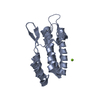
| Title | Visualization of a missing link in retrovirus capsid assembly | |||||||||
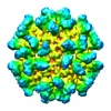
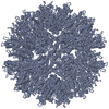
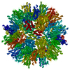
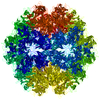
 Map data Map data | Icosahedral assembly of Rous sarcoma virus capsid proteins | |||||||||
 Sample Sample |
| |||||||||
 Keywords Keywords | Retrovirus / Capsid / Icosahedral | |||||||||
| Biological species |  Avian leukosis virus Avian leukosis virus | |||||||||
| Method | single particle reconstruction / cryo EM / Resolution: 10.4 Å | |||||||||
 Authors Authors | Cardone G / Purdy JG / Cheng N / Craven RC / Steven AC | |||||||||
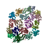
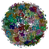
 Citation Citation |  Journal: Nature / Year: 2009 Journal: Nature / Year: 2009Title: Visualization of a missing link in retrovirus capsid assembly. Authors: Giovanni Cardone / John G Purdy / Naiqian Cheng / Rebecca C Craven / Alasdair C Steven /  Abstract: For a retrovirus such as HIV to be infectious, a properly formed capsid is needed; however, unusually among viruses, retrovirus capsids are highly variable in structure. According to the fullerene ...For a retrovirus such as HIV to be infectious, a properly formed capsid is needed; however, unusually among viruses, retrovirus capsids are highly variable in structure. According to the fullerene conjecture, they are composed of hexamers and pentamers of capsid protein (CA), with the shape of a capsid varying according to how the twelve pentamers are distributed and its size depending on the number of hexamers. Hexamers have been studied in planar and tubular arrays, but the predicted pentamers have not been observed. Here we report cryo-electron microscopic analyses of two in-vitro-assembled capsids of Rous sarcoma virus. Both are icosahedrally symmetric: one is composed of 12 pentamers, and the other of 12 pentamers and 20 hexamers. Fitting of atomic models of the two CA domains into the reconstructions shows three distinct inter-subunit interactions. These observations substantiate the fullerene conjecture, show how pentamers are accommodated at vertices, support the inference that nucleation is a crucial morphologic determinant, and imply that electrostatic interactions govern the differential assembly of pentamers and hexamers. | |||||||||
| History |
|
- Structure visualization
| Movie |
 Movie viewer Movie viewer |
|---|---|
| Structure viewer | EM map:  SurfView SurfView Molmil Molmil Jmol/JSmol Jmol/JSmol |
| Supplemental images |
- Downloads & links
-EMDB archive
| Map data |  emd_1862.map.gz emd_1862.map.gz | 23.5 MB |  EMDB map data format EMDB map data format | |
|---|---|---|---|---|
| Header (meta data) |  emd-1862-v30.xml emd-1862-v30.xml emd-1862.xml emd-1862.xml | 11 KB 11 KB | Display Display |  EMDB header EMDB header |
| Images |  1862.png 1862.png emd_1862.png emd_1862.png | 285.5 KB 473.4 KB | ||
| Archive directory |  http://ftp.pdbj.org/pub/emdb/structures/EMD-1862 http://ftp.pdbj.org/pub/emdb/structures/EMD-1862 ftp://ftp.pdbj.org/pub/emdb/structures/EMD-1862 ftp://ftp.pdbj.org/pub/emdb/structures/EMD-1862 | HTTPS FTP |
-Validation report
| Summary document |  emd_1862_validation.pdf.gz emd_1862_validation.pdf.gz | 250.1 KB | Display |  EMDB validaton report EMDB validaton report |
|---|---|---|---|---|
| Full document |  emd_1862_full_validation.pdf.gz emd_1862_full_validation.pdf.gz | 249.3 KB | Display | |
| Data in XML |  emd_1862_validation.xml.gz emd_1862_validation.xml.gz | 5.9 KB | Display | |
| Arichive directory |  https://ftp.pdbj.org/pub/emdb/validation_reports/EMD-1862 https://ftp.pdbj.org/pub/emdb/validation_reports/EMD-1862 ftp://ftp.pdbj.org/pub/emdb/validation_reports/EMD-1862 ftp://ftp.pdbj.org/pub/emdb/validation_reports/EMD-1862 | HTTPS FTP |
-Related structure data
| Similar structure data |
|---|
- Links
Links
| EMDB pages |  EMDB (EBI/PDBe) / EMDB (EBI/PDBe) /  EMDataResource EMDataResource |
|---|
- Map
Map
| File |  Download / File: emd_1862.map.gz / Format: CCP4 / Size: 30.9 MB / Type: IMAGE STORED AS SIGNED INTEGER (2 BYTES) Download / File: emd_1862.map.gz / Format: CCP4 / Size: 30.9 MB / Type: IMAGE STORED AS SIGNED INTEGER (2 BYTES) | ||||||||||||||||||||||||||||||||||||||||||||||||||||||||||||||||||||
|---|---|---|---|---|---|---|---|---|---|---|---|---|---|---|---|---|---|---|---|---|---|---|---|---|---|---|---|---|---|---|---|---|---|---|---|---|---|---|---|---|---|---|---|---|---|---|---|---|---|---|---|---|---|---|---|---|---|---|---|---|---|---|---|---|---|---|---|---|---|
| Annotation | Icosahedral assembly of Rous sarcoma virus capsid proteins | ||||||||||||||||||||||||||||||||||||||||||||||||||||||||||||||||||||
| Projections & slices | Image control
Images are generated by Spider. | ||||||||||||||||||||||||||||||||||||||||||||||||||||||||||||||||||||
| Voxel size | X=Y=Z: 1.27 Å | ||||||||||||||||||||||||||||||||||||||||||||||||||||||||||||||||||||
| Density |
| ||||||||||||||||||||||||||||||||||||||||||||||||||||||||||||||||||||
| Symmetry | Space group: 1 | ||||||||||||||||||||||||||||||||||||||||||||||||||||||||||||||||||||
| Details | EMDB XML:
CCP4 map header:
| ||||||||||||||||||||||||||||||||||||||||||||||||||||||||||||||||||||
-Supplemental data
- Sample components
-Entire : Icosahedral assembly of Rous sarcoma virus capsid proteins
| Entire | Name: Icosahedral assembly of Rous sarcoma virus capsid proteins |
|---|---|
| Components |
|
-Supramolecule #1000: Icosahedral assembly of Rous sarcoma virus capsid proteins
| Supramolecule | Name: Icosahedral assembly of Rous sarcoma virus capsid proteins type: sample / ID: 1000 Oligomeric state: 60 capsid protein subunits form 12 pentamers Number unique components: 1 |
|---|---|
| Molecular weight | Theoretical: 1.53 MDa |
-Macromolecule #1: Avian leukosis virus capsid protein
| Macromolecule | Name: Avian leukosis virus capsid protein / type: protein_or_peptide / ID: 1 / Name.synonym: RSV CA protein / Number of copies: 60 / Oligomeric state: Icosahedral composed of 12 pentamers / Recombinant expression: Yes |
|---|---|
| Source (natural) | Organism:  Avian leukosis virus / Strain: Prague C Avian leukosis virus / Strain: Prague C |
| Molecular weight | Theoretical: 1.53 MDa |
| Recombinant expression | Organism:  |
-Experimental details
-Structure determination
| Method | cryo EM |
|---|---|
 Processing Processing | single particle reconstruction |
| Aggregation state | particle |
- Sample preparation
| Concentration | 2 mg/mL |
|---|---|
| Buffer | pH: 7.5 Details: 10 mM Tris-HCl, 75 mM NaCl, 0.05 mM ETDA, 0.5 M NaPO4 |
| Grid | Details: Holey carbon film on 400 mesh gold grid |
| Vitrification | Cryogen name: ETHANE / Chamber temperature: 93.15 K / Instrument: LEICA KF80 Details: Vitrification instrument: Reichert-Jung KF80 plunger. Vitrification carried out in nitrogen atmosphere Method: 4.5 microliter sample dropped onto grid, blotted on one side for 1 second, then plunged |
- Electron microscopy
| Microscope | FEI/PHILIPS CM200FEG |
|---|---|
| Temperature | Min: 88.15 K / Max: 98.15 K / Average: 93.15 K |
| Image recording | Category: FILM / Film or detector model: KODAK SO-163 FILM / Digitization - Scanner: NIKON SUPER COOLSCAN 9000 / Digitization - Sampling interval: 6.35 µm / Number real images: 6 / Average electron dose: 14 e/Å2 / Od range: 4.8 / Bits/pixel: 16 |
| Electron beam | Acceleration voltage: 120 kV / Electron source:  FIELD EMISSION GUN FIELD EMISSION GUN |
| Electron optics | Illumination mode: FLOOD BEAM / Imaging mode: BRIGHT FIELD / Cs: 2.0 mm / Nominal defocus max: 1.5 µm / Nominal defocus min: 1.0 µm / Nominal magnification: 50000 |
| Sample stage | Specimen holder: Eucentric / Specimen holder model: GATAN LIQUID NITROGEN |
- Image processing
| CTF correction | Details: Each particle, phase reversal and baseline subtraction |
|---|---|
| Final reconstruction | Applied symmetry - Point group: I (icosahedral) / Resolution.type: BY AUTHOR / Resolution: 10.4 Å / Resolution method: FSC 0.5 CUT-OFF / Software - Name: EMAN, Bsoft, PFT2, EM3DR2 Details: Particles were classified using EMAN and initial template generated using 3-fold class average. Preprocessing was done using Bsoft. For orientation assignment and 3D reconstruction PFT2 and ...Details: Particles were classified using EMAN and initial template generated using 3-fold class average. Preprocessing was done using Bsoft. For orientation assignment and 3D reconstruction PFT2 and EM3DR2 were used, repsectively Number images used: 1478 |
-Atomic model buiding 1
| Initial model | PDB ID: Chain - Chain ID: A |
|---|---|
| Software | Name: SITUS |
| Details | PDBEntryID_givenInChain. Protocol: Rigid Body. The NTD domain was fitted automatically using colores |
| Refinement | Space: RECIPROCAL / Protocol: RIGID BODY FIT / Target criteria: Laplacian correlation |
-Atomic model buiding 2
| Initial model | PDB ID: Chain - Chain ID: A |
|---|---|
| Software | Name: SITUS |
| Details | PDBEntryID_givenInChain. Protocol: Rigid Body. The CTD domain (aa 152-230) was extracted and fitted using colacor. Model 14 was selected as the best fit. |
| Refinement | Space: RECIPROCAL / Protocol: RIGID BODY FIT / Target criteria: Laplacian correlation |
 Movie
Movie Controller





 Z (Sec.)
 Y (Row.)
 X (Col.)


