 Movie
Movie Controller


+ Open data

- Basic information
| Entry |  | |||||||||
|---|---|---|---|---|---|---|---|---|---|---|
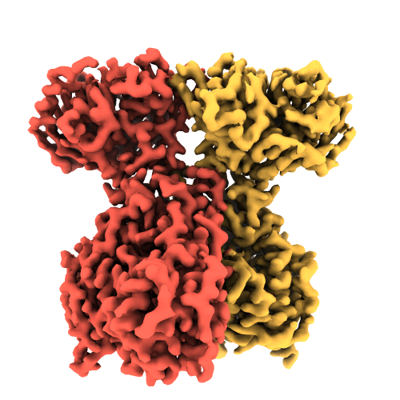
| Title | Cryo-EM KSB domain of RhiE from Burkholderia rhizoxinica | |||||||||
 Map data Map data | ||||||||||
 Sample Sample |
| |||||||||
 Keywords Keywords | Polyketide / TRANSFERASE | |||||||||
| Function / homology |  Function and homology information Function and homology informationDIM/DIP cell wall layer assembly / fatty acid synthase activity / phosphopantetheine binding / 3-oxoacyl-[acyl-carrier-protein] synthase activity / fatty acid biosynthetic process / plasma membrane / cytoplasm Similarity search - Function | |||||||||
| Biological species |  Mycetohabitans rhizoxinica (bacteria) Mycetohabitans rhizoxinica (bacteria) | |||||||||
| Method | single particle reconstruction / cryo EM / Resolution: 2.84 Å | |||||||||
 Authors Authors | Capper MJ / Koehnke J | |||||||||
| Funding support |  United Kingdom, 1 items United Kingdom, 1 items
| |||||||||
 Citation Citation | Journal: To Be Published Title: Structural Analysis of a Chain-Branching Polyketide Synthase Module Authors: Dell M / Tran MA / Capper MJ / Sundaram S / Fiedler J / Koehnke J / Hellmich U / Hertwick C | |||||||||
| History |
|
- Structure visualization
| Supplemental images |
|---|
- Downloads & links
-EMDB archive
| Map data | emd_16892.map.gz | 32.2 MB | EMDB map data format | |
|---|---|---|---|---|
| Header (meta data) | emd-16892-v30.xmlemd-16892.xml | 18.1 KB 18.1 KB | Display Display | EMDB header |
| FSC (resolution estimation) | emd_16892_fsc.xml | 8.4 KB | Display | FSC data file |
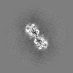
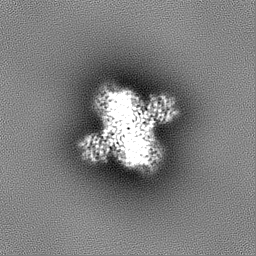
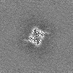
| Images |  emd_16892.png emd_16892.png | 149.1 KB | ||
| Masks | emd_16892_msk_1.map | 64 MB | Mask map | |
| Filedesc metadata | emd-16892.cif.gz | 6.5 KB | ||
| Others | emd_16892_additional_1.map.gzemd_16892_half_map_1.map.gzemd_16892_half_map_2.map.gz | 56.2 MB 59.3 MB 59.3 MB | ||
| Archive directory |  http://ftp.pdbj.org/pub/emdb/structures/EMD-16892ftp://ftp.pdbj.org/pub/emdb/structures/EMD-16892 http://ftp.pdbj.org/pub/emdb/structures/EMD-16892ftp://ftp.pdbj.org/pub/emdb/structures/EMD-16892 | HTTPS FTP |
-Related structure data
| Related structure data |  8oiiMC M: atomic model generated by this map C: citing same article ( |
|---|---|
| Similar structure data |

-Links
| EMDB pages | EMDB (EBI/PDBe) / EMDataResource |
|---|---|
| Related items in Molecule of the Month |


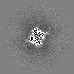
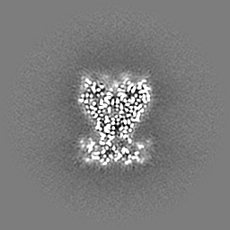
-Map
| File | Download / File: emd_16892.map.gz / Format: CCP4 / Size: 64 MB / Type: IMAGE STORED AS FLOATING POINT NUMBER (4 BYTES) | ||||||||||||||||||||||||||||||||||||
|---|---|---|---|---|---|---|---|---|---|---|---|---|---|---|---|---|---|---|---|---|---|---|---|---|---|---|---|---|---|---|---|---|---|---|---|---|---|
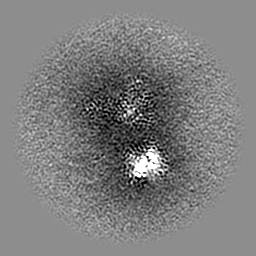
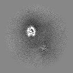
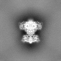
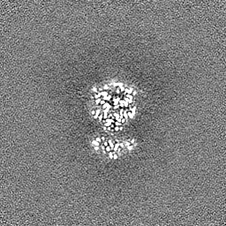
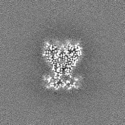
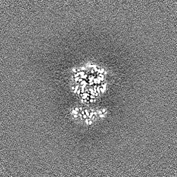
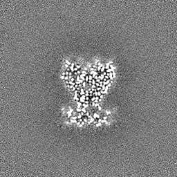
| Projections & slices | Image control
Images are generated by Spider. | ||||||||||||||||||||||||||||||||||||
| Voxel size | X=Y=Z: 1.015 Å | ||||||||||||||||||||||||||||||||||||
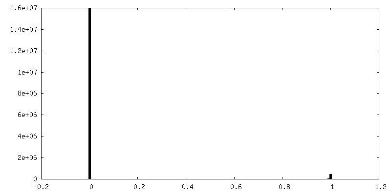
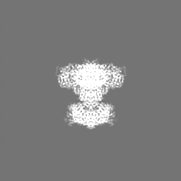
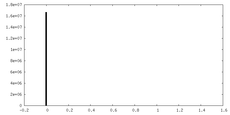
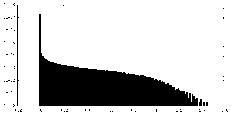
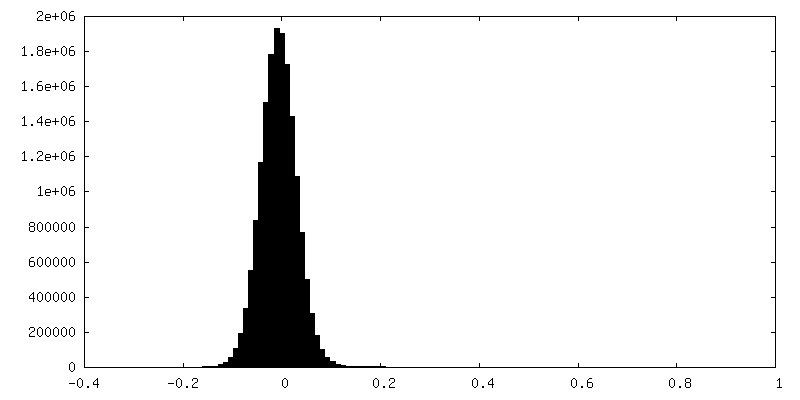
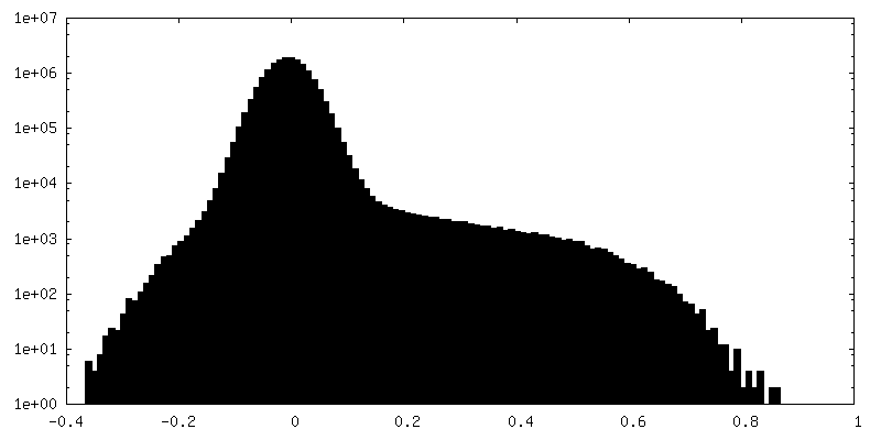
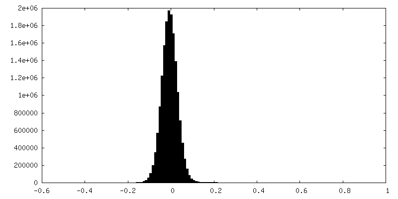
| Density |
| ||||||||||||||||||||||||||||||||||||
| Symmetry | Space group: 1 | ||||||||||||||||||||||||||||||||||||
| Details | EMDB XML:
|
 Z (Sec.)
 Y (Row.)
 X (Col.)


-Supplemental data
-Mask #1
| File | emd_16892_msk_1.map | ||||||||||||
|---|---|---|---|---|---|---|---|---|---|---|---|---|---|
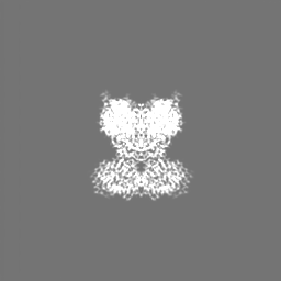
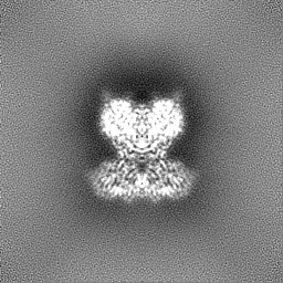
| Projections & Slices |
| ||||||||||||
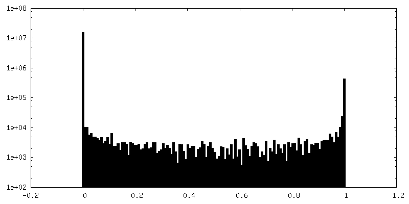
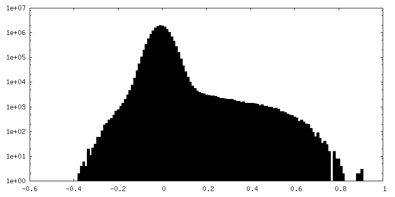
| Density Histograms |


-Additional map: #1
| File | emd_16892_additional_1.map | ||||||||||||
|---|---|---|---|---|---|---|---|---|---|---|---|---|---|
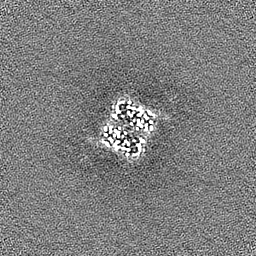
| Projections & Slices |
| ||||||||||||
| Density Histograms |

-Half map: #2
| File | emd_16892_half_map_1.map | ||||||||||||
|---|---|---|---|---|---|---|---|---|---|---|---|---|---|
| Projections & Slices |
| ||||||||||||
| Density Histograms |
-Half map: #1
| File | emd_16892_half_map_2.map | ||||||||||||
|---|---|---|---|---|---|---|---|---|---|---|---|---|---|
| Projections & Slices |
| ||||||||||||
| Density Histograms |


- Sample components
-Entire : Homodimeric complex of RhiE KSB domains
| Entire | Name: Homodimeric complex of RhiE KSB domains |
|---|---|
| Components |
|
-Supramolecule #1: Homodimeric complex of RhiE KSB domains
| Supramolecule | Name: Homodimeric complex of RhiE KSB domains / type: complex / ID: 1 / Parent: 0 / Macromolecule list: all |
|---|---|
| Source (natural) | Organism: Mycetohabitans rhizoxinica (bacteria) |
| Molecular weight | Theoretical: 230 KDa |
-Macromolecule #1: RhiE protein,Polyketide synthase domain protein RhiE
| Macromolecule | Name: RhiE protein,Polyketide synthase domain protein RhiE / type: protein_or_peptide / ID: 1 / Number of copies: 2 / Enantiomer: LEVO |
|---|---|
| Source (natural) | Organism: Mycetohabitans rhizoxinica (bacteria) |
| Molecular weight | Theoretical: 115.527039 KDa |
| Recombinant expression | Organism: |
| Sequence | String: MKHHHHHHHH GGLVPRGSHG GSSGERVEDN ELANYIAVIG LGGYYPGADS IDELWQNLAN GVDCMSDFPA DRWDHSKIYY KNRKVLGKT TCINGSFIKD VDKFDYSYFK MPKVYADHMS PEVRLFLQVA VHTFEDAGYS KETLLSRYNG DVGVLLGTMS N DYHYYGFE ...String: MKHHHHHHHH GGLVPRGSHG GSSGERVEDN ELANYIAVIG LGGYYPGADS IDELWQNLAN GVDCMSDFPA DRWDHSKIYY KNRKVLGKT TCINGSFIKD VDKFDYSYFK MPKVYADHMS PEVRLFLQVA VHTFEDAGYS KETLLSRYNG DVGVLLGTMS N DYHYYGFE SNVFRGSMAS GSGMATIPMT VSYFYGLTGP SLFIDTMCSS SSTCIHTACQ MLKHDETKMV LAGGLNLMYH PY TTVNTSQ GNFTSITSES VNSYGVGADG TVIGEGIGAV LLKRLDRAIA DRDQIYGVIK GSAMTNAGER NGFNVPNPDL QTL AIRQAM DQAKVHPSSI SYIEGHGSGT KLGDPIEVLG LNNAFRWATD DKQFCYLGSI KSNIGHLLAA SGIAGLTKTL LQFK HKQIA PSIHSSQLNQ DIDFADTPFV VPQQLIEWRQ PERIINGRKQ VFPRRAGLTS IAAGGMNAHM IVEEYPEPAD SAGQI SEDQ LVFVFSVHKL ALLAQNLTSF RDWLASSEAP LAQIAYTLQV GKNNLRNRLA IRCRTRQALS RALNACIDGH YQSSAD SKI FYRFQESDAV QPLESDLNDP LAPLLTQWLN GDSQVDWASL YAQPPVRISL PAYRFEKTRC WYTEEGYESS IVNPLMF KN KLHPLVAKNC STPQPGAIFR TDFVEDELLD YVYSGRGGRR LSAFNFADVA LAMPALASRF DGRTLSVSCA FEHYIADW T TVTGLEYRLF EIDSEQLELE FDFRRSGEQP THLGFAVINP LTSDEPPLPQ QWLDDARELL NRQALQAGRQ LSAAEVSQR LAQAGYDFAP YLDHDGELTI GRSGLVLKGR PPVNRHNHYA DNVQLSPYLA TTIDKALYLL LDELGLPQGR VIVRNIERLC CYHTPAGGF SVVLSGIGLN DNELSLSLLV LDEREQICVK LDKVSLYLGK QEVASVDRKH SLLTGEERMA EFKQEAKPQA D DSEAGFGE KILAFIQQEL QDKLGFAADI GESTQVHDLG LDSIMVVQLT DSVNKRFGTK LMPDLFYEKQ QLGELVARLE AA A UniProtKB: RhiE protein, Polyketide synthase domain protein RhiE |
-Experimental details
-Structure determination
| Method | cryo EM |
|---|---|
 Processing Processing | single particle reconstruction |
| Aggregation state | particle |
-Sample preparation
| Concentration | 6 mg/mL |
|---|---|
| Buffer | pH: 8 / Component - Concentration: 20.0 mM / Component - Name: Tris-HCl / Details: 20 mM Tris-HCl pH 8 |
| Grid | Model: Quantifoil R1.2/1.3 / Material: COPPER / Support film - Material: CARBON / Support film - topology: HOLEY ARRAY / Pretreatment - Type: GLOW DISCHARGE / Pretreatment - Time: 30 sec. |
| Vitrification | Cryogen name: ETHANE / Chamber humidity: 95 % / Chamber temperature: 288 K / Instrument: FEI VITROBOT MARK III |
| Details | Monodisperse sample in minimal buffer |
- Electron microscopy
| Microscope | JEOL CRYO ARM 300 |
|---|---|
| Image recording | Film or detector model: DIRECT ELECTRON DE-64 (8k x 8k) / Detector mode: COUNTING / Number grids imaged: 1 / Number real images: 3579 / Average electron dose: 53.9 e/Å2 |
| Electron beam | Acceleration voltage: 300 kV / Electron source:  FIELD EMISSION GUN FIELD EMISSION GUN |
| Electron optics | C2 aperture diameter: 100.0 µm / Illumination mode: FLOOD BEAM / Imaging mode: BRIGHT FIELD / Cs: 2.7 mm / Nominal defocus max: 2.5 µm / Nominal defocus min: 1.5 µm / Nominal magnification: 120000 |
| Sample stage | Specimen holder model: JEOL / Cooling holder cryogen: NITROGEN |

