Name: MAGNESIUM ION / type: ligand / ID: 3 / Number of copies: 1 / Formula: MG
Molecular weight
Theoretical: 24.305 Da
-
Experimental details
-
Structure determination
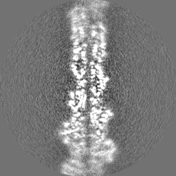
Method
cryo EM
Processing
single particle reconstruction
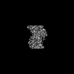
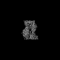
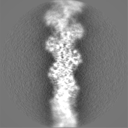
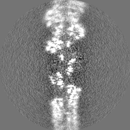
Aggregation state
helical array
-
Sample preparation
Buffer
pH: 7.4 Details: Actin for EM was polymerized by mixing, 20 ul 20 uM G-actin, 8 ul 10x MKE and 52 ul 5 mM HEPES-KOH pH 7.4 containing 0.2 mM ATP and 0.5 mM DTT and incubating at RT for 1 hour.
Grid
Model: Quantifoil R3.5/1 / Material: COPPER / Mesh: 200 / Support film - Material: CARBON / Support film - topology: HOLEY ARRAY / Pretreatment - Type: GLOW DISCHARGE / Pretreatment - Time: 60 sec. / Pretreatment - Atmosphere: AIR
Vitrification
Cryogen name: ETHANE-PROPANE / Chamber humidity: 95 % / Chamber temperature: 277 K / Instrument: LEICA EM GP
-
Electron microscopy
Microscope
FEI TITAN KRIOS
Image recording
Film or detector model: GATAN K2 SUMMIT (4k x 4k) / Detector mode: COUNTING / Number grids imaged: 1 / Average exposure time: 60.0 sec. / Average electron dose: 42.0 e/Å2
Electron beam
Acceleration voltage: 300 kV / Electron source: FIELD EMISSION GUN
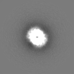
Type of model: OTHER Details: Used featureless cylinder as starting model for 3D classification.
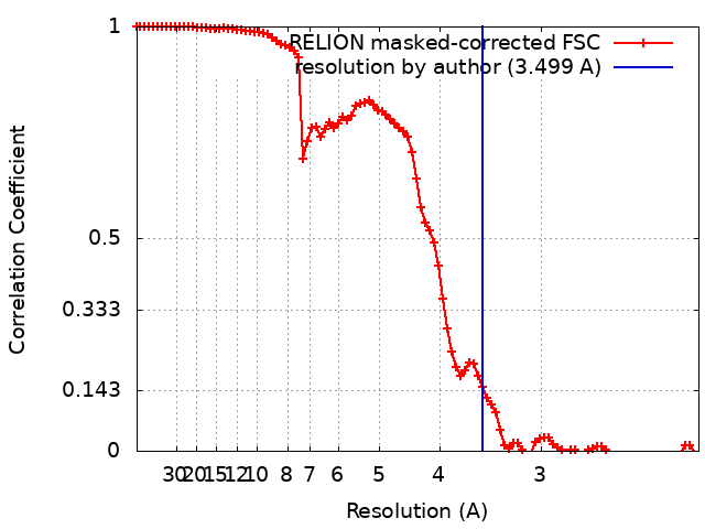
Final reconstruction
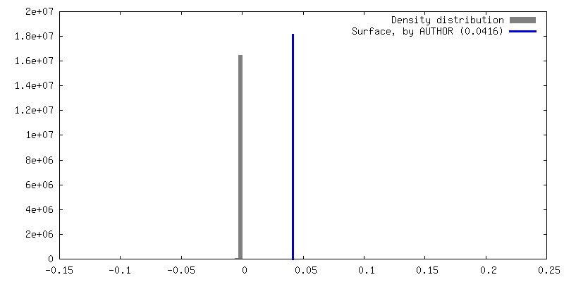
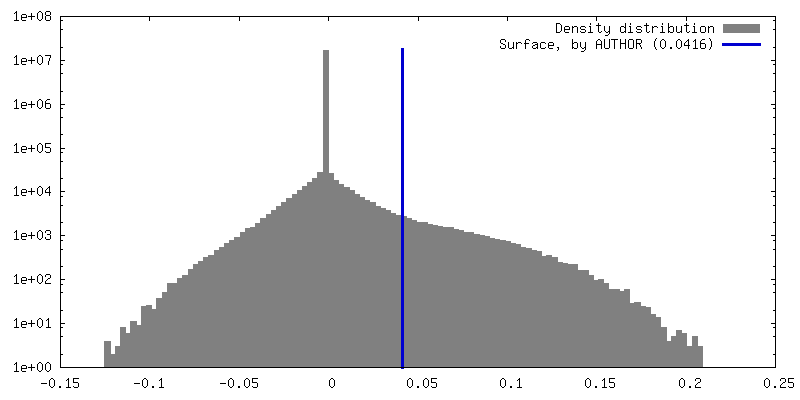
Algorithm: FOURIER SPACE / Resolution.type: BY AUTHOR / Resolution: 3.499 Å / Resolution method: FSC 0.143 CUT-OFF Details: RELION 3.09 was used throughout using helical reconstruction. Number images used: 42701
Initial angle assignment
Type: MAXIMUM LIKELIHOOD
Final angle assignment
Type: MAXIMUM LIKELIHOOD
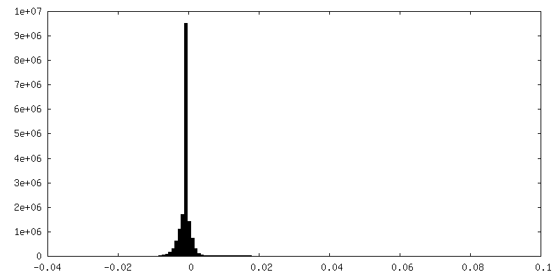
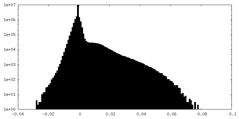
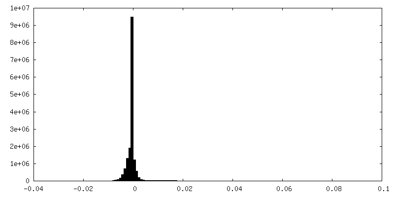
FSC plot (resolution estimation)
+
About Yorodumi
-
News
-
Feb 9, 2022. New format data for meta-information of EMDB entries
New format data for meta-information of EMDB entries
Version 3 of the EMDB header file is now the official format.
The previous official version 1.9 will be removed from the archive.
In the structure databanks used in Yorodumi, some data are registered as the other names, "COVID-19 virus" and "2019-nCoV". Here are the details of the virus and the list of structure data.
Jan 31, 2019. EMDB accession codes are about to change! (news from PDBe EMDB page)
EMDB accession codes are about to change! (news from PDBe EMDB page)
The allocation of 4 digits for EMDB accession codes will soon come to an end. Whilst these codes will remain in use, new EMDB accession codes will include an additional digit and will expand incrementally as the available range of codes is exhausted. The current 4-digit format prefixed with “EMD-” (i.e. EMD-XXXX) will advance to a 5-digit format (i.e. EMD-XXXXX), and so on. It is currently estimated that the 4-digit codes will be depleted around Spring 2019, at which point the 5-digit format will come into force.
The EM Navigator/Yorodumi systems omit the EMD- prefix.
Related info.:Q: What is EMD? / ID/Accession-code notation in Yorodumi/EM Navigator
Yorodumi is a browser for structure data from EMDB, PDB, SASBDB, etc.
This page is also the successor to EM Navigator detail page, and also detail information page/front-end page for Omokage search.
The word "yorodu" (or yorozu) is an old Japanese word meaning "ten thousand". "mi" (miru) is to see.
Related info.:EMDB / PDB / SASBDB / Comparison of 3 databanks / Yorodumi Search / Aug 31, 2016. New EM Navigator & Yorodumi / Yorodumi Papers / Jmol/JSmol / Function and homology information / Changes in new EM Navigator and Yorodumi
 Movie
Movie Controller


 Open data
 Basic information
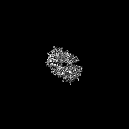
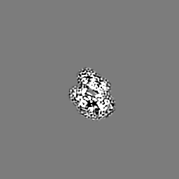
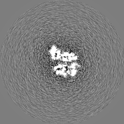
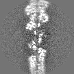
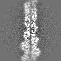
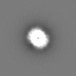
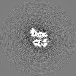
 Map data
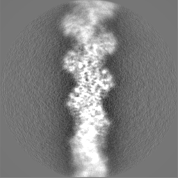
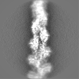
 Sample
 Keywords
 Function and homology information
 Homo sapiens (human)
 Authors
 United Kingdom, European Union, 4 items
  Citation
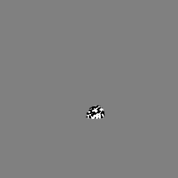
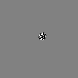
 Structure visualization
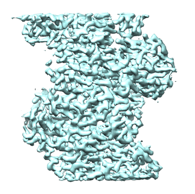
 Downloads & links
 emd_16776.png
 http://ftp.pdbj.org/pub/emdb/structures/EMD-16776
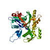


 Z (Sec.)
 Y (Row.)
 X (Col.)


 Sample components
 Komagataella pastoris (fungus)
 Processing
 Electron microscopy
 FIELD EMISSION GUN