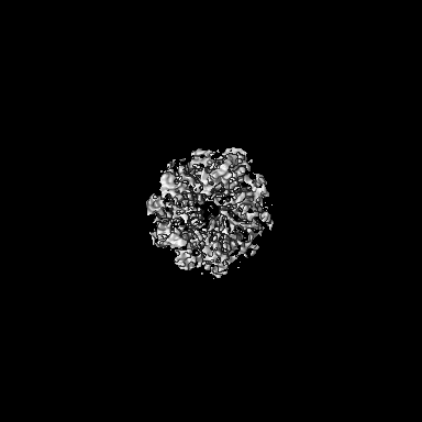
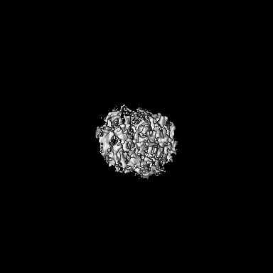
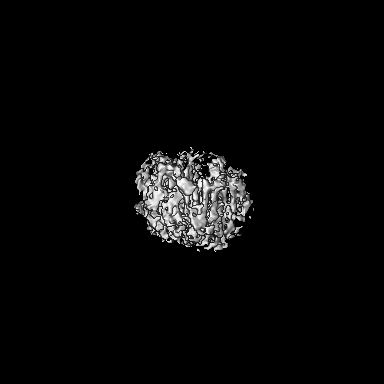
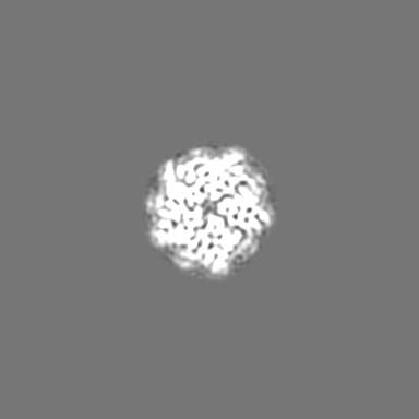
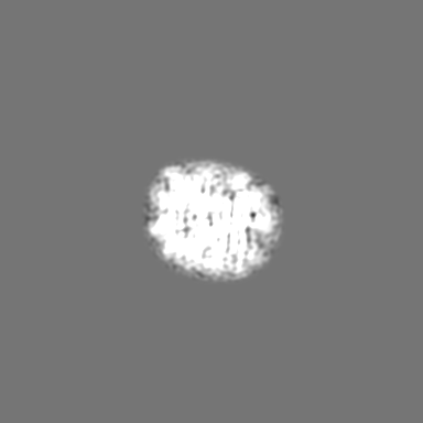
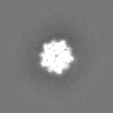
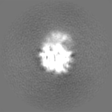
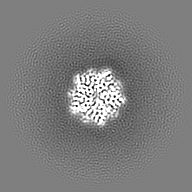
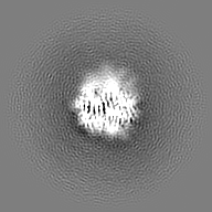
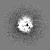
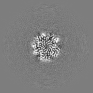
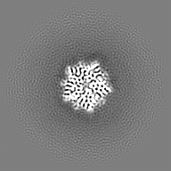
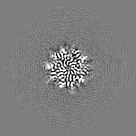
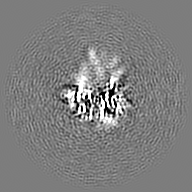
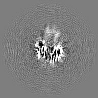
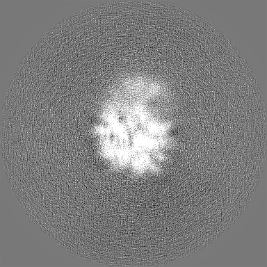
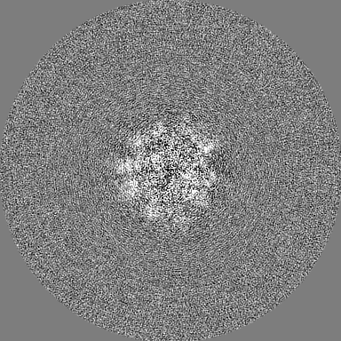
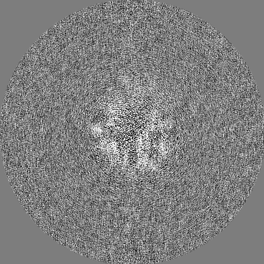
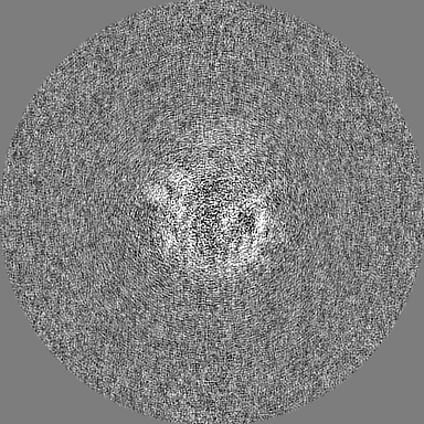
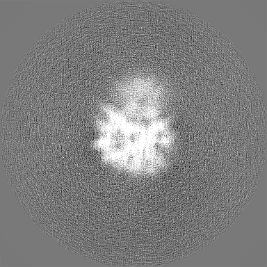
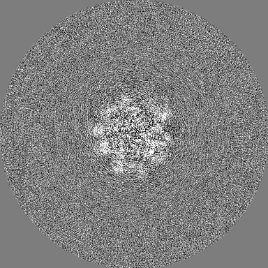
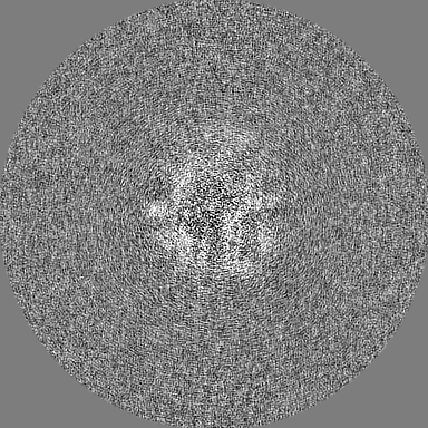
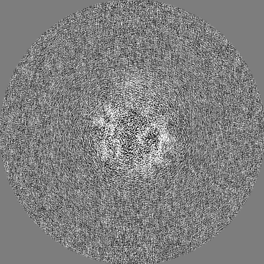
- EMDB-15606: CryoEM structure of the human Nucleophosmin 1 core -
+
Open data
ID or keywords:
Loading...
-
Basic information
Entry
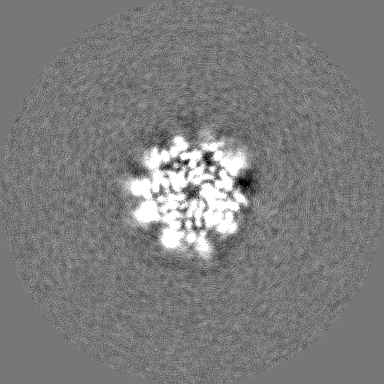
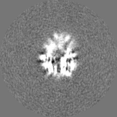
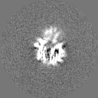
Database: EMDB / ID: EMD-15606
Title
CryoEM structure of the human Nucleophosmin 1 core
Map data
Sample
Complex: Nucleophosmin 1
Protein or peptide: Nucleophosmin
Keywords
Phase separation / CHAPERONE
Function / homology
Function and homology information
regulation of eIF2 alpha phosphorylation by dsRNA / regulation of mRNA stability involved in cellular response to UV / regulation of endoribonuclease activity / negative regulation of centrosome duplication / regulation of endodeoxyribonuclease activity / positive regulation of cell cycle G2/M phase transition / regulation of centriole replication / granular component / TFAP2A acts as a transcriptional repressor during retinoic acid induced cell differentiation / negative regulation of protein kinase activity by regulation of protein phosphorylation ...regulation of eIF2 alpha phosphorylation by dsRNA / regulation of mRNA stability involved in cellular response to UV / regulation of endoribonuclease activity / negative regulation of centrosome duplication / regulation of endodeoxyribonuclease activity / positive regulation of cell cycle G2/M phase transition / regulation of centriole replication / granular component / TFAP2A acts as a transcriptional repressor during retinoic acid induced cell differentiation / negative regulation of protein kinase activity by regulation of protein phosphorylation / SARS-CoV-1-host interactions / Tat protein binding / regulation of centrosome duplication / ALK mutants bind TKIs / spindle pole centrosome / Nuclear import of Rev protein / centrosome cycle / nucleocytoplasmic transport / TP53 regulates transcription of additional cell cycle genes whose exact role in the p53 pathway remain uncertain / protein kinase inhibitor activity / ribosomal large subunit binding / ribosomal small subunit binding / NF-kappaB binding / ribosomal large subunit export from nucleus / ribosomal small subunit export from nucleus / Nuclear events stimulated by ALK signaling in cancer / ribosomal subunit export from nucleus / core promoter sequence-specific DNA binding / Deposition of new CENPA-containing nucleosomes at the centromere / ribosome assembly / SUMOylation of transcription cofactors / ribosomal large subunit biogenesis / positive regulation of translation / protein-DNA complex / intracellular protein transport / PKR-mediated signaling / protein localization / : / cellular response to UV / cellular senescence / Signaling by ALK fusions and activated point mutants / unfolded protein binding / nucleosome assembly / positive regulation of NF-kappaB transcription factor activity / ribosomal small subunit biogenesis / histone binding / DNA-binding transcription factor binding / transcription coactivator activity / rRNA binding / chromatin remodeling / ribonucleoprotein complex / negative regulation of cell population proliferation / DNA repair / focal adhesion / centrosome / chromatin binding / positive regulation of cell population proliferation / nucleolus / negative regulation of apoptotic process / protein kinase binding / positive regulation of DNA-templated transcription / signal transduction / protein homodimerization activity / positive regulation of transcription by RNA polymerase II / protein-containing complex / RNA binding / nucleoplasm / membrane / nucleus / cytoplasm / cytosol Similarity search - Function
Journal: PNAS Nexus / Year: 2023 Title: A "grappling hook" interaction connects self-assembly and chaperone activity of Nucleophosmin 1. Authors: Mihkel Saluri / Axel Leppert / Genis Valentin Gese / Cagla Sahin / Dilraj Lama / Margit Kaldmäe / Gefei Chen / Arne Elofsson / Timothy M Allison / Marie Arsenian-Henriksson / Jan Johansson ...Authors: Mihkel Saluri / Axel Leppert / Genis Valentin Gese / Cagla Sahin / Dilraj Lama / Margit Kaldmäe / Gefei Chen / Arne Elofsson / Timothy M Allison / Marie Arsenian-Henriksson / Jan Johansson / David P Lane / B Martin Hällberg / Michael Landreh / Abstract: How the self-assembly of partially disordered proteins generates functional compartments in the cytoplasm and particularly in the nucleus is poorly understood. Nucleophosmin 1 (NPM1) is an abundant ...How the self-assembly of partially disordered proteins generates functional compartments in the cytoplasm and particularly in the nucleus is poorly understood. Nucleophosmin 1 (NPM1) is an abundant nucleolar protein that forms large oligomers and undergoes liquid-liquid phase separation by binding RNA or ribosomal proteins. It provides the scaffold for ribosome assembly but also prevents protein aggregation as part of the cellular stress response. Here, we use aggregation assays and native mass spectrometry (MS) to examine the relationship between the self-assembly and chaperone activity of NPM1. We find that oligomerization of full-length NPM1 modulates its ability to retard amyloid formation in vitro. Machine learning-based structure prediction and cryo-electron microscopy reveal fuzzy interactions between the acidic disordered region and the C-terminal nucleotide-binding domain, which cross-link NPM1 pentamers into partially disordered oligomers. The addition of basic peptides results in a tighter association within the oligomers, reducing their capacity to prevent amyloid formation. Together, our findings show that NPM1 uses a "grappling hook" mechanism to form a network-like structure that traps aggregation-prone proteins. Nucleolar proteins and RNAs simultaneously modulate the association strength and chaperone activity, suggesting a mechanism by which nucleolar composition regulates the chaperone activity of NPM1.
In the structure databanks used in Yorodumi, some data are registered as the other names, "COVID-19 virus" and "2019-nCoV". Here are the details of the virus and the list of structure data.
Jan 31, 2019. EMDB accession codes are about to change! (news from PDBe EMDB page)
EMDB accession codes are about to change! (news from PDBe EMDB page)
The allocation of 4 digits for EMDB accession codes will soon come to an end. Whilst these codes will remain in use, new EMDB accession codes will include an additional digit and will expand incrementally as the available range of codes is exhausted. The current 4-digit format prefixed with “EMD-” (i.e. EMD-XXXX) will advance to a 5-digit format (i.e. EMD-XXXXX), and so on. It is currently estimated that the 4-digit codes will be depleted around Spring 2019, at which point the 5-digit format will come into force.
The EM Navigator/Yorodumi systems omit the EMD- prefix.
Related info.:Q: What is EMD? / ID/Accession-code notation in Yorodumi/EM Navigator
Yorodumi is a browser for structure data from EMDB, PDB, SASBDB, etc.
This page is also the successor to EM Navigator detail page, and also detail information page/front-end page for Omokage search.
The word "yorodu" (or yorozu) is an old Japanese word meaning "ten thousand". "mi" (miru) is to see.
Related info.:EMDB / PDB / SASBDB / Comparison of 3 databanks / Yorodumi Search / Aug 31, 2016. New EM Navigator & Yorodumi / Yorodumi Papers / Jmol/JSmol / Function and homology information / Changes in new EM Navigator and Yorodumi
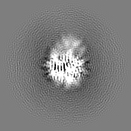
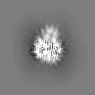
 Movie
Movie Controller


 Open data
 Basic information
 Map data
 Sample
 Keywords
 Function and homology information
 Homo sapiens (human)
 Authors
 Sweden, 1 items
  Citation

 Structure visualization
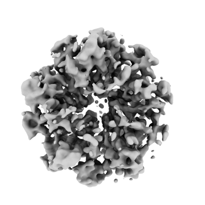
 Downloads & links
 emd_15606.png
 http://ftp.pdbj.org/pub/emdb/structures/EMD-15606


 Z (Sec.)
 Y (Row.)
 X (Col.)


 Sample components
 Processing
 Electron microscopy
 FIELD EMISSION GUN