 Movie
Movie Controller


+ Open data

- Basic information
| Entry | 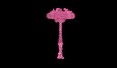 | |||||||||
|---|---|---|---|---|---|---|---|---|---|---|
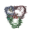
| Title | SDBC and SOD assembly, composite map | |||||||||
 Map data Map data | Composite map | |||||||||
 Sample Sample |
| |||||||||
| Biological species |  Deinococcus radiodurans R1 (radioresistant) Deinococcus radiodurans R1 (radioresistant) | |||||||||
| Method | single particle reconstruction / cryo EM / Resolution: 3.5 Å | |||||||||
 Authors Authors | Farci D / Piano D | |||||||||
| Funding support |  Poland, 1 items Poland, 1 items
| |||||||||
 Citation Citation | Journal: J Biol Chem / Year: 2023 Title: The SDBC is active in quenching oxidative conditions and bridges the cell envelope layers in Deinococcus radiodurans. Authors: Domenica Farci / André T Graça / Luca Iesu / Daniele de Sanctis / Dario Piano /    Abstract: Deinococcus radiodurans is known for its remarkable ability to withstand harsh stressful conditions. The outermost layer of its cell envelope is a proteinaceous coat, the S-layer, essential for ...Deinococcus radiodurans is known for its remarkable ability to withstand harsh stressful conditions. The outermost layer of its cell envelope is a proteinaceous coat, the S-layer, essential for resistance to and interactions with the environment. The S-layer Deinoxanthin-binding complex (SDBC), one of the main units of the characteristic multilayered cell envelope of this bacterium, protects against environmental stressors and allows exchanges with the environment. So far, specific regions of this complex, the collar and the stalk, remained unassigned. Here, these regions are resolved by cryo-EM and locally refined. The resulting 3D map shows that the collar region of this multiprotein complex is a trimer of the protein DR_0644, a Cu-only superoxide dismutase (SOD) identified here to be efficient in quenching reactive oxygen species. The same data also showed that the stalk region consists of a coiled coil that extends into the cell envelope for ∼280 Å, reaching the inner membrane. Finally, the orientation and localization of the complex are defined by in situ cryo-electron crystallography. The structural organization of the SDBC couples fundamental UV antenna properties with the presence of a Cu-only SOD, showing here coexisting photoprotective and chemoprotective functions. These features suggests how the SDBC and similar protein complexes, might have played a primary role as evolutive templates for the origin of photoautotrophic processes by combining primary protective needs with more independent energetic strategies. | |||||||||
| History |
|
- Structure visualization
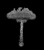
| Supplemental images |
|---|

- Downloads & links
-EMDB archive
| Map data | emd_15487.map.gz | 483.6 MB |  EMDB map data format EMDB map data format | |
|---|---|---|---|---|
| Header (meta data) | emd-15487-v30.xmlemd-15487.xml | 9.3 KB 9.3 KB | Display Display | EMDB header |
| Images | 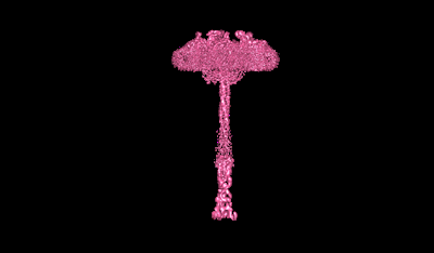 emd_15487.png emd_15487.png | 15.6 KB | ||
| Archive directory |  http://ftp.pdbj.org/pub/emdb/structures/EMD-15487ftp://ftp.pdbj.org/pub/emdb/structures/EMD-15487 http://ftp.pdbj.org/pub/emdb/structures/EMD-15487ftp://ftp.pdbj.org/pub/emdb/structures/EMD-15487 | HTTPS FTP |
-Validation report
| Summary document | emd_15487_validation.pdf.gz | 405.4 KB | Display | EMDB validaton report |
|---|---|---|---|---|
| Full document | emd_15487_full_validation.pdf.gz | 404.9 KB | Display | |
| Data in XML | emd_15487_validation.xml.gz | 4.4 KB | Display | |
| Data in CIF | emd_15487_validation.cif.gz | 5 KB | Display | |
| Arichive directory | https://ftp.pdbj.org/pub/emdb/validation_reports/EMD-15487ftp://ftp.pdbj.org/pub/emdb/validation_reports/EMD-15487 | HTTPS FTP |
-Related structure data

-Links
| EMDB pages | EMDB (EBI/PDBe) / EMDataResource |
|---|
-Map
| File | Download / File: emd_15487.map.gz / Format: CCP4 / Size: 1.1 GB / Type: IMAGE STORED AS FLOATING POINT NUMBER (4 BYTES) | ||||||||||||||||||||
|---|---|---|---|---|---|---|---|---|---|---|---|---|---|---|---|---|---|---|---|---|---|
| Annotation | Composite map | ||||||||||||||||||||
| Voxel size | X=Y=Z: 0.818 Å | ||||||||||||||||||||
| Density |
| ||||||||||||||||||||
| Symmetry | Space group: 1 | ||||||||||||||||||||
| Details | EMDB XML:
|
-Supplemental data
- Sample components
-Entire : SDBC and SOD assembly, composite map
| Entire | Name: SDBC and SOD assembly, composite map |
|---|---|
| Components |
|
-Supramolecule #1: SDBC and SOD assembly, composite map
| Supramolecule | Name: SDBC and SOD assembly, composite map / type: complex / ID: 1 / Chimera: Yes / Parent: 0 / Macromolecule list: #1 Details: This map is a composite map of the maps with deposition codes EMD-15382 and EMD-15384, and it is the composite map for the model with deposition code PDB-8AGD. The map has to be used as a ...Details: This map is a composite map of the maps with deposition codes EMD-15382 and EMD-15384, and it is the composite map for the model with deposition code PDB-8AGD. The map has to be used as a consensus map of the full structure (PDB-8AGD). |
|---|---|
| Source (natural) | Organism: Deinococcus radiodurans R1 (radioresistant) |
| Molecular weight | Theoretical: 800 KDa |
-Experimental details
-Structure determination
| Method | cryo EM |
|---|---|
 Processing Processing | single particle reconstruction |
| Aggregation state | particle |
-Sample preparation
| Buffer | pH: 7.4 |
|---|---|
| Vitrification | Cryogen name: ETHANE / Instrument: FEI VITROBOT MARK IV |
- Electron microscopy
| Microscope | FEI TITAN KRIOS |
|---|---|
| Image recording | Film or detector model: GATAN K2 QUANTUM (4k x 4k) / Average electron dose: 1.3 e/Å2 |
| Electron beam | Acceleration voltage: 300 kV / Electron source:  FIELD EMISSION GUN FIELD EMISSION GUN |
| Electron optics | Illumination mode: SPOT SCAN / Imaging mode: BRIGHT FIELD / Nominal defocus max: 1.0 µm / Nominal defocus min: 0.5 µm |
| Experimental equipment |  Model: Titan Krios / Image courtesy: FEI Company |
-Image processing
| Final reconstruction | Resolution.type: BY AUTHOR / Resolution: 3.5 Å / Resolution method: FSC 0.143 CUT-OFF Details: This map is a composite map of the maps with deposition codes EMD-15382 and EMD-15384, and it is the composite map for the model with deposition code PDB-8AGD. This map is a composite map of ...Details: This map is a composite map of the maps with deposition codes EMD-15382 and EMD-15384, and it is the composite map for the model with deposition code PDB-8AGD. This map is a composite map of the maps with deposition codes EMD-15382 and EMD-15384, and it is the composite map for the model with deposition code PDB-8AGD. The map has to be used as a consensus map of the full structure (PDB-8AGD). Number images used: 252122 |
|---|---|
| Initial angle assignment | Type: MAXIMUM LIKELIHOOD |
| Final angle assignment | Type: MAXIMUM LIKELIHOOD |
