- EMDB-14321: Human nuclear pore complex (dilated) -
+
データを開く
IDまたはキーワード:
読み込み中...
-
基本情報
登録情報
データベース: EMDB / ID: EMD-14321
タイトル
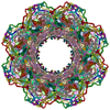
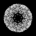
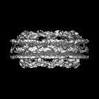
Human nuclear pore complex (dilated)
マップデータ
Structure of the human nuclear pore complex from HEK cells
試料
細胞器官・細胞要素: Human nuclear pore complex from HEK cells
タンパク質・ペプチド: x 25種
キーワード
nucleocytoplasmic transport / multiprotein complex / TRANSPORT PROTEIN
機能・相同性
機能・相同性情報
cytoplasmic side of nuclear pore / nuclear pore transmembrane ring / GATOR2 complex / nephron development / centriole assembly / positive regulation of centriole replication / cytoplasmic periphery of the nuclear pore complex / regulation of protein import into nucleus / Seh1-associated complex / regulation of Ras protein signal transduction ...cytoplasmic side of nuclear pore / nuclear pore transmembrane ring / GATOR2 complex / nephron development / centriole assembly / positive regulation of centriole replication / cytoplasmic periphery of the nuclear pore complex / regulation of protein import into nucleus / Seh1-associated complex / regulation of Ras protein signal transduction / HuR (ELAVL1) binds and stabilizes mRNA / positive regulation of mitotic cytokinetic process / SUMO ligase complex / SUMO ligase activity / protein exit from endoplasmic reticulum / COPII-coated vesicle budding / protein localization to nuclear inner membrane / nuclear pore inner ring / annulate lamellae / nuclear envelope organization / nuclear pore localization / regulation of nucleocytoplasmic transport / nuclear pore central transport channel / transcription-dependent tethering of RNA polymerase II gene DNA at nuclear periphery / COPII-coated vesicle cargo loading / nuclear pore outer ring / nuclear pore complex assembly / telomere tethering at nuclear periphery / atrial cardiac muscle cell action potential / nuclear pore organization / homologous chromosome pairing at meiosis / somite development / COPII vesicle coat / positive regulation of protein localization to centrosome / nuclear pore cytoplasmic filaments / post-transcriptional tethering of RNA polymerase II gene DNA at nuclear periphery / nuclear export signal receptor activity / paraxial mesoderm development / Nuclear Pore Complex (NPC) Disassembly / nuclear inclusion body / nuclear pore nuclear basket / Amino acids regulate mTORC1 / Transport of Ribonucleoproteins into the Host Nucleus / Regulation of Glucokinase by Glucokinase Regulatory Protein / Defective TPR may confer susceptibility towards thyroid papillary carcinoma (TPC) / miRNA processing / attachment of mitotic spindle microtubules to kinetochore / Transport of the SLBP independent Mature mRNA / Transport of the SLBP Dependant Mature mRNA / NS1 Mediated Effects on Host Pathways / SUMOylation of SUMOylation proteins / negative regulation of Ras protein signal transduction / protein-containing complex localization / microtubule bundle formation / Transport of Mature mRNA Derived from an Intronless Transcript / structural constituent of nuclear pore / nuclear localization sequence binding / positive regulation of mRNA splicing, via spliceosome / Rev-mediated nuclear export of HIV RNA / 転移酵素; アシル基を移すもの; アミノアシル基を移すもの / Nuclear import of Rev protein / Flemming body / SUMOylation of RNA binding proteins / nuclear export / fertilization / NLS-bearing protein import into nucleus / mitotic centrosome separation / NEP/NS2 Interacts with the Cellular Export Machinery / Transport of Mature mRNA derived from an Intron-Containing Transcript / RNA export from nucleus / centrosome cycle / tRNA processing in the nucleus / Postmitotic nuclear pore complex (NPC) reformation / SUMO transferase activity / negative regulation of programmed cell death / COPII-mediated vesicle transport / nucleocytoplasmic transport / neural tube development / lamellipodium assembly / centrosome localization / positive regulation of epidermal growth factor receptor signaling pathway / Viral Messenger RNA Synthesis / poly(A)+ mRNA export from nucleus / PTB domain binding / regulation of gluconeogenesis / mitotic metaphase chromosome alignment / SUMOylation of ubiquitinylation proteins / female gonad development / Vpr-mediated nuclear import of PICs / negative regulation of epidermal growth factor receptor signaling pathway / macrophage chemotaxis / SUMOylation of DNA replication proteins / protein sumoylation / 加水分解酵素; プロテアーゼ; ペプチド結合加水分解酵素; セリンエンドペプチターゼ / positive regulation of SMAD protein signal transduction / ribosomal large subunit export from nucleus / RHOA GTPase cycle / positive regulation of TOR signaling / mitotic spindle assembly / mRNA transport 類似検索 - 分子機能
German Federal Ministry for Education and Research
031L0100
ドイツ
引用
ジャーナル: Science / 年: 2022 タイトル: AI-based structure prediction empowers integrative structural analysis of human nuclear pores. 著者: Shyamal Mosalaganti / Agnieszka Obarska-Kosinska / Marc Siggel / Reiya Taniguchi / Beata Turoňová / Christian E Zimmerli / Katarzyna Buczak / Florian H Schmidt / Erica Margiotta / Marie- ...著者: Shyamal Mosalaganti / Agnieszka Obarska-Kosinska / Marc Siggel / Reiya Taniguchi / Beata Turoňová / Christian E Zimmerli / Katarzyna Buczak / Florian H Schmidt / Erica Margiotta / Marie-Therese Mackmull / Wim J H Hagen / Gerhard Hummer / Jan Kosinski / Martin Beck / 要旨: INTRODUCTION The eukaryotic nucleus pro-tects the genome and is enclosed by the two membranes of the nuclear envelope. Nuclear pore complexes (NPCs) perforate the nuclear envelope to facilitate ...INTRODUCTION The eukaryotic nucleus pro-tects the genome and is enclosed by the two membranes of the nuclear envelope. Nuclear pore complexes (NPCs) perforate the nuclear envelope to facilitate nucleocytoplasmic transport. With a molecular weight of ∼120 MDa, the human NPC is one of the larg-est protein complexes. Its ~1000 proteins are taken in multiple copies from a set of about 30 distinct nucleoporins (NUPs). They can be roughly categorized into two classes. Scaf-fold NUPs contain folded domains and form a cylindrical scaffold architecture around a central channel. Intrinsically disordered NUPs line the scaffold and extend into the central channel, where they interact with cargo complexes. The NPC architecture is highly dynamic. It responds to changes in nuclear envelope tension with conforma-tional breathing that manifests in dilation and constriction movements. Elucidating the scaffold architecture, ultimately at atomic resolution, will be important for gaining a more precise understanding of NPC function and dynamics but imposes a substantial chal-lenge for structural biologists. RATIONALE Considerable progress has been made toward this goal by a joint effort in the field. A synergistic combination of complementary approaches has turned out to be critical. In situ structural biology techniques were used to reveal the overall layout of the NPC scaffold that defines the spatial reference for molecular modeling. High-resolution structures of many NUPs were determined in vitro. Proteomic analysis and extensive biochemical work unraveled the interaction network of NUPs. Integra-tive modeling has been used to combine the different types of data, resulting in a rough outline of the NPC scaffold. Previous struc-tural models of the human NPC, however, were patchy and limited in accuracy owing to several challenges: (i) Many of the high-resolution structures of individual NUPs have been solved from distantly related species and, consequently, do not comprehensively cover their human counterparts. (ii) The scaf-fold is interconnected by a set of intrinsically disordered linker NUPs that are not straight-forwardly accessible to common structural biology techniques. (iii) The NPC scaffold intimately embraces the fused inner and outer nuclear membranes in a distinctive topol-ogy and cannot be studied in isolation. (iv) The conformational dynamics of scaffold NUPs limits the resolution achievable in structure determination. RESULTS In this study, we used artificial intelligence (AI)-based prediction to generate an exten-sive repertoire of structural models of human NUPs and their subcomplexes. The resulting models cover various domains and interfaces that so far remained structurally uncharac-terized. Benchmarking against previous and unpublished x-ray and cryo-electron micros-copy structures revealed unprecedented accu-racy. We obtained well-resolved cryo-electron tomographic maps of both the constricted and dilated conformational states of the hu-man NPC. Using integrative modeling, we fit-ted the structural models of individual NUPs into the cryo-electron microscopy maps. We explicitly included several linker NUPs and traced their trajectory through the NPC scaf-fold. We elucidated in great detail how mem-brane-associated and transmembrane NUPs are distributed across the fusion topology of both nuclear membranes. The resulting architectural model increases the structural coverage of the human NPC scaffold by about twofold. We extensively validated our model against both earlier and new experimental data. The completeness of our model has enabled microsecond-long coarse-grained molecular dynamics simulations of the NPC scaffold within an explicit membrane en-vironment and solvent. These simulations reveal that the NPC scaffold prevents the constriction of the otherwise stable double-membrane fusion pore to small diameters in the absence of membrane tension. CONCLUSION Our 70-MDa atomically re-solved model covers >90% of the human NPC scaffold. It captures conforma-tional changes that occur during dilation and constriction. It also reveals the precise anchoring sites for intrinsically disordered NUPs, the identification of which is a prerequisite for a complete and dy-namic model of the NPC. Our study exempli-fies how AI-based structure prediction may accelerate the elucidation of subcellular ar-chitecture at atomic resolution. [Figure: see text].
 ムービー
ムービー コントローラー


 データを開く
 基本情報
基本情報
 マップデータ
 試料
試料 キーワード
キーワード 機能・相同性情報
 Homo sapiens (ヒト)
 データ登録者
 ドイツ, 2件
  引用
引用
 構造の表示
構造の表示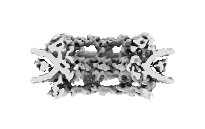
 ダウンロードとリンク
 emd_14321.png
 http://ftp.pdbj.org/pub/emdb/structures/EMD-14321
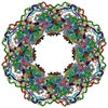


 Z (Sec.)
 Y (Row.)
 X (Col.)


 試料の構成要素
 解析
解析 電子顕微鏡法
 FIELD EMISSION GUN
