 Movie
Movie Controller


+ Open data

- Basic information
| Entry |  | ||||||||||||
|---|---|---|---|---|---|---|---|---|---|---|---|---|---|
| Title | Spinach 26S proteasome | ||||||||||||
 Map data Map data | |||||||||||||
 Sample Sample |
| ||||||||||||
 Keywords Keywords | PROTEASOME / UPS / PLANT / SPINACH / PLANT PROTEIN | ||||||||||||
| Function / homology |  Function and homology information Function and homology information: / : / nuclear proteasome complex / proteasome regulatory particle / cytosolic proteasome complex / proteasome-activating activity / proteasome regulatory particle, lid subcomplex / proteasome regulatory particle, base subcomplex / metal-dependent deubiquitinase activity / proteasome core complex ...: / : / nuclear proteasome complex / proteasome regulatory particle / cytosolic proteasome complex / proteasome-activating activity / proteasome regulatory particle, lid subcomplex / proteasome regulatory particle, base subcomplex / metal-dependent deubiquitinase activity / proteasome core complex / K63-linked deubiquitinase activity / proteasome binding / regulation of protein catabolic process / proteasome storage granule / positive regulation of RNA polymerase II transcription preinitiation complex assembly / membrane => GO:0016020 / protein deubiquitination / polyubiquitin modification-dependent protein binding / proteasome endopeptidase complex / proteasome core complex, beta-subunit complex / threonine-type endopeptidase activity / proteasome core complex, alpha-subunit complex / enzyme regulator activity / proteasomal protein catabolic process / proteasome complex / metallopeptidase activity / peptidase activity / endopeptidase activity / ubiquitin-dependent protein catabolic process / proteasome-mediated ubiquitin-dependent protein catabolic process / structural molecule activity / ATP hydrolysis activity / ATP binding / nucleus / cytoplasm / cytosol Similarity search - Function | ||||||||||||
| Biological species |  Spinacia oleracea (spinach) Spinacia oleracea (spinach) | ||||||||||||
| Method | single particle reconstruction / cryo EM / Resolution: 3.3 Å | ||||||||||||
 Authors Authors | Kandolf S / Grishkovskaya I | ||||||||||||
| Funding support | 1 items
| ||||||||||||
 Citation Citation | Journal: Plant Commun / Year: 2022 Title: Cryo-EM structure of the plant 26S proteasome. Authors: Susanne Kandolf / Irina Grishkovskaya / Katarina Belačić / Derek L Bolhuis / Sascha Amann / Brent Foster / Richard Imre / Karl Mechtler / Alexander Schleiffer / Hemant D Tagare / Ellen D ...Authors: Susanne Kandolf / Irina Grishkovskaya / Katarina Belačić / Derek L Bolhuis / Sascha Amann / Brent Foster / Richard Imre / Karl Mechtler / Alexander Schleiffer / Hemant D Tagare / Ellen D Zhong / Anton Meinhart / Nicholas G Brown / David Haselbach /    Abstract: Targeted proteolysis is a hallmark of life. It is especially important in long-lived cells that can be found in higher eukaryotes, like plants. This task is mainly fulfilled by the ubiquitin- ...Targeted proteolysis is a hallmark of life. It is especially important in long-lived cells that can be found in higher eukaryotes, like plants. This task is mainly fulfilled by the ubiquitin-proteasome system. Thus, proteolysis by the 26S proteasome is vital to development, immunity, and cell division. Although the yeast and animal proteasomes are well characterized, there is only limited information on the plant proteasome. We determined the first plant 26S proteasome structure from Spinacia oleracea by single-particle electron cryogenic microscopy at an overall resolution of 3.3 Å. We found an almost identical overall architecture of the spinach proteasome compared with the known structures from mammals and yeast. Nevertheless, we noticed a structural difference in the proteolytic active β1 subunit. Furthermore, we uncovered an unseen compression state by characterizing the proteasome's conformational landscape. We suspect that this new conformation of the 20S core protease, in correlation with a partial opening of the unoccupied gate, may contribute to peptide release after proteolysis. Our data provide a structural basis for the plant proteasome, which is crucial for further studies. | ||||||||||||
| History |
|
- Structure visualization
| Supplemental images |
|---|

- Downloads & links
-EMDB archive
| Map data | emd_14175.map.gz | 22.4 MB | EMDB map data format | |
|---|---|---|---|---|
| Header (meta data) | emd-14175-v30.xmlemd-14175.xml | 30.1 KB 30.1 KB | Display Display | EMDB header |
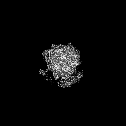
| Images | 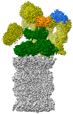 emd_14175.png emd_14175.png | 130.9 KB | ||
| Filedesc metadata | emd-14175.cif.gz | 8.5 KB | ||
| Archive directory |  http://ftp.pdbj.org/pub/emdb/structures/EMD-14175ftp://ftp.pdbj.org/pub/emdb/structures/EMD-14175 http://ftp.pdbj.org/pub/emdb/structures/EMD-14175ftp://ftp.pdbj.org/pub/emdb/structures/EMD-14175 | HTTPS FTP |
-Related structure data
| Related structure data |  7qveMC  8amzM  7qvg M: atomic model generated by this map C: citing same article ( |
|---|---|
| Similar structure data | |
| EM raw data | EMPIAR-10974 (Title: Cryo-EM structure of the plant 26S proteasome / Data size: 7.9 TB Data #1: Unaligned multi-frame micrographs of spinach 26S proteasome [micrographs - multiframe] Data #2: Unaligned multi-frame micrographs of spinach 26S proteasome [micrographs - multiframe] Data #3: Unaligned multi-frame micrographs of spinach 26S proteasome [micrographs - multiframe]) |
-Links
| EMDB pages | EMDB (EBI/PDBe) / EMDataResource |
|---|---|
| Related items in Molecule of the Month |


-Map
| File | Download / File: emd_14175.map.gz / Format: CCP4 / Size: 325 MB / Type: IMAGE STORED AS FLOATING POINT NUMBER (4 BYTES) | ||||||||||||||||||||||||||||||||||||
|---|---|---|---|---|---|---|---|---|---|---|---|---|---|---|---|---|---|---|---|---|---|---|---|---|---|---|---|---|---|---|---|---|---|---|---|---|---|
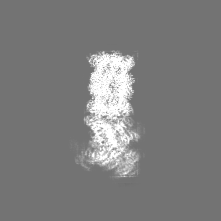
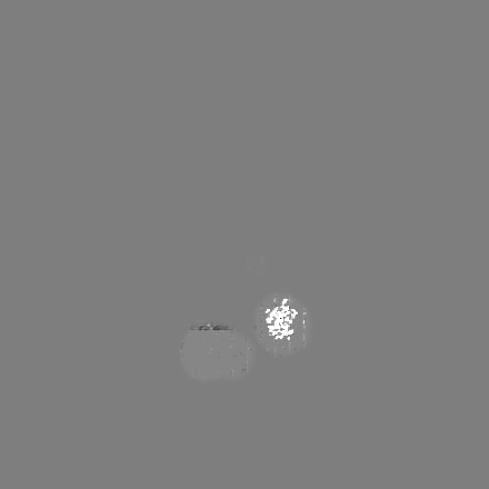
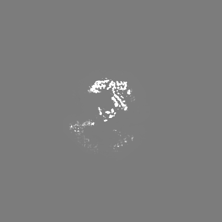
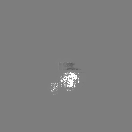
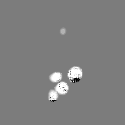
| Projections & slices | Image control
Images are generated by Spider. | ||||||||||||||||||||||||||||||||||||
| Voxel size | X=Y=Z: 1.23113 Å | ||||||||||||||||||||||||||||||||||||
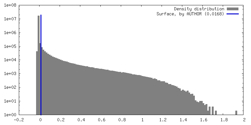
| Density |
| ||||||||||||||||||||||||||||||||||||
| Symmetry | Space group: 1 | ||||||||||||||||||||||||||||||||||||
| Details | EMDB XML:
|
 Z (Sec.)
 Y (Row.)
 X (Col.)


-Supplemental data
- Sample components
+Entire : Spinach 26S proteasome
+Supramolecule #1: Spinach 26S proteasome
+Macromolecule #1: Proteasome subunit alpha type
+Macromolecule #2: Proteasome subunit alpha type
+Macromolecule #3: Proteasome subunit alpha type
+Macromolecule #4: Proteasome subunit alpha type
+Macromolecule #5: Proteasome subunit alpha type
+Macromolecule #6: Proteasome subunit alpha type-3
+Macromolecule #7: Proteasome subunit alpha type
+Macromolecule #8: Proteasome subunit beta
+Macromolecule #9: Proteasome subunit beta
+Macromolecule #10: Proteasome subunit beta
+Macromolecule #11: Proteasome subunit beta
+Macromolecule #12: Proteasome subunit beta type-5
+Macromolecule #13: Proteasome subunit beta
+Macromolecule #14: Proteasome subunit beta
-Experimental details
-Structure determination
| Method | cryo EM |
|---|---|
 Processing Processing | single particle reconstruction |
| Aggregation state | particle |
-Sample preparation
| Concentration | 0.02 mg/mL | |||||||||||||||||||||||||||
|---|---|---|---|---|---|---|---|---|---|---|---|---|---|---|---|---|---|---|---|---|---|---|---|---|---|---|---|---|
| Buffer | pH: 6.5 Component:
| |||||||||||||||||||||||||||
| Grid | Model: Quantifoil R3.5/1 / Material: COPPER / Mesh: 200 / Support film - Material: CARBON / Support film - topology: CONTINUOUS / Support film - Film thickness: 2 / Pretreatment - Type: GLOW DISCHARGE / Pretreatment - Time: 60 sec. / Pretreatment - Atmosphere: AIR | |||||||||||||||||||||||||||
| Vitrification | Cryogen name: ETHANE / Chamber humidity: 80 % / Chamber temperature: 277 K / Instrument: LEICA EM GP |
- Electron microscopy
| Microscope | FEI TITAN KRIOS |
|---|---|
| Image recording | #0 - Image recording ID: 1 / #0 - Film or detector model: FEI FALCON III (4k x 4k) / #0 - Detector mode: INTEGRATING / #0 - Number real images: 24769 / #0 - Average electron dose: 80.0 e/Å2 / #1 - Image recording ID: 2 / #1 - Film or detector model: FEI FALCON III (4k x 4k) / #1 - Detector mode: INTEGRATING / #1 - Digitization - Dimensions - Width: 4096 pixel / #1 - Digitization - Dimensions - Height: 4096 pixel / #1 - Number real images: 8089 / #1 - Average electron dose: 50.0 e/Å2 |
| Electron beam | Acceleration voltage: 300 kV / Electron source:  FIELD EMISSION GUN FIELD EMISSION GUN |
| Electron optics | C2 aperture diameter: 70.0 µm / Illumination mode: FLOOD BEAM / Imaging mode: BRIGHT FIELD / Cs: 2.7 mm / Nominal defocus max: 4.0 µm / Nominal defocus min: 1.0 µm |
| Sample stage | Specimen holder model: FEI TITAN KRIOS AUTOGRID HOLDER / Cooling holder cryogen: NITROGEN |
| Experimental equipment |  Model: Titan Krios / Image courtesy: FEI Company |

