- EMDB-14098: Structure of the human inner kinetochore CCAN complex -
+
Open data
ID or keywords:
Loading...
-
Basic information
Entry
Database: EMDB / ID: EMD-14098
Title
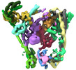
Structure of the human inner kinetochore CCAN complex
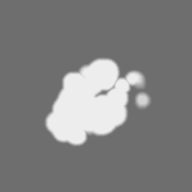
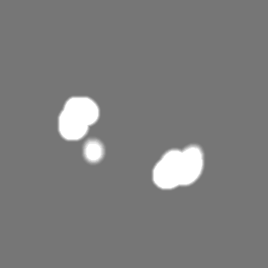
Map data
main map
Sample
Complex: human inner kinetochore CCAN complex
Protein or peptide: x 15 types
Keywords
INNER KINETOCHORE / CCAN / COMPLEX / DNA BINDING PROTEIN / CELL CYCLE
Function / homology
Function and homology information
positive regulation of protein localization to kinetochore / Mis6-Sim4 complex / centromere complex assembly / kinetochore organization / spindle attachment to meiosis I kinetochore / metaphase chromosome alignment / kinetochore binding / sex differentiation / centromeric DNA binding / CENP-A containing chromatin assembly ...positive regulation of protein localization to kinetochore / Mis6-Sim4 complex / centromere complex assembly / kinetochore organization / spindle attachment to meiosis I kinetochore / metaphase chromosome alignment / kinetochore binding / sex differentiation / centromeric DNA binding / CENP-A containing chromatin assembly / chordate embryonic development / negative regulation of epithelial cell apoptotic process / kinetochore assembly / inner kinetochore / condensed chromosome, centromeric region / attachment of mitotic spindle microtubules to kinetochore / mitotic sister chromatid segregation / chromosome, centromeric region / centriolar satellite / chromosome organization / Amplification of signal from unattached kinetochores via a MAD2 inhibitory signal / pericentric heterochromatin / Mitotic Prometaphase / EML4 and NUDC in mitotic spindle formation / Deposition of new CENPA-containing nucleosomes at the centromere / Resolution of Sister Chromatid Cohesion / NRIF signals cell death from the nucleus / mitotic spindle organization / positive regulation of epithelial cell proliferation / chromosome segregation / RHO GTPases Activate Formins / kinetochore / nuclear matrix / Separation of Sister Chromatids / actin cytoskeleton / chromosome / mitotic cell cycle / midbody / nuclear body / cell adhesion / protein heterodimerization activity / cell division / apoptotic process / regulation of DNA-templated transcription / nucleolus / signal transduction / DNA binding / nucleoplasm / identical protein binding / membrane / nucleus / cytoplasm / cytosol Similarity search - Function
Centromere protein W / CENP-W protein / Centromere protein T / Centromere kinetochore component CENP-T, N-terminal domain / Centromere kinetochore component CENP-T N-terminus / Centromere subunit L / Kinetochore complex Sim4 subunit Fta1 / Centromere protein R / Centromere protein Q / Centromere protein U ...Centromere protein W / CENP-W protein / Centromere protein T / Centromere kinetochore component CENP-T, N-terminal domain / Centromere kinetochore component CENP-T N-terminus / Centromere subunit L / Kinetochore complex Sim4 subunit Fta1 / Centromere protein R / Centromere protein Q / Centromere protein U / Centromere protein P / Centromere protein H / Kinetochore component, CENP-R / CENP-Q, a CENPA-CAD centromere complex subunit / CENP-A-nucleosome distal (CAD) centromere subunit, CENP-P / CENP-A nucleosome associated complex (NAC) subunit / Centromere protein H, C-terminal / Centromere protein Cenp-M / Centromere protein Cenp-K / Centromere protein H (CENP-H) / Centromere protein M (CENP-M) / Centromere-associated protein K / Centromere protein I / Mis6 / CENP-C, middle DNMT3B-binding domain / Centromere assembly component CENP-C middle DNMT3B-binding region / : / Centromere protein O / Cenp-O kinetochore centromere component / Kinetochore assembly subunit CENP-C, N-terminal domain / Kinetochore assembly subunit CENP-C N-terminal / Mif2/CENP-C cupin domain / Centromere protein C/Mif2/cnp3 / Mif2/CENP-C like / Centromere protein Chl4/mis15/CENP-N / Kinetochore protein CHL4 like / RmlC-like cupin domain superfamily / RmlC-like jelly roll fold / CENP-T/Histone H4, histone fold / Centromere kinetochore component CENP-T histone fold / Histone-fold / P-loop containing nucleoside triphosphate hydrolase Similarity search - Domain/homology
Centromere protein C / Centromere protein R / Centromere protein W / Centromere protein P / Centromere protein U / Centromere protein Q / Centromere protein L / Centromere protein I / Centromere protein T / Centromere protein N ...Centromere protein C / Centromere protein R / Centromere protein W / Centromere protein P / Centromere protein U / Centromere protein Q / Centromere protein L / Centromere protein I / Centromere protein T / Centromere protein N / Centromere protein K / Centromere protein O / Centromere protein H / Centromere protein M Similarity search - Component
Biological species
Homo sapiens (human)
Method
single particle reconstruction / cryo EM / Resolution: 4.6 Å
Journal: Mol Cell / Year: 2022 Title: Structure of the human inner kinetochore CCAN complex and its significance for human centromere organization. Authors: Marion E Pesenti / Tobias Raisch / Duccio Conti / Kai Walstein / Ingrid Hoffmann / Dorothee Vogt / Daniel Prumbaum / Ingrid R Vetter / Stefan Raunser / Andrea Musacchio / Abstract: Centromeres are specialized chromosome loci that seed the kinetochore, a large protein complex that effects chromosome segregation. A 16-subunit complex, the constitutive centromere associated ...Centromeres are specialized chromosome loci that seed the kinetochore, a large protein complex that effects chromosome segregation. A 16-subunit complex, the constitutive centromere associated network (CCAN), connects between the specialized centromeric chromatin, marked by the histone H3 variant CENP-A, and the spindle-binding moiety of the kinetochore. Here, we report a cryo-electron microscopy structure of human CCAN. We highlight unique features such as the pseudo GTPase CENP-M and report how a crucial CENP-C motif binds the CENP-LN complex. The CCAN structure has implications for the mechanism of specific recognition of the CENP-A nucleosome. A model consistent with our structure depicts the CENP-C-bound nucleosome as connected to the CCAN through extended, flexible regions of CENP-C. An alternative model identifies both CENP-C and CENP-N as specificity determinants but requires CENP-N to bind CENP-A in a mode distinct from the classical nucleosome octamer.
Cryogen name: ETHANE / Chamber humidity: 100 % / Chamber temperature: 286 K / Instrument: FEI VITROBOT MARK IV / Details: blot for 3.5 seconds at blot force -3.
-
Electron microscopy
Microscope
FEI TITAN KRIOS
Specialist optics
Phase plate: VOLTA PHASE PLATE / Energy filter - Name: GIF Bioquantum / Energy filter - Slit width: 20 eV
Image recording
Film or detector model: GATAN K3 (6k x 4k) / Number real images: 1540 / Average electron dose: 76.8 e/Å2 Details: Images were collected in movie mode with 80 frames per image in superresolution mode with 0.35 A/px (0.7A native)
Electron beam
Acceleration voltage: 300 kV / Electron source: FIELD EMISSION GUN
In the structure databanks used in Yorodumi, some data are registered as the other names, "COVID-19 virus" and "2019-nCoV". Here are the details of the virus and the list of structure data.
Jan 31, 2019. EMDB accession codes are about to change! (news from PDBe EMDB page)
EMDB accession codes are about to change! (news from PDBe EMDB page)
The allocation of 4 digits for EMDB accession codes will soon come to an end. Whilst these codes will remain in use, new EMDB accession codes will include an additional digit and will expand incrementally as the available range of codes is exhausted. The current 4-digit format prefixed with “EMD-” (i.e. EMD-XXXX) will advance to a 5-digit format (i.e. EMD-XXXXX), and so on. It is currently estimated that the 4-digit codes will be depleted around Spring 2019, at which point the 5-digit format will come into force.
The EM Navigator/Yorodumi systems omit the EMD- prefix.
Related info.:Q: What is EMD? / ID/Accession-code notation in Yorodumi/EM Navigator
Yorodumi is a browser for structure data from EMDB, PDB, SASBDB, etc.
This page is also the successor to EM Navigator detail page, and also detail information page/front-end page for Omokage search.
The word "yorodu" (or yorozu) is an old Japanese word meaning "ten thousand". "mi" (miru) is to see.
Related info.:EMDB / PDB / SASBDB / Comparison of 3 databanks / Yorodumi Search / Aug 31, 2016. New EM Navigator & Yorodumi / Yorodumi Papers / Jmol/JSmol / Function and homology information / Changes in new EM Navigator and Yorodumi
 Movie
Movie Controller


 Open data
 Basic information
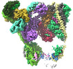
 Map data
 Sample
 Keywords
 Function and homology information
 Homo sapiens (human)
 Authors
 Citation
 Structure visualization
 Downloads & links
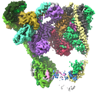 emd_14098.png
 http://ftp.pdbj.org/pub/emdb/structures/EMD-14098
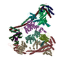


 Z
Z Y
Y X
X


 Sample components
 Trichoplusia ni (cabbage looper)
 Processing
 Electron microscopy
 FIELD EMISSION GUN

