[English] 日本語
Yorodumi
- EMDB-13493: CryoEM reconstruction of pyruvate ferredoxin oxidoreductase (PFOR... -

+
Open data


ID or keywords:

Loading...

-
Basic information

Entry
Database: EMDB / ID: EMD-13493
TitleCryoEM reconstruction of pyruvate ferredoxin oxidoreductase (PFOR) in anaerobic conditions
Map data
Sample
  • Complex: PYRUVATE FERREDOXIN OXIDOREDUCTASE
    • Protein or peptide: Pyruvate:ferredoxin oxidoreductase
  • Ligand: IRON/SULFUR CLUSTER
  • Ligand: THIAMINE DIPHOSPHATE
  • Ligand: CALCIUM ION
  • Ligand: MAGNESIUM ION
  • Ligand: water
Function / homology
Function and homology information


pyruvate synthase / pyruvate synthase activity / thiamine pyrophosphate binding / electron transport chain / 4 iron, 4 sulfur cluster binding / response to oxidative stress / iron ion binding / cytoplasm
Similarity search - Function
Pyruvate-flavodoxin oxidoreductase / Pyruvate-flavodoxin oxidoreductase, EKR domain / Pyruvate-flavodoxin oxidoreductase, EKR domain superfamily / Domain of unknown function / Domain of unknown function / Pyruvate:ferredoxin oxidoreductase, core domain II / Pyruvate:ferredoxin oxidoreductase core domain II / Pyruvate flavodoxin/ferredoxin oxidoreductase, pyrimidine binding domain / Pyruvate flavodoxin/ferredoxin oxidoreductase, thiamine diP-bdg / Pyruvate-flavodoxin oxidoreductase, central domain ...Pyruvate-flavodoxin oxidoreductase / Pyruvate-flavodoxin oxidoreductase, EKR domain / Pyruvate-flavodoxin oxidoreductase, EKR domain superfamily / Domain of unknown function / Domain of unknown function / Pyruvate:ferredoxin oxidoreductase, core domain II / Pyruvate:ferredoxin oxidoreductase core domain II / Pyruvate flavodoxin/ferredoxin oxidoreductase, pyrimidine binding domain / Pyruvate flavodoxin/ferredoxin oxidoreductase, thiamine diP-bdg / Pyruvate-flavodoxin oxidoreductase, central domain / Pyruvate/ketoisovalerate oxidoreductase, catalytic domain / Pyruvate ferredoxin/flavodoxin oxidoreductase / Transketolase C-terminal/Pyruvate-ferredoxin oxidoreductase domain II / Thiamine pyrophosphate enzyme, C-terminal TPP-binding / Thiamine pyrophosphate enzyme, C-terminal TPP binding domain / Thiamin diphosphate-binding fold / 4Fe-4S dicluster domain / 4Fe-4S ferredoxin, iron-sulphur binding, conserved site / 4Fe-4S ferredoxin-type iron-sulfur binding region signature. / 4Fe-4S ferredoxin-type iron-sulfur binding domain profile. / 4Fe-4S ferredoxin-type, iron-sulphur binding domain
Similarity search - Domain/homology
Pyruvate:ferredoxin oxidoreductase
Similarity search - Component
Biological speciesDesulfocurvibacter africanus (bacteria)
Methodsingle particle reconstruction / cryo EM / Resolution: 2.9 Å
AuthorsCherrier MV / Vernede X / Fenel D / Martin L / Arragain B / Neumann E / Fontecilla Camps JC / Schoehn G / Nicolet Y
Funding support France, 4 items
OrganizationGrant numberCountry
Agence Nationale de la Recherche (ANR)ANR-15-IDEX-02 France
Grenoble Instruct-ERIC Center (ISBG)UMS 3518 CNRS-CEA-UGA-EMBL France
French Infrastructure for Integrated Structural Biology (FRISBI)ANR-10-INSB-05-02 France
Grenoble Alliance for Integrated Structural Cell Biology (GRAL)ANR-10-LABX-49-01 France
Citation
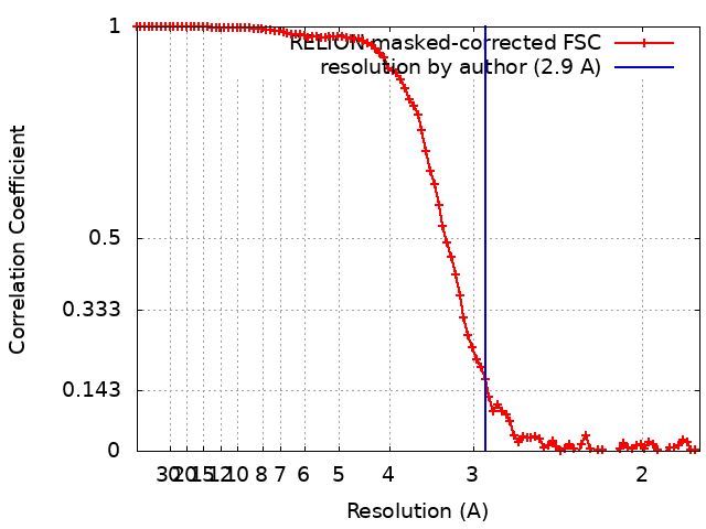
Journal: Biomolecules / Year: 2022
Title: Oxygen-Sensitive Metalloprotein Structure Determination by Cryo-Electron Microscopy.
Authors: Mickaël V Cherrier / Xavier Vernède / Daphna Fenel / Lydie Martin / Benoit Arragain / Emmanuelle Neumann / Juan C Fontecilla-Camps / Guy Schoehn / Yvain Nicolet /
Abstract: Metalloproteins are involved in key cell processes such as photosynthesis, respiration, and oxygen transport. However, the presence of transition metals (notably iron as a component of [Fe-S] ...Metalloproteins are involved in key cell processes such as photosynthesis, respiration, and oxygen transport. However, the presence of transition metals (notably iron as a component of [Fe-S] clusters) often makes these proteins sensitive to oxygen-induced degradation. Consequently, their study usually requires strict anaerobic conditions. Although X-ray crystallography has been the method of choice for solving macromolecular structures for many years, recently electron microscopy has also become an increasingly powerful structure-solving technique. We have used our previous experience with cryo-crystallography to develop a method to prepare cryo-EM grids in an anaerobic chamber and have applied it to solve the structures of apoferritin and the 3 [FeS]-containing pyruvate ferredoxin oxidoreductase (PFOR) at 2.40 Å and 2.90 Å resolution, respectively. The maps are of similar quality to the ones obtained under air, thereby validating our method as an improvement in the structural investigation of oxygen-sensitive metalloproteins by cryo-EM.
History
DepositionAug 31, 2021-
Header (metadata) releaseMar 23, 2022-
Map releaseMar 23, 2022-
UpdateApr 6, 2022-
Current statusApr 6, 2022Processing site: PDBe / Status: Released

-
Structure visualization
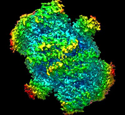
Supplemental images

Downloads & links

-
Map

FileDownload / File: emd_13493.map.gz / Format: CCP4 / Size: 73.4 MB / Type: IMAGE STORED AS FLOATING POINT NUMBER (4 BYTES)
Voxel sizeX=Y=Z: 0.899 Å
Density
Contour LevelBy AUTHOR: 0.0194
Minimum - Maximum-0.11396713 - 0.18406008
Average (Standard dev.)4.554769e-05 (±0.007088646)
SymmetrySpace group: 1
Details

EMDB XML:

Map geometry
Axis orderXYZ
Origin000
Dimensions268268268
Spacing268268268
CellA=B=C: 240.93199 Å
α=β=γ: 90.0 °

-
Supplemental data

-
Sample components

-
Entire : PYRUVATE FERREDOXIN OXIDOREDUCTASE

EntireName: PYRUVATE FERREDOXIN OXIDOREDUCTASE
Components
  • Complex: PYRUVATE FERREDOXIN OXIDOREDUCTASE
    • Protein or peptide: Pyruvate:ferredoxin oxidoreductase
  • Ligand: IRON/SULFUR CLUSTER
  • Ligand: THIAMINE DIPHOSPHATE
  • Ligand: CALCIUM ION
  • Ligand: MAGNESIUM ION
  • Ligand: water

-
Supramolecule #1: PYRUVATE FERREDOXIN OXIDOREDUCTASE

SupramoleculeName: PYRUVATE FERREDOXIN OXIDOREDUCTASE / type: complex / ID: 1 / Parent: 0 / Macromolecule list: #1
Source (natural)Organism: Desulfocurvibacter africanus (bacteria)
Molecular weightTheoretical: 267.36 KDa

-
Macromolecule #1: Pyruvate:ferredoxin oxidoreductase

MacromoleculeName: Pyruvate:ferredoxin oxidoreductase / type: protein_or_peptide / ID: 1 / Number of copies: 2 / Enantiomer: LEVO / EC number: pyruvate synthase
Source (natural)Organism: Desulfocurvibacter africanus (bacteria)
Molecular weightTheoretical: 133.835109 KDa
Recombinant expressionOrganism: Desulfocurvibacter africanus (bacteria)
SequenceString: MGKKMMTTDG NTATAHVAYA MSEVAAIYPI TPSSTMGEEA DDWAAQGRKN IFGQTLTIRE MQSEAGAAGA VHGALAAGAL TTTFTASQG LLLMIPNMYK ISGELLPGVF HVTARAIAAH ALSIFGDHQD IYAARQTGFA MLASSSVQEA HDMALVAHLA A IESNVPFM ...String:
MGKKMMTTDG NTATAHVAYA MSEVAAIYPI TPSSTMGEEA DDWAAQGRKN IFGQTLTIRE MQSEAGAAGA VHGALAAGAL TTTFTASQG LLLMIPNMYK ISGELLPGVF HVTARAIAAH ALSIFGDHQD IYAARQTGFA MLASSSVQEA HDMALVAHLA A IESNVPFM HFFDGFRTSH EIQKIEVLDY ADMASLVNQK ALAEFRAKSM NPEHPHVRGT AQNPDIYFQG REAANPYYLK VP GIVAEYM QKVASLTGRS YKLFDYVGAP DAERVIVSMG SSCETIEEVI NHLAAKGEKI GLIKVRLYRP FVSEAFFAAL PAS AKVITV LDRTKEPGAP GDPLYLDVCS AFVERGEAMP KILAGRYGLG SKEFSPAMVK SVYDNMSGAK KNHFTVGIED DVTG TSLPV DNAFADTTPK GTIQCQFWGL GADGTVGANK QAIKIIGDNT DLFAQGYFSY DSKKSGGITI SHLRFGEKPI QSTYL VNRA DYVACHNPAY VGIYDILEGI KDGGTFVLNS PWSSLEDMDK HLPSGIKRTI ANKKLKFYNI DAVKIATDVG LGGRIN MIM QTAFFKLAGV LPFEKAVDLL KKSIHKAYGK KGEKIVKMNT DAVDQAVTSL QEFKYPDSWK DAPAETKAEP MTNEFFK NV VKPILTQQGD KLPVSAFEAD GRFPLGTSQF EKRGVAINVP QWVPENCIQC NQCAFVCPHS AILPVLAKEE ELVGAPAN F TALEAKGKEL KGYKFRIQIN TLDCMGCGNC ADICPPKEKA LVMQPLDTQR DAQVPNLEYA ARIPVKSEVL PRDSLKGSQ FQEPLMEFSG ACSGCGETPY VRVITQLFGE RMFIANATGC SSIWGASAPS MPYKTNRLGQ GPAWGNSLFE DAAEYGFGMN MSMFARRTH LADLAAKALE SDASGDVKEA LQGWLAGKND PIKSKEYGDK LKKLLAGQKD GLLGQIAAMS DLYTKKSVWI F GGDGWAYD IGYGGLDHVL ASGEDVNVFV MDTEVYSNTG GQSSKATPTG AVAKFAAAGK RTGKKDLARM VMTYGYVYVA TV SMGYSKQ QFLKVLKEAE SFPGPSLVIA YATCINQGLR KGMGKSQDVM NTAVKSGYWP LFRYDPRLAA QGKNPFQLDS KAP DGSVEE FLMAQNRFAV LDRSFPEDAK RLRAQVAHEL DVRFKELEHM AATNIFESFA PAGGKADGSV DFGEGAEFCT RDDT PMMAR PDSGEACDQN RAGTSEQQGD LSKRTKK

-
Macromolecule #2: IRON/SULFUR CLUSTER

MacromoleculeName: IRON/SULFUR CLUSTER / type: ligand / ID: 2 / Number of copies: 6 / Formula: SF4
Molecular weightTheoretical: 351.64 Da
Chemical component information

ChemComp-FS1:
IRON/SULFUR CLUSTER

-
Macromolecule #3: THIAMINE DIPHOSPHATE

MacromoleculeName: THIAMINE DIPHOSPHATE / type: ligand / ID: 3 / Number of copies: 2 / Formula: TPP
Molecular weightTheoretical: 425.314 Da
Chemical component information

ChemComp-TPP:
THIAMINE DIPHOSPHATE

-
Macromolecule #4: CALCIUM ION

MacromoleculeName: CALCIUM ION / type: ligand / ID: 4 / Number of copies: 2 / Formula: CA
Molecular weightTheoretical: 40.078 Da

-
Macromolecule #5: MAGNESIUM ION

MacromoleculeName: MAGNESIUM ION / type: ligand / ID: 5 / Number of copies: 2 / Formula: MG
Molecular weightTheoretical: 24.305 Da

-
Macromolecule #6: water

MacromoleculeName: water / type: ligand / ID: 6 / Number of copies: 114 / Formula: HOH
Molecular weightTheoretical: 18.015 Da
Chemical component information

ChemComp-HOH:
WATER

-
Experimental details

-
Structure determination

Methodcryo EM
Processingsingle particle reconstruction
Aggregation stateparticle

-
Sample preparation

Concentration2.3 mg/mL
BufferpH: 8.4 / Component - Concentration: 10.0 mM / Component - Name: Tris
GridModel: Quantifoil R2/1 / Material: COPPER/RHODIUM / Mesh: 300 / Pretreatment - Type: GLOW DISCHARGE / Pretreatment - Atmosphere: AIR
VitrificationCryogen name: PROPANE / Chamber humidity: 100 % / Chamber temperature: 295 K / Instrument: FEI VITROBOT MARK IV
DetailsMonodisperse

-
Electron microscopy

MicroscopeTFS GLACIOS
Image recordingFilm or detector model: GATAN K2 SUMMIT (4k x 4k) / Digitization - Frames/image: 2-60 / Number grids imaged: 1 / Number real images: 1304 / Average electron dose: 60.0 e/Å2
Electron beamAcceleration voltage: 200 kV / Electron source: FIELD EMISSION GUN
Electron opticsIllumination mode: FLOOD BEAM / Imaging mode: BRIGHT FIELD / Nominal defocus max: 3.5 µm / Nominal defocus min: 0.5 µm

+
Image processing

CTF correctionSoftware - Name: CTFFIND (ver. 4.1)
Final reconstructionApplied symmetry - Point group: C2 (2 fold cyclic) / Resolution.type: BY AUTHOR / Resolution: 2.9 Å / Resolution method: FSC 0.143 CUT-OFF / Software - Name: RELION (ver. 3.1) / Number images used: 180868
Initial angle assignmentType: MAXIMUM LIKELIHOOD / Software - Name: RELION (ver. 3.1)
Final angle assignmentType: MAXIMUM LIKELIHOOD / Software - Name: RELION (ver. 3.1)
FSC plot (resolution estimation)

-
Atomic model buiding 1

Initial modelPDB ID:

Chain - Chain ID: A
RefinementSpace: RECIPROCAL / Protocol: RIGID BODY FIT
Output model

PDB-7plm:
CryoEM reconstruction of pyruvate ferredoxin oxidoreductase (PFOR) in anaerobic conditions

+
About Yorodumi

-
News

-
Feb 9, 2022. New format data for meta-information of EMDB entries

New format data for meta-information of EMDB entries

  • Version 3 of the EMDB header file is now the official format.
  • The previous official version 1.9 will be removed from the archive.

Related info.:EMDB header

External links:wwPDB to switch to version 3 of the EMDB data model

-
Aug 12, 2020. Covid-19 info

Covid-19 info

URL: https://pdbjlvh1.pdbj.org/emnavi/covid19.php

New page: Covid-19 featured information page in EM Navigator.

Related info.:Covid-19 info / Mar 5, 2020. Novel coronavirus structure data

+
Mar 5, 2020. Novel coronavirus structure data

Novel coronavirus structure data

Related info.:Yorodumi Speices / Aug 12, 2020. Covid-19 info

External links:COVID-19 featured content - PDBj / Molecule of the Month (242):Coronavirus Proteases

+
Jan 31, 2019. EMDB accession codes are about to change! (news from PDBe EMDB page)

EMDB accession codes are about to change! (news from PDBe EMDB page)

  • The allocation of 4 digits for EMDB accession codes will soon come to an end. Whilst these codes will remain in use, new EMDB accession codes will include an additional digit and will expand incrementally as the available range of codes is exhausted. The current 4-digit format prefixed with “EMD-” (i.e. EMD-XXXX) will advance to a 5-digit format (i.e. EMD-XXXXX), and so on. It is currently estimated that the 4-digit codes will be depleted around Spring 2019, at which point the 5-digit format will come into force.
  • The EM Navigator/Yorodumi systems omit the EMD- prefix.

Related info.:Q: What is EMD? / ID/Accession-code notation in Yorodumi/EM Navigator

External links:EMDB Accession Codes are Changing Soon! / Contact to PDBj

+
Jul 12, 2017. Major update of PDB

Major update of PDB

  • wwPDB released updated PDB data conforming to the new PDBx/mmCIF dictionary.
  • This is a major update changing the version number from 4 to 5, and with Remediation, in which all the entries are updated.
  • In this update, many items about electron microscopy experimental information are reorganized (e.g. em_software).
  • Now, EM Navigator and Yorodumi are based on the updated data.

External links:wwPDB Remediation / Enriched Model Files Conforming to OneDep Data Standards Now Available in the PDB FTP Archive

-
Yorodumi

Thousand views of thousand structures

  • Yorodumi is a browser for structure data from EMDB, PDB, SASBDB, etc.
  • This page is also the successor to EM Navigator detail page, and also detail information page/front-end page for Omokage search.
  • The word "yorodu" (or yorozu) is an old Japanese word meaning "ten thousand". "mi" (miru) is to see.

Related info.:EMDB / PDB / SASBDB / Comparison of 3 databanks / Yorodumi Search / Aug 31, 2016. New EM Navigator & Yorodumi / Yorodumi Papers / Jmol/JSmol / Function and homology information / Changes in new EM Navigator and Yorodumi

Read more