Movie
Movie Controller

[English] 日本語
 Yorodumi
- EMDB-13164: Cryo-EM structure of encapsulin from Mycobacterium tuberculosis -
+ Open data

- Basic information
| Entry |  | |||||||||
|---|---|---|---|---|---|---|---|---|---|---|
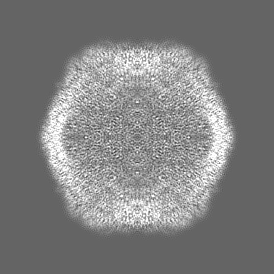
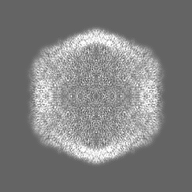
| Title | Cryo-EM structure of encapsulin from Mycobacterium tuberculosis | |||||||||
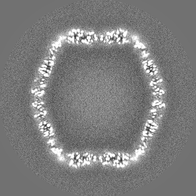
 Map data Map data | LocSpiral map | |||||||||
 Sample Sample |
| |||||||||
 Keywords Keywords | encapsulin / cfp29 / nanocompartment / VIRUS LIKE PARTICLE | |||||||||
| Function / homology | Type 1 encapsulin shell protein / Encapsulating protein for peroxidase / : / encapsulin nanocompartment / hydrolase activity / plasma membrane / Type 1 encapsulin shell protein Function and homology information Function and homology information | |||||||||
| Biological species |  Mycobacterium tuberculosis H37Rv (bacteria) / Mycobacterium tuberculosis H37Rv (bacteria) /  Mycobacterium tuberculosis (bacteria) Mycobacterium tuberculosis (bacteria) | |||||||||
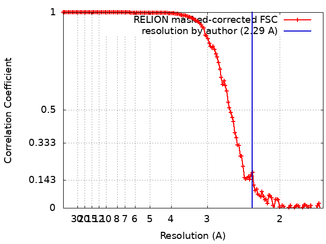
| Method | single particle reconstruction / cryo EM / Resolution: 2.29 Å | |||||||||
 Authors Authors | Lewis CJ / Berger C | |||||||||
| Funding support |  Netherlands, 2 items Netherlands, 2 items
| |||||||||
 Citation Citation | Journal: Commun Biol / Year: 2025 Title: In situ and in vitro cryo-EM reveal structures of mycobacterial encapsulin assembly intermediates. Authors: Casper Berger / Chris Lewis / Ye Gao / Kèvin Knoops / Carmen López-Iglesias / Peter J Peters / Raimond B G Ravelli /  Abstract: Prokaryotes rely on proteinaceous compartments such as encapsulin to isolate harmful reactions. Encapsulin are widely expressed by bacteria, including the Mycobacteriaceae, which include the human ...Prokaryotes rely on proteinaceous compartments such as encapsulin to isolate harmful reactions. Encapsulin are widely expressed by bacteria, including the Mycobacteriaceae, which include the human pathogens Mycobacterium tuberculosis and Mycobacterium leprae. Structures of fully assembled encapsulin shells have been determined for several species, but encapsulin assembly and cargo encapsulation are still poorly characterised, because of the absence of encapsulin structures in intermediate assembly states. We combine in situ and in vitro structural electron microscopy to show that encapsulins are dynamic assemblies with intermediate states of cargo encapsulation and shell assembly. Using cryo-focused ion beam (FIB) lamella preparation and cryo-electron tomography (CET), we directly visualise encapsulins in Mycobacterium marinum, and observed ribbon-like attachments to the shell, encapsulin shells with and without cargoes, and encapsulin shells in partially assembled states. In vitro cryo-electron microscopy (EM) single-particle analysis of the Mycobacterium tuberculosis encapsulin was used to obtain three structures of the encapsulin shell in intermediate states, as well as a 2.3 Å structure of the fully assembled shell. Based on the analysis of the intermediate encapsulin shell structures, we propose a model of encapsulin self-assembly via the pairwise addition of monomers. | |||||||||
| History |
|
- Structure visualization
| Supplemental images |
|---|

- Downloads & links
-EMDB archive
| Map data | emd_13164.map.gz | 41.9 MB | EMDB map data format | |
|---|---|---|---|---|
| Header (meta data) | emd-13164-v30.xmlemd-13164.xml | 23 KB 23 KB | Display Display | EMDB header |
| FSC (resolution estimation) | emd_13164_fsc.xml | 13.5 KB | Display | FSC data file |
| Images | 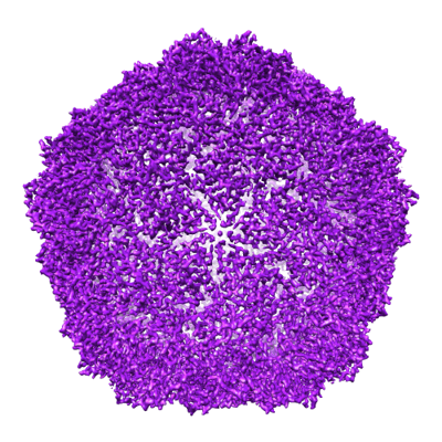 emd_13164.png emd_13164.png | 237.9 KB | ||
| Masks | emd_13164_msk_1.map | 216 MB | Mask map | |
| Filedesc metadata | emd-13164.cif.gz | 6.9 KB | ||
| Others | emd_13164_additional_1.map.gzemd_13164_half_map_1.map.gzemd_13164_half_map_2.map.gz | 40.4 MB 170.7 MB 170.7 MB | ||
| Archive directory |  http://ftp.pdbj.org/pub/emdb/structures/EMD-13164ftp://ftp.pdbj.org/pub/emdb/structures/EMD-13164 http://ftp.pdbj.org/pub/emdb/structures/EMD-13164ftp://ftp.pdbj.org/pub/emdb/structures/EMD-13164 | HTTPS FTP |
-Related structure data
| Related structure data |  7p1tMC  9gotC  9hq7C  9hqcC M: atomic model generated by this map C: citing same article ( |
|---|---|
| Similar structure data |


-Links
| EMDB pages | EMDB (EBI/PDBe) / EMDataResource |
|---|---|
| Related items in Molecule of the Month |

-Map
| File | Download / File: emd_13164.map.gz / Format: CCP4 / Size: 216 MB / Type: IMAGE STORED AS FLOATING POINT NUMBER (4 BYTES) | ||||||||||||||||||||||||||||||||||||
|---|---|---|---|---|---|---|---|---|---|---|---|---|---|---|---|---|---|---|---|---|---|---|---|---|---|---|---|---|---|---|---|---|---|---|---|---|---|
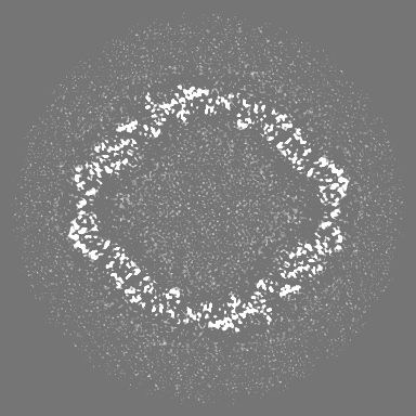
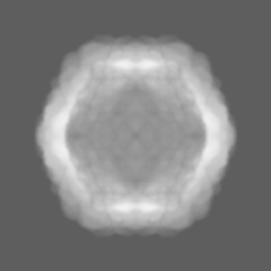
| Annotation | LocSpiral map | ||||||||||||||||||||||||||||||||||||
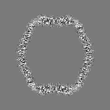
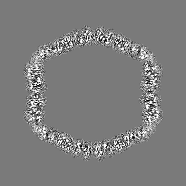
| Projections & slices | Image control
Images are generated by Spider. | ||||||||||||||||||||||||||||||||||||
| Voxel size | X=Y=Z: 0.834 Å | ||||||||||||||||||||||||||||||||||||
| Density |
| ||||||||||||||||||||||||||||||||||||
| Symmetry | Space group: 1 | ||||||||||||||||||||||||||||||||||||
| Details | EMDB XML:
|
 Z (Sec.)
 Y (Row.)
 X (Col.)


-Supplemental data
-Mask #1
| File | emd_13164_msk_1.map | ||||||||||||
|---|---|---|---|---|---|---|---|---|---|---|---|---|---|
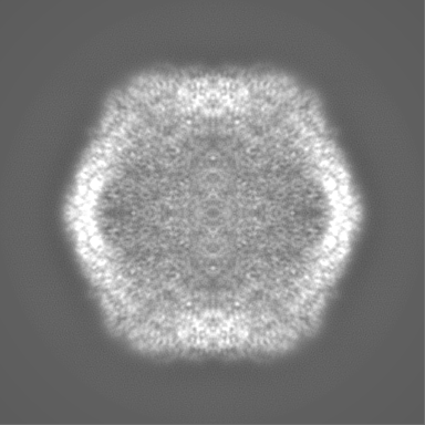
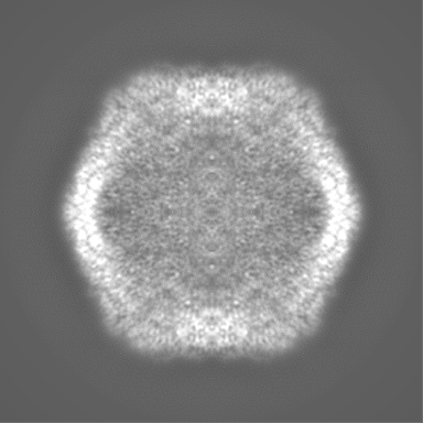
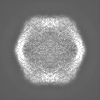
| Projections & Slices |
| ||||||||||||
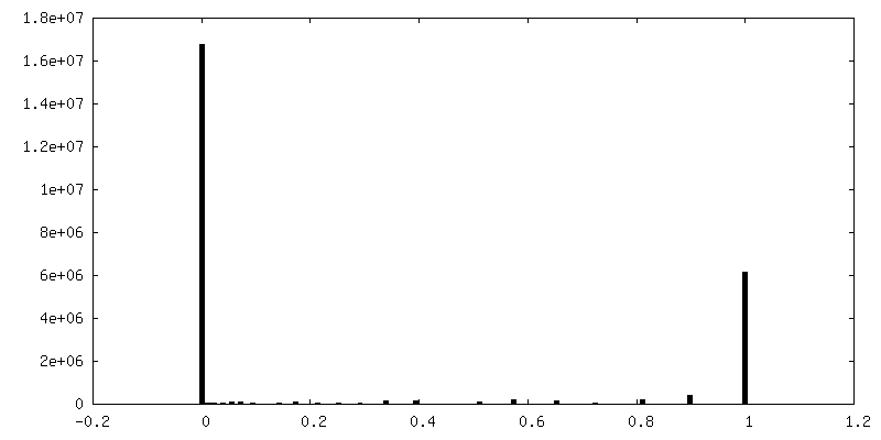
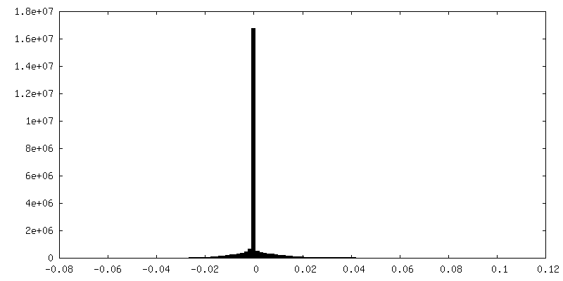
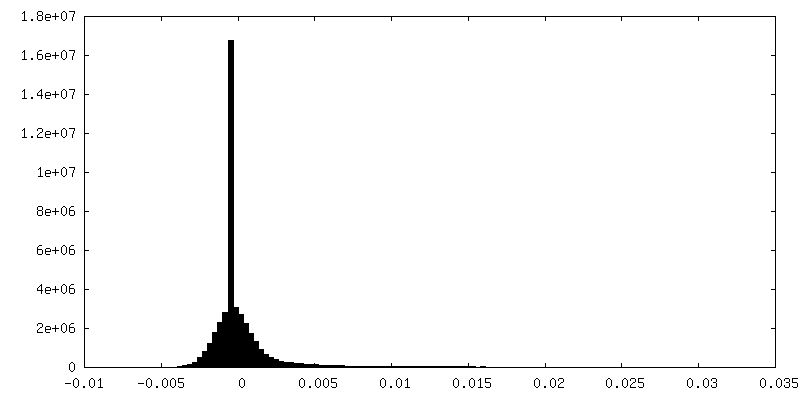
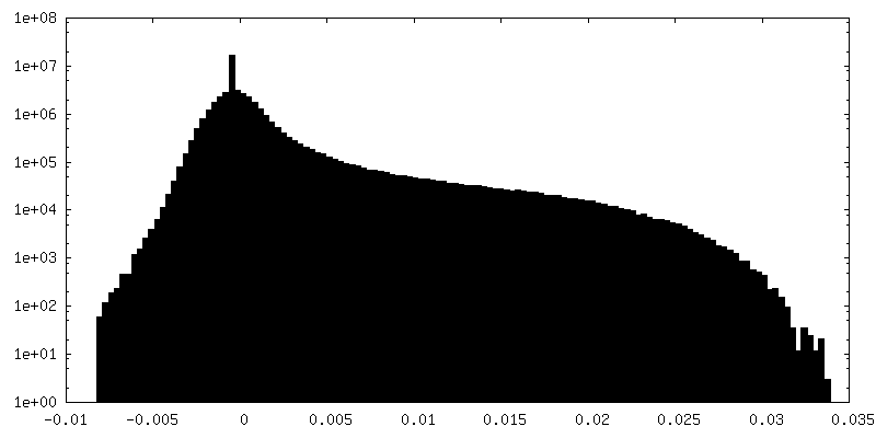
| Density Histograms |


-Additional map: relion postprocess masked
| File | emd_13164_additional_1.map | ||||||||||||
|---|---|---|---|---|---|---|---|---|---|---|---|---|---|
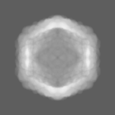
| Annotation | relion postprocess masked | ||||||||||||
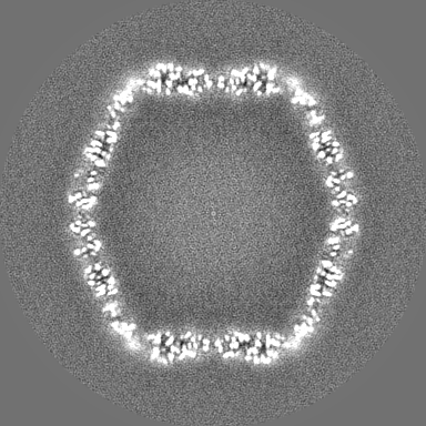
| Projections & Slices |
| ||||||||||||
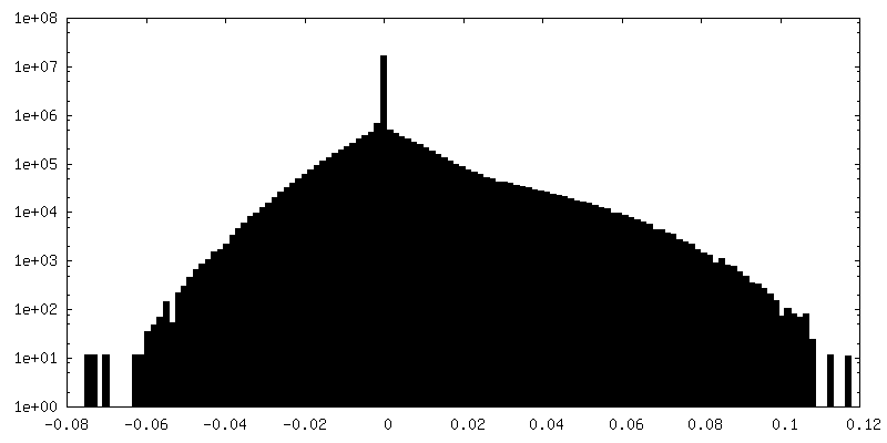
| Density Histograms |


-Half map: halfmap
| File | emd_13164_half_map_1.map | ||||||||||||
|---|---|---|---|---|---|---|---|---|---|---|---|---|---|
| Annotation | halfmap | ||||||||||||
| Projections & Slices |
| ||||||||||||
| Density Histograms |


-Half map: halfmap
| File | emd_13164_half_map_2.map | ||||||||||||
|---|---|---|---|---|---|---|---|---|---|---|---|---|---|
| Annotation | halfmap | ||||||||||||
| Projections & Slices |
| ||||||||||||
| Density Histograms |


- Sample components
-Entire : encapsulin shell from Mycobacterium tuberculosis
| Entire | Name: encapsulin shell from Mycobacterium tuberculosis |
|---|---|
| Components |
|
-Supramolecule #1: encapsulin shell from Mycobacterium tuberculosis
| Supramolecule | Name: encapsulin shell from Mycobacterium tuberculosis / type: organelle_or_cellular_component / ID: 1 / Parent: 0 / Macromolecule list: all |
|---|---|
| Source (natural) | Organism: Mycobacterium tuberculosis H37Rv (bacteria) |
| Molecular weight | Theoretical: 1.729 MDa |
-Macromolecule #1: 29 kDa antigen, Cfp29
| Macromolecule | Name: 29 kDa antigen, Cfp29 / type: protein_or_peptide / ID: 1 / Number of copies: 60 / Enantiomer: LEVO / EC number: Hydrolases; Acting on peptide bonds (peptidases) |
|---|---|
| Source (natural) | Organism: Mycobacterium tuberculosis (bacteria) |
| Molecular weight | Theoretical: 28.863346 KDa |
| Recombinant expression | Organism: |
| Sequence | String: MNNLYRDLAP VTEAAWAEIE LEAARTFKRH IAGRRVVDVS DPGGPVTAAV STGRLIDVKA PTNGVIAHLR ASKPLVRLRV PFTLSRNEI DDVERGSKDS DWEPVKEAAK KLAFVEDRTI FEGYSAASIE GIRSASSNPA LTLPEDPREI PDVISQALSE L RLAGVDGP ...String: MNNLYRDLAP VTEAAWAEIE LEAARTFKRH IAGRRVVDVS DPGGPVTAAV STGRLIDVKA PTNGVIAHLR ASKPLVRLRV PFTLSRNEI DDVERGSKDS DWEPVKEAAK KLAFVEDRTI FEGYSAASIE GIRSASSNPA LTLPEDPREI PDVISQALSE L RLAGVDGP YSVLLSADVY TKVSETSDHG YPIREHLNRL VDGDIIWAPA IDGAFVLTTR GGDFDLQLGT DVAIGYASHD TD TVRLYLQ ETLTFLCYTA EASVALSH UniProtKB: Type 1 encapsulin shell protein |
-Experimental details
-Structure determination
| Method | cryo EM |
|---|---|
 Processing Processing | single particle reconstruction |
| Aggregation state | particle |
-Sample preparation
| Concentration | 2.5 mg/mL |
|---|---|
| Buffer | pH: 7 |
| Grid | Model: Quantifoil R1.2/1.3 / Material: GOLD / Mesh: 300 / Support film - Material: GOLD / Support film - topology: HOLEY / Pretreatment - Type: GLOW DISCHARGE / Pretreatment - Time: 40 sec. |
| Vitrification | Cryogen name: ETHANE / Chamber humidity: 90 % / Instrument: FEI VITROBOT MARK IV |
- Electron microscopy
| Microscope | FEI TITAN KRIOS |
|---|---|
| Image recording | Film or detector model: GATAN K3 (6k x 4k) / Average exposure time: 1.8 sec. / Average electron dose: 40.0 e/Å2 |
| Electron beam | Acceleration voltage: 300 kV / Electron source:  FIELD EMISSION GUN FIELD EMISSION GUN |
| Electron optics | Illumination mode: FLOOD BEAM / Imaging mode: BRIGHT FIELD / Nominal defocus max: 2.0 µm / Nominal defocus min: 0.7000000000000001 µm / Nominal magnification: 163000 |
| Sample stage | Specimen holder model: FEI TITAN KRIOS AUTOGRID HOLDER / Cooling holder cryogen: NITROGEN |
| Experimental equipment |  Model: Titan Krios / Image courtesy: FEI Company |