ムービー
ムービー コントローラー


+ データを開く

- 基本情報
基本情報
| 登録情報 |  | |||||||||
|---|---|---|---|---|---|---|---|---|---|---|
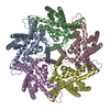
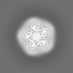
| タイトル | Structure of the AI-2 exporter family protein YdiK from E. coli | |||||||||
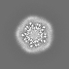
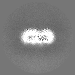
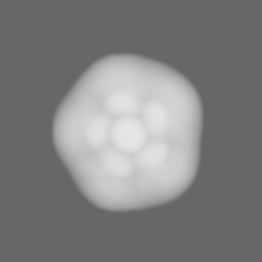
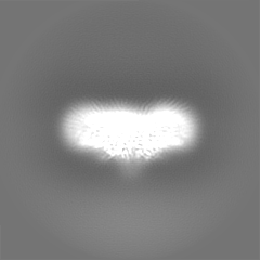
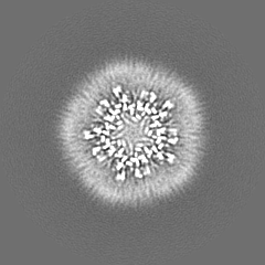
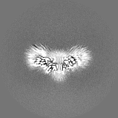
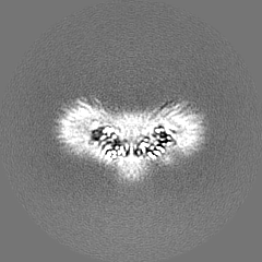
 マップデータ マップデータ | Final single particle EM map | |||||||||
 試料 試料 |
| |||||||||
 キーワード キーワード | cryo-EM / membrane protein / autoinducer-2 exporter / quorum sensing / pentamer / protein oligomerization / STRUCTURAL PROTEIN / AI-2E / transporter | |||||||||
| 機能・相同性 | Transmembrane protein TqsA-like / AI-2E family transporter / plasma membrane / Putative transport protein YdiK 機能・相同性情報 機能・相同性情報 | |||||||||
| 生物種 |  | |||||||||
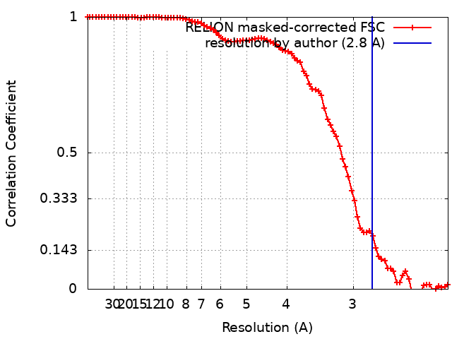
| 手法 | 単粒子再構成法 / クライオ電子顕微鏡法 / 解像度: 2.8 Å | |||||||||
 データ登録者 データ登録者 | Khera R / Xie H | |||||||||
| 資金援助 |  ドイツ, 1件 ドイツ, 1件
| |||||||||
 引用 引用 | ジャーナル: EMBO J / 年: 2022 タイトル: Cryo-EM structures of pentameric autoinducer-2 exporter from Escherichia coli reveal its transport mechanism. 著者: Radhika Khera / Ahmad R Mehdipour / Jani R Bolla / Joerg Kahnt / Sonja Welsch / Ulrich Ermler / Cornelia Muenke / Carol V Robinson / Gerhard Hummer / Hao Xie / Hartmut Michel /   要旨: Bacteria utilize small extracellular molecules to communicate in order to collectively coordinate their behaviors in response to the population density. Autoinducer-2 (AI-2), a universal molecule for ...Bacteria utilize small extracellular molecules to communicate in order to collectively coordinate their behaviors in response to the population density. Autoinducer-2 (AI-2), a universal molecule for both intra- and inter-species communication, is involved in the regulation of biofilm formation, virulence, motility, chemotaxis, and antibiotic resistance. While many studies have been devoted to understanding the biosynthesis and sensing of AI-2, very little information is available on its export. The protein TqsA from Escherichia coli, which belongs to the AI-2 exporter superfamily, has been shown to export AI-2. Here, we report the cryogenic electron microscopic structures of two AI-2 exporters (TqsA and YdiK) from E. coli at 3.35 Å and 2.80 Å resolutions, respectively. Our structures suggest that the AI-2 exporter exists as a homo-pentameric complex. In silico molecular docking and native mass spectrometry experiments were employed to demonstrate the interaction between AI-2 and TqsA, and the results highlight the functional importance of two helical hairpins in substrate binding. We propose that each monomer works as an independent functional unit utilizing an elevator-type transport mechanism. | |||||||||
| 履歴 |
|
- 構造の表示
構造の表示
| 添付画像 |
|---|

- ダウンロードとリンク
-EMDBアーカイブ
| マップデータ | emd_13057.map.gz | 40 MB | EMDBマップデータ形式 | |
|---|---|---|---|---|
| ヘッダ (付随情報) | emd-13057-v30.xmlemd-13057.xml | 16.6 KB 16.6 KB | 表示 表示 | EMDBヘッダ |
| FSC (解像度算出) | emd_13057_fsc.xml | 8.5 KB | 表示 | FSCデータファイル |
| 画像 | 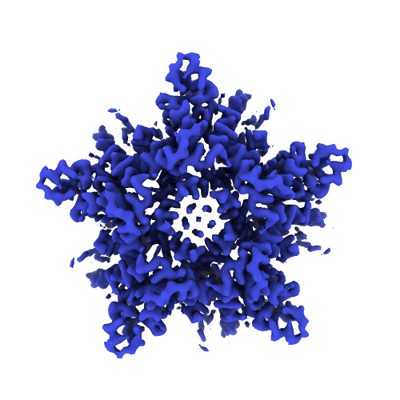 emd_13057.png emd_13057.png | 111.9 KB | ||
| マスクデータ | emd_13057_msk_1.map | 52.7 MB | マスクマップ | |
| Filedesc metadata | emd-13057.cif.gz | 5.7 KB | ||
| その他 | emd_13057_half_map_1.map.gzemd_13057_half_map_2.map.gz | 40.4 MB 40.4 MB | ||
| アーカイブディレクトリ |  http://ftp.pdbj.org/pub/emdb/structures/EMD-13057ftp://ftp.pdbj.org/pub/emdb/structures/EMD-13057 http://ftp.pdbj.org/pub/emdb/structures/EMD-13057ftp://ftp.pdbj.org/pub/emdb/structures/EMD-13057 | HTTPS FTP |
-関連構造データ
-リンク
| EMDBのページ | EMDB (EBI/PDBe) / EMDataResource |
|---|
-マップ
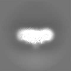
| ファイル | ダウンロード / ファイル: emd_13057.map.gz / 形式: CCP4 / 大きさ: 52.7 MB / タイプ: IMAGE STORED AS FLOATING POINT NUMBER (4 BYTES) | ||||||||||||||||||||||||||||||||||||
|---|---|---|---|---|---|---|---|---|---|---|---|---|---|---|---|---|---|---|---|---|---|---|---|---|---|---|---|---|---|---|---|---|---|---|---|---|---|
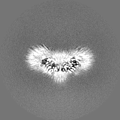
| 注釈 | Final single particle EM map | ||||||||||||||||||||||||||||||||||||
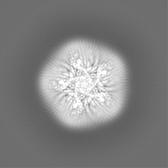
| 投影像・断面図 | 画像のコントロール
画像は Spider により作成 | ||||||||||||||||||||||||||||||||||||
| ボクセルのサイズ | X=Y=Z: 1.108 Å | ||||||||||||||||||||||||||||||||||||
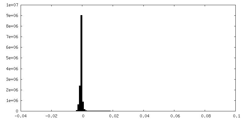
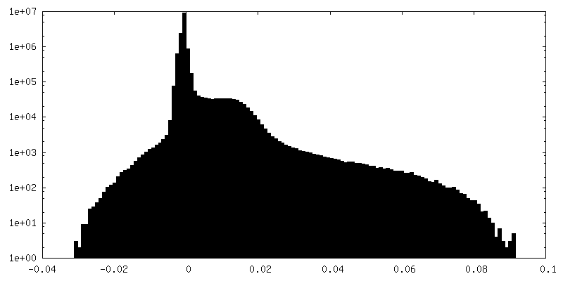
| 密度 |
| ||||||||||||||||||||||||||||||||||||
| 対称性 | 空間群: 1 | ||||||||||||||||||||||||||||||||||||
| 詳細 | EMDB XML:
|
 Z (Sec.)
 Y (Row.)
 X (Col.)


-添付データ
-マスク #1
| ファイル | emd_13057_msk_1.map | ||||||||||||
|---|---|---|---|---|---|---|---|---|---|---|---|---|---|
| 投影像・断面図 |
| ||||||||||||
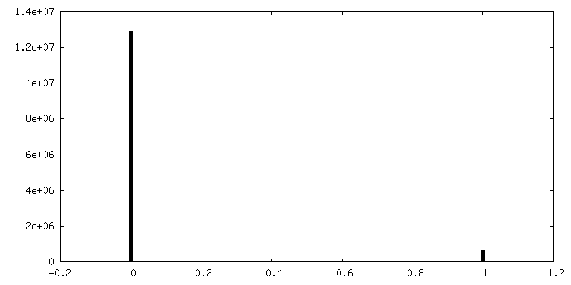
| 密度ヒストグラム |


-ハーフマップ: Half map1
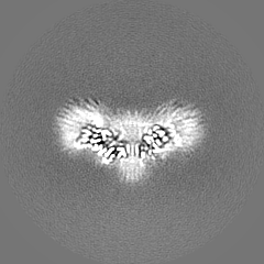
| ファイル | emd_13057_half_map_1.map | ||||||||||||
|---|---|---|---|---|---|---|---|---|---|---|---|---|---|
| 注釈 | Half map1 | ||||||||||||
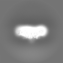
| 投影像・断面図 |
| ||||||||||||
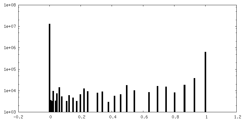
| 密度ヒストグラム |


-ハーフマップ: Half map2
| ファイル | emd_13057_half_map_2.map | ||||||||||||
|---|---|---|---|---|---|---|---|---|---|---|---|---|---|
| 注釈 | Half map2 | ||||||||||||
| 投影像・断面図 |
| ||||||||||||
| 密度ヒストグラム |

- 試料の構成要素
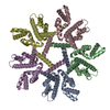
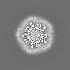
-全体 : Pentameric YdiK
| 全体 | 名称: Pentameric YdiK |
|---|---|
| 要素 |
|
-超分子 #1: Pentameric YdiK
| 超分子 | 名称: Pentameric YdiK / タイプ: complex / ID: 1 / 親要素: 0 / 含まれる分子: all |
|---|---|
| 由来(天然) | 生物種: |
| 分子量 | 理論値: 199 KDa |
-分子 #1: AI-2E member YdiK
| 分子 | 名称: AI-2E member YdiK / タイプ: protein_or_peptide / ID: 1 / コピー数: 5 / 光学異性体: LEVO |
|---|---|
| 由来(天然) | 生物種: |
| 分子量 | 理論値: 39.865891 KDa |
| 組換発現 | 生物種: |
| 配列 | 文字列: MVNVRQPRDV AQILLSVLFL AIMIVACLWI VQPFILGFAW AGTVVIATWP VLLRLQKIMF GRRSLAVLVM TLLLVMVFII PIALLVNSI VDGSGPLIKA ISSGDMTLPD LAWLNTIPVI GAKLYAGWHN LLDMGGTAIM AKVRPYIGTT TTWFVGQAAH I GRFMVHCA ...文字列: MVNVRQPRDV AQILLSVLFL AIMIVACLWI VQPFILGFAW AGTVVIATWP VLLRLQKIMF GRRSLAVLVM TLLLVMVFII PIALLVNSI VDGSGPLIKA ISSGDMTLPD LAWLNTIPVI GAKLYAGWHN LLDMGGTAIM AKVRPYIGTT TTWFVGQAAH I GRFMVHCA LMLLFSALLY WRGEQVAQGI RHFATRLAGV RGDAAVLLAA QAIRAVALGV VVTALVQAVL GGIGLAVSGV PY ATLLTVL MILSCLVQLG PLPVLIPAII WLYWTGDTTW GTVLLVWSGV VGTLDNVIRP MLIRMGADLP LILILSGVIG GLI AFGMIG LFIGPVLLAV SWRLFAAWVE EVPPPTDQPE EILEELGEIE KPNK UniProtKB: Putative transport protein YdiK |
-実験情報
-構造解析
| 手法 | クライオ電子顕微鏡法 |
|---|---|
 解析 解析 | 単粒子再構成法 |
| 試料の集合状態 | particle |
-試料調製
| 濃度 | 3.8 mg/mL |
|---|---|
| 緩衝液 | pH: 7.5 / 詳細: 50 mM Tris (pH 7.5), 150 mM NaCl and 0.006% GDN |
| 凍結 | 凍結剤: ETHANE / チャンバー内湿度: 100 % / チャンバー内温度: 277.15 K / 装置: FEI VITROBOT MARK III / 詳細: blot time 4 s, blot force +20. |
| 詳細 | Its a membrane protein and for purification, glyco-diosgenin was used. |
- 電子顕微鏡法
| 顕微鏡 | FEI TITAN KRIOS |
|---|---|
| 撮影 | フィルム・検出器のモデル: GATAN K3 BIOQUANTUM (6k x 4k) デジタル化 - サイズ - 横: 5760 pixel / デジタル化 - サイズ - 縦: 4092 pixel / 撮影したグリッド数: 1 / 実像数: 7378 / 平均電子線量: 80.0 e/Å2 |
| 電子線 | 加速電圧: 300 kV / 電子線源:  FIELD EMISSION GUN FIELD EMISSION GUN |
| 電子光学系 | 照射モード: OTHER / 撮影モード: OTHER / Cs: 2.7 mm |
| 試料ステージ | 試料ホルダーモデル: FEI TITAN KRIOS AUTOGRID HOLDER ホルダー冷却材: NITROGEN |
| 実験機器 |  モデル: Titan Krios / 画像提供: FEI Company |