Movie
Movie Controller

[English] 日本語
 Yorodumi
- EMDB-13038: La Crosse virus polymerase at replication late-elongation stage -
+ Open data

- Basic information
| Entry | Database: EMDB / ID: EMD-13038 | |||||||||
|---|---|---|---|---|---|---|---|---|---|---|
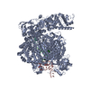
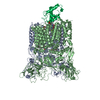
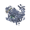
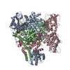
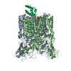
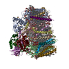
| Title | La Crosse virus polymerase at replication late-elongation stage | |||||||||
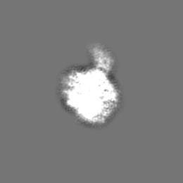
 Map data Map data | ||||||||||
 Sample Sample |
| |||||||||
 Keywords Keywords | RNA-dependent RNA polymerase / VIRAL PROTEIN | |||||||||
| Function / homology |  Function and homology information Function and homology informationhost cell endoplasmic reticulum / virion component / host cell endoplasmic reticulum-Golgi intermediate compartment / Hydrolases; Acting on ester bonds / host cell Golgi apparatus / RNA-directed RNA polymerase / nucleotide binding / viral RNA genome replication / hydrolase activity / RNA-directed RNA polymerase activity ...host cell endoplasmic reticulum / virion component / host cell endoplasmic reticulum-Golgi intermediate compartment / Hydrolases; Acting on ester bonds / host cell Golgi apparatus / RNA-directed RNA polymerase / nucleotide binding / viral RNA genome replication / hydrolase activity / RNA-directed RNA polymerase activity / DNA-templated transcription / RNA binding / metal ion binding Similarity search - Function | |||||||||
| Biological species |  La Crosse orthobunyavirus La Crosse orthobunyavirus | |||||||||
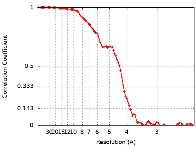
| Method | single particle reconstruction / cryo EM / Resolution: 3.9 Å | |||||||||
 Authors Authors | Arragain B / Durieux Trouilleton Q | |||||||||
| Funding support |  France, 1 items France, 1 items
| |||||||||
 Citation Citation | Journal: Nat Commun / Year: 2022 Title: Structural snapshots of La Crosse virus polymerase reveal the mechanisms underlying Peribunyaviridae replication and transcription. Authors: Benoît Arragain / Quentin Durieux Trouilleton / Florence Baudin / Jan Provaznik / Nayara Azevedo / Stephen Cusack / Guy Schoehn / Hélène Malet /  Abstract: Segmented negative-strand RNA bunyaviruses encode a multi-functional polymerase that performs genome replication and transcription. Here, we establish conditions for in vitro activity of La Crosse ...Segmented negative-strand RNA bunyaviruses encode a multi-functional polymerase that performs genome replication and transcription. Here, we establish conditions for in vitro activity of La Crosse virus polymerase and visualize its conformational dynamics by cryo-electron microscopy, unveiling the precise molecular mechanics underlying its essential activities. We find that replication initiation is coupled to distal duplex promoter formation, endonuclease movement, prime-and-realign loop extension and closure of the polymerase core that direct the template towards the active site. Transcription initiation depends on C-terminal region closure and endonuclease movements that prompt primer cleavage prior to primer entry in the active site. Product realignment after priming, observed in replication and transcription, is triggered by the prime-and-realign loop. Switch to elongation results in polymerase reorganization and core region opening to facilitate template-product duplex formation in the active site cavity. The uncovered detailed mechanics should be helpful for the future design of antivirals counteracting bunyaviral life threatening pathogens. | |||||||||
| History |
|
- Structure visualization
| Movie |
Movie viewer |
|---|---|
| Structure viewer | EM map: SurfViewMolmilJmol/JSmol |
| Supplemental images |

- Downloads & links
-EMDB archive
| Map data | emd_13038.map.gz | 5.9 MB | EMDB map data format | |
|---|---|---|---|---|
| Header (meta data) | emd-13038-v30.xmlemd-13038.xml | 22.8 KB 22.8 KB | Display Display | EMDB header |
| FSC (resolution estimation) | emd_13038_fsc.xml | 9.3 KB | Display | FSC data file |
| Images | 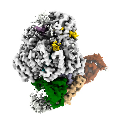 emd_13038.png emd_13038.png | 132.3 KB | ||
| Filedesc metadata | emd-13038.cif.gz | 8.4 KB | ||
| Archive directory |  http://ftp.pdbj.org/pub/emdb/structures/EMD-13038ftp://ftp.pdbj.org/pub/emdb/structures/EMD-13038 http://ftp.pdbj.org/pub/emdb/structures/EMD-13038ftp://ftp.pdbj.org/pub/emdb/structures/EMD-13038 | HTTPS FTP |
-Related structure data
| Related structure data |  7oriMC  7orjC  7orkC  7orlC  7ormC  7ornC  7oroC M: atomic model generated by this map C: citing same article ( |
|---|---|
| Similar structure data |


-Links
| EMDB pages | EMDB (EBI/PDBe) / EMDataResource |
|---|
-Map
| File | Download / File: emd_13038.map.gz / Format: CCP4 / Size: 67 MB / Type: IMAGE STORED AS FLOATING POINT NUMBER (4 BYTES) | ||||||||||||||||||||||||||||||||||||||||||||||||||||||||||||
|---|---|---|---|---|---|---|---|---|---|---|---|---|---|---|---|---|---|---|---|---|---|---|---|---|---|---|---|---|---|---|---|---|---|---|---|---|---|---|---|---|---|---|---|---|---|---|---|---|---|---|---|---|---|---|---|---|---|---|---|---|---|
| Projections & slices | Image control
Images are generated by Spider. | ||||||||||||||||||||||||||||||||||||||||||||||||||||||||||||
| Voxel size | X=Y=Z: 1.145 Å | ||||||||||||||||||||||||||||||||||||||||||||||||||||||||||||
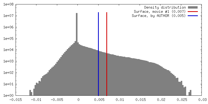
| Density |
| ||||||||||||||||||||||||||||||||||||||||||||||||||||||||||||
| Symmetry | Space group: 1 | ||||||||||||||||||||||||||||||||||||||||||||||||||||||||||||
| Details | EMDB XML:
CCP4 map header:
| ||||||||||||||||||||||||||||||||||||||||||||||||||||||||||||
 Z (Sec.)
 Y (Row.)
 X (Col.)


-Supplemental data
- Sample components
+Entire : La Crosse virus polymerase at late elongation stage
+Supramolecule #1: La Crosse virus polymerase at late elongation stage
+Supramolecule #2: RNA
+Supramolecule #3: RNA
+Supramolecule #4: La Crosse virus polymerase
+Macromolecule #1: RNA (5'-R(P*AP*CP*GP*AP*GP*UP*GP*UP*CP*GP*U)-3')
+Macromolecule #2: RNA (5'-R(P*AP*AP*CP*GP*UP*UP*AP*UP*CP*UP*AP*UP*AP*AP*CP*AP*CP*UP...
+Macromolecule #3: RNA (5'-R(P*AP*UP*AP*GP*AP*UP*AP*AP*CP*GP*UP*U)-3')
+Macromolecule #4: La Crosse virus polymerase
 Trichoplusia ni (cabbage looper)
+Macromolecule #5: ZINC ION
+Macromolecule #6: MAGNESIUM ION
+Macromolecule #7: PYROPHOSPHATE 2-

-Experimental details
-Structure determination
| Method | cryo EM |
|---|---|
 Processing Processing | single particle reconstruction |
| Aggregation state | particle |
-Sample preparation
| Concentration | 0.45 mg/mL | ||||||||||||||||||
|---|---|---|---|---|---|---|---|---|---|---|---|---|---|---|---|---|---|---|---|
| Buffer | pH: 8 Component:
| ||||||||||||||||||
| Grid | Model: UltrAuFoil R1.2/1.3 / Material: GOLD / Mesh: 300 / Support film - Material: GOLD / Support film - topology: HOLEY / Pretreatment - Type: GLOW DISCHARGE / Pretreatment - Time: 45 sec. / Details: 25 mA | ||||||||||||||||||
| Vitrification | Cryogen name: ETHANE / Chamber humidity: 100 % / Chamber temperature: 293 K / Instrument: FEI VITROBOT MARK IV | ||||||||||||||||||
| Details | 1.7 uM LACV-LCItag_H34K were sequentially incubated for 1h at 4degree with (i) 1.9 uM 5prime 1-17 BPm, (ii) 1.9 uM 3prime vRNA 1-30. LACV-LCItag_H34K bound to vRNAs was incubated with 100 uM ATP/GTP/UTP/CTP and 5mM MgCl2 for 4h at 30degree |
- Electron microscopy
| Microscope | TFS GLACIOS |
|---|---|
| Temperature | Min: 70.0 K / Max: 70.0 K |
| Image recording | Film or detector model: GATAN K2 SUMMIT (4k x 4k) / Detector mode: COUNTING / Digitization - Frames/image: 3-50 / Number grids imaged: 1 / Number real images: 1848 / Average exposure time: 6.6 sec. / Average electron dose: 60.0 e/Å2 |
| Electron beam | Acceleration voltage: 200 kV / Electron source:  FIELD EMISSION GUN FIELD EMISSION GUN |
| Electron optics | C2 aperture diameter: 100.0 µm / Calibrated defocus max: 2.0 µm / Calibrated defocus min: 0.8 µm / Calibrated magnification: 36000 / Illumination mode: FLOOD BEAM / Imaging mode: BRIGHT FIELD / Cs: 2.7 mm / Nominal defocus max: 2.0 µm / Nominal defocus min: 0.8 µm / Nominal magnification: 36000 |
| Sample stage | Specimen holder model: FEI TITAN KRIOS AUTOGRID HOLDER / Cooling holder cryogen: NITROGEN |