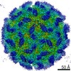
Journal: J Mol Biol / Year: 2006 Title: Structure of the dodecahedral penton particle from human adenovirus type 3. Authors: P Fuschiotti / G Schoehn / P Fender / C M S Fabry / E A Hewat / J Chroboczek / R W H Ruigrok / J F Conway / Abstract: The sub-viral dodecahedral particle of human adenovirus type 3, composed of the viral penton base and fiber proteins, shares an important characteristic of the entire virus: it can attach to cells ...The sub-viral dodecahedral particle of human adenovirus type 3, composed of the viral penton base and fiber proteins, shares an important characteristic of the entire virus: it can attach to cells and penetrate them. Structure determination of the fiberless dodecahedron by cryo-electron microscopy to 9 Angstroms resolution reveals tightly bound pentamer subunits, with only minimal interfaces between penton bases stabilizing the fragile dodecahedron. The internal cavity of the dodecahedron is approximately 80 Angstroms in diameter, and the interior surface is accessible to solvent through perforations of approximately 20 Angstroms diameter between the pentamer towers. We observe weak density beneath pentamers that we attribute to a penton base peptide including residues 38-48. The intact amino-terminal domain appears to interfere with pentamer-pentamer interactions and its absence by mutation or proteolysis is essential for dodecamer assembly. Differences between the 9 Angstroms dodecahedron structure and the adenovirus serotype 2 (Ad2) crystallographic model correlate closely with differences in sequence. The 3D structure of the dodecahedron including fibers at 16 Angstroms resolution reveals extra density on the top of the penton base that can be attributed to the fiber N terminus. The fiber itself exhibits striations that correlate with features of the atomic structure of the partial Ad2 fiber and that represent a repeat motif present in the amino acid sequence. These new observations offer important insights into particle assembly and stability, as well as the practicality of using the dodecahedron in targeted drug delivery. The structural work provides a sound basis for manipulating the properties of this particle and thereby enhancing its value for such therapeutic use.
History
Deposition
Nov 4, 2005
-
Header (metadata) release
Nov 4, 2005
-
Map release
Nov 7, 2005
-
Update
Oct 24, 2012
-
Current status
Oct 24, 2012
Processing site: PDBe / Status: Released
-
Structure visualization
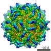
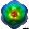
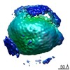
Movie
Surface view with section colored by density value
Supramolecule #1000: adenovirus 3 penton base dodecahedron
Supramolecule
Name: adenovirus 3 penton base dodecahedron / type: sample / ID: 1000 Details: The penton base was expressed in baculovirus abd the penton base self-assemble into dodecahedrons Oligomeric state: 12 pentamers / Number unique components: 1
Molecular weight
Experimental: 3.5 MDa
-
Macromolecule #1: Ad3 penton base
Macromolecule
Name: Ad3 penton base / type: protein_or_peptide / ID: 1 / Number of copies: 60 / Oligomeric state: dodecamer of pentamer / Recombinant expression: Yes
Source (natural)
Organism: Human adenovirus 3 / synonym: human adenovirus 3
pH: 6.6 / Details: 25 mM phosphate buffer at pH 6.6
Staining
Type: NEGATIVE Details: Quantifoil R2 1 grids (Quantifoil Micro Tools GmbH, Germany) were loaded with 4 ul of sample at 1 mg ml, blotted and rapidly frozen in liquid ethane within a liquid nitrogen bath using a Zeiss cryoplunger
Grid
Details: Quantifoil R2/1 grids
Vitrification
Cryogen name: ETHANE / Chamber temperature: 100 K / Instrument: OTHER / Details: Vitrification instrument: Zeiss cryoplunger
-
Electron microscopy
Microscope
JEOL 2010F
Temperature
Average: 100 K
Alignment procedure
Legacy - Astigmatism: objective lens astigmatism was corrected at 100,000
Image recording
Category: FILM / Film or detector model: KODAK SO-163 FILM / Digitization - Scanner: ZEISS SCAI / Digitization - Sampling interval: 7 µm / Number real images: 14
Electron beam
Acceleration voltage: 200 kV / Electron source: FIELD EMISSION GUN
In the structure databanks used in Yorodumi, some data are registered as the other names, "COVID-19 virus" and "2019-nCoV". Here are the details of the virus and the list of structure data.
Jan 31, 2019. EMDB accession codes are about to change! (news from PDBe EMDB page)
EMDB accession codes are about to change! (news from PDBe EMDB page)
The allocation of 4 digits for EMDB accession codes will soon come to an end. Whilst these codes will remain in use, new EMDB accession codes will include an additional digit and will expand incrementally as the available range of codes is exhausted. The current 4-digit format prefixed with “EMD-” (i.e. EMD-XXXX) will advance to a 5-digit format (i.e. EMD-XXXXX), and so on. It is currently estimated that the 4-digit codes will be depleted around Spring 2019, at which point the 5-digit format will come into force.
The EM Navigator/Yorodumi systems omit the EMD- prefix.
Related info.:Q: What is EMD? / ID/Accession-code notation in Yorodumi/EM Navigator
Yorodumi is a browser for structure data from EMDB, PDB, SASBDB, etc.
This page is also the successor to EM Navigator detail page, and also detail information page/front-end page for Omokage search.
The word "yorodu" (or yorozu) is an old Japanese word meaning "ten thousand". "mi" (miru) is to see.
Related info.:EMDB / PDB / SASBDB / Comparison of 3 databanks / Yorodumi Search / Aug 31, 2016. New EM Navigator & Yorodumi / Yorodumi Papers / Jmol/JSmol / Function and homology information / Changes in new EM Navigator and Yorodumi
 Movie
Movie Controller

 Yorodumi
 Open data
 Basic information
 Map data
 Sample
 Function and homology information
 Human adenovirus 3
 Authors
 Citation
 Structure visualization
 Downloads & links
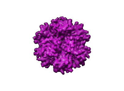 1178.gif
 http://ftp.pdbj.org/pub/emdb/structures/EMD-1178


 Z (Sec.)
 Y (Row.)
 X (Col.)


 Sample components
 Processing
 Electron microscopy
 FIELD EMISSION GUN