ムービー
ムービー コントローラー


+ データを開く

- 基本情報
基本情報
| 登録情報 | データベース: EMDB / ID: EMD-1088 | |||||||||
|---|---|---|---|---|---|---|---|---|---|---|
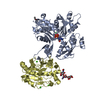
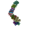
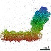
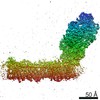
| タイトル | Structure of the acrosomal bundle. | |||||||||
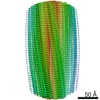
 マップデータ マップデータ | One unit cell of the acrosomal bundle, pseudo-centered to provide a orthogonal map from the pseudo-hexagonal packing | |||||||||
 試料 試料 |
| |||||||||
| 機能・相同性 |  機能・相同性情報 機能・相同性情報anatomical structure formation involved in morphogenesis / cell development / cytoskeletal motor activator activity / myosin heavy chain binding / tropomyosin binding / actin filament bundle / troponin I binding / filamentous actin / mesenchyme migration / skeletal muscle myofibril ...anatomical structure formation involved in morphogenesis / cell development / cytoskeletal motor activator activity / myosin heavy chain binding / tropomyosin binding / actin filament bundle / troponin I binding / filamentous actin / mesenchyme migration / skeletal muscle myofibril / actin filament bundle assembly / striated muscle thin filament / skeletal muscle thin filament assembly / actin monomer binding / skeletal muscle fiber development / stress fiber / titin binding / actin filament polymerization / filopodium / actin filament / 加水分解酵素; 酸無水物に作用; 酸無水物に作用・細胞または細胞小器官の運動に関与 / calcium-dependent protein binding / lamellipodium / cell body / cytoskeleton / protein domain specific binding / hydrolase activity / calcium ion binding / positive regulation of gene expression / magnesium ion binding / ATP binding / identical protein binding / cytoplasm 類似検索 - 分子機能 | |||||||||
| 生物種 |  Limulus polyphemus (カブトガニ) Limulus polyphemus (カブトガニ) | |||||||||
| 手法 | らせん対称体再構成法 / クライオ電子顕微鏡法 / 解像度: 9.5 Å | |||||||||
 データ登録者 データ登録者 | Schmid MF / Sherman MB / Matsudaira P / Chiu W | |||||||||
 引用 引用 | ジャーナル: Nature / 年: 2004 タイトル: Structure of the acrosomal bundle. 著者: Michael F Schmid / Michael B Sherman / Paul Matsudaira / Wah Chiu /  要旨: In the unactivated Limulus sperm, a 60- micro m-long bundle of actin filaments crosslinked by the protein scruin is bent and twisted into a coil around the base of the nucleus. At fertilization, the ...In the unactivated Limulus sperm, a 60- micro m-long bundle of actin filaments crosslinked by the protein scruin is bent and twisted into a coil around the base of the nucleus. At fertilization, the bundle uncoils and fully extends in five seconds to support a finger of membrane known as the acrosomal process. This biological spring is powered by stored elastic energy and does not require the action of motor proteins or actin polymerization. In a 9.5-A electron cryomicroscopic structure of the extended bundle, we show that twist, tilt and rotation of actin-scruin subunits deviate widely from a 'standard' F-actin filament. This variability in structural organization allows filaments to pack into a highly ordered and rigid bundle in the extended state and suggests a mechanism for storing and releasing energy between coiled and extended states without disassembly. | |||||||||
| 履歴 |
|
- 構造の表示
構造の表示
| ムービー |
ムービービューア |
|---|---|
| 構造ビューア | EMマップ: SurfViewMolmilJmol/JSmol |
| 添付画像 |
 UCSF Chimera
- ダウンロードとリンク
-EMDBアーカイブ
| マップデータ | emd_1088.map.gz | 43.6 MB | EMDBマップデータ形式 | |
|---|---|---|---|---|
| ヘッダ (付随情報) | emd-1088-v30.xmlemd-1088.xml | 12.6 KB 12.6 KB | 表示 表示 | EMDBヘッダ |
| 画像 |  1088.gif 1088.gif | 13.6 KB | ||
| アーカイブディレクトリ |  http://ftp.pdbj.org/pub/emdb/structures/EMD-1088ftp://ftp.pdbj.org/pub/emdb/structures/EMD-1088 http://ftp.pdbj.org/pub/emdb/structures/EMD-1088ftp://ftp.pdbj.org/pub/emdb/structures/EMD-1088 | HTTPS FTP |
-関連構造データ


-リンク
| EMDBのページ | EMDB (EBI/PDBe) / EMDataResource |
|---|---|
| 「今月の分子」の関連する項目 |


-マップ
| ファイル | ダウンロード / ファイル: emd_1088.map.gz / 形式: CCP4 / 大きさ: 46.1 MB / タイプ: IMAGE STORED AS FLOATING POINT NUMBER (4 BYTES) | ||||||||||||||||||||||||||||||||||||||||||||||||||||||||||||||||||||
|---|---|---|---|---|---|---|---|---|---|---|---|---|---|---|---|---|---|---|---|---|---|---|---|---|---|---|---|---|---|---|---|---|---|---|---|---|---|---|---|---|---|---|---|---|---|---|---|---|---|---|---|---|---|---|---|---|---|---|---|---|---|---|---|---|---|---|---|---|---|
| 注釈 | One unit cell of the acrosomal bundle, pseudo-centered to provide a orthogonal map from the pseudo-hexagonal packing | ||||||||||||||||||||||||||||||||||||||||||||||||||||||||||||||||||||
| 投影像・断面図 | 画像のコントロール
画像は Spider により作成 これらの図は立方格子座標系で作成されたものです | ||||||||||||||||||||||||||||||||||||||||||||||||||||||||||||||||||||
| ボクセルのサイズ | X: 1.33333 Å / Y: 1.32143 Å / Z: 1.32951 Å | ||||||||||||||||||||||||||||||||||||||||||||||||||||||||||||||||||||
| 密度 |
| ||||||||||||||||||||||||||||||||||||||||||||||||||||||||||||||||||||
| 対称性 | 空間群: 1 | ||||||||||||||||||||||||||||||||||||||||||||||||||||||||||||||||||||
| 詳細 | EMDB XML:
CCP4マップ ヘッダ情報:
| ||||||||||||||||||||||||||||||||||||||||||||||||||||||||||||||||||||
 Y (Sec.)
 X (Row.)
 Z (Col.)


-添付データ
- 試料の構成要素
-全体 : Acrosomal bundle from Limulus sperm
| 全体 | 名称: Acrosomal bundle from Limulus sperm |
|---|---|
| 要素 |
|
-超分子 #1000: Acrosomal bundle from Limulus sperm
| 超分子 | 名称: Acrosomal bundle from Limulus sperm / タイプ: sample / ID: 1000 集合状態: helix of actin and scruin in a 1 to 1 stoichiometry Number unique components: 2 |
|---|---|
| 分子量 | 実験値: 4.2 MDa / 理論値: 4.2 MDa / 手法: calculated from molecular weight |
-分子 #1: actin
| 分子 | 名称: actin / タイプ: protein_or_peptide / ID: 1 / Name.synonym: F-actin / 詳細: 28 of these per unit cell / コピー数: 28 / 集合状態: helical / 組換発現: No / データベース: NCBI |
|---|---|
| 由来(天然) | 生物種: Limulus polyphemus (カブトガニ) / 別称: horseshoe crab / 組織: sperm / 細胞: sperm / Organelle: acrosomal process / 細胞中の位置: acrosomal bundle |
| 分子量 | 実験値: 1.176 MDa / 理論値: 1.176 MDa |
-分子 #2: scruin
| 分子 | 名称: scruin / タイプ: protein_or_peptide / ID: 2 / Name.synonym: scruin / 詳細: 28 of these per unit cell / コピー数: 28 / 集合状態: helical / 組換発現: No / データベース: NCBI |
|---|---|
| 由来(天然) | 生物種: Limulus polyphemus (カブトガニ) / 別称: horseshoe crab / 組織: sperm / 細胞: sperm / Organelle: acrosomal process / 細胞中の位置: acrosomal bundle |
| 分子量 | 実験値: 3.08 MDa / 理論値: 3.08 MDa |
-実験情報
-構造解析
| 手法 | クライオ電子顕微鏡法 |
|---|---|
 解析 解析 | らせん対称体再構成法 |
| 試料の集合状態 | 2D array |
-試料調製
| 濃度 | 10 mg/mL |
|---|---|
| 緩衝液 | pH: 7.4 / 詳細: see J Mol Biol, 221: 711-725 (1991) |
| 凍結 | 凍結剤: ETHANE / チャンバー内湿度: 100 % / チャンバー内温度: 90 K / 装置: HOMEMADE PLUNGER / 詳細: Vitrification instrument: homemade guillotine |
| 詳細 | naturally occurring intracellular ordered array. Space group is P2sub1 or P1 21 1. P 1 21 refers to a 2-fold screw axis in the plane of the 2D crystal, whereas I have defined the 2-fold screw along z (second setting monoclinic). |
- 電子顕微鏡法
| 顕微鏡 | JEOL 4000EX |
|---|---|
| 温度 | 平均: 90 K |
| 撮影 | カテゴリ: FILM / フィルム・検出器のモデル: KODAK SO-163 FILM / デジタル化 - スキャナー: ZEISS SCAI / デジタル化 - サンプリング間隔: 7 µm / 実像数: 90 / 平均電子線量: 15 e/Å2 / 詳細: 153 bundles selected / Od range: 1.5 |
| 電子線 | 加速電圧: 400 kV / 電子線源: LAB6 |
| 電子光学系 | 照射モード: FLOOD BEAM / 撮影モード: BRIGHT FIELD / 最大 デフォーカス(公称値): 3.0 µm / 最小 デフォーカス(公称値): 0.8 µm / 倍率(公称値): 40000 |
| 試料ステージ | 試料ホルダー: side entry / 試料ホルダーモデル: GATAN LIQUID NITROGEN |
-画像解析
| 最終 再構成 | アルゴリズム: OTHER / 解像度のタイプ: BY AUTHOR / 解像度: 9.5 Å / 解像度の算出法: OTHER / ソフトウェア - 名称: MRC, CCP4, homemade 詳細: 1000 2 unit cell segments from 153 bundles (ref:Schmid, MF, J,. Struct Biol. 2003, 144:195-208. |
|---|---|
| CTF補正 | 詳細: each bundle |
| 結晶パラメータ | 単位格子 - A: 145 Å / 単位格子 - B: 145 Å / 単位格子 - C: 765 Å / 単位格子 - γ: 120 ° / 単位格子 - α: 90 ° / 単位格子 - β: 90 ° / 面群: P 1 21 |
-原子モデル構築 1
| 初期モデル | PDB ID: |
|---|---|
| ソフトウェア | 名称: foldhunter and homemade software |
| 詳細 | Protocol: cross-correlation. each of the 28 actins was fitted and averaged, scruin was density-correlated and averaged because there is no crystal structure. overall B is the NEGATIVE B factor applied as a high-pass filter in the calculation of the map (-500). |
| 精密化 | 空間: REAL / 温度因子: -500 / 当てはまり具合の基準: cross-correlation |
| 得られたモデル |  PDB-3b5u:  PDB-3b63: |