 Movie
Movie Controller


+ Open data

- Basic information
| Entry | Database: EMDB / ID: EMD-1052 | |||||||||
|---|---|---|---|---|---|---|---|---|---|---|
| Title | Untangling desmosomal knots with electron tomography. | |||||||||
 Map data Map data | This is a 3D reconstruction map of an desmosome based on high pressure freezing/freeze-subsitution electron tomography | |||||||||
 Sample Sample |
| |||||||||
| Biological species |  | |||||||||
| Method | electron tomography / cryo EM / negative staining / Resolution: 30.0 Å | |||||||||
 Authors Authors | He W / Cowin P / Stokes DL | |||||||||
 Citation Citation | Journal: Science / Year: 2003 Title: Untangling desmosomal knots with electron tomography. Authors: Wanzhong He / Pamela Cowin / David L Stokes /  Abstract: Cell adhesion by adherens junctions and desmosomes relies on interactions between cadherin molecules. However, the molecular interfaces that define molecular specificity and that mediate adhesion ...Cell adhesion by adherens junctions and desmosomes relies on interactions between cadherin molecules. However, the molecular interfaces that define molecular specificity and that mediate adhesion remain controversial. We used electron tomography of plastic sections from neonatal mouse skin to visualize the organization of desmosomes in situ. The resulting three-dimensional maps reveal individual cadherin molecules forming discrete groups and interacting through their tips. Fitting of an x-ray crystal structure for C-cadherin to these maps is consistent with a flexible intermolecular interface mediated by an exchange of amino-terminal tryptophans. This flexibility suggests a novel mechanism for generating both cis and trans interactions and for propagating these adhesive interactions along the junction. | |||||||||
| History |
|
- Structure visualization
| Movie |
 Movie viewer Movie viewer |
|---|---|
| Structure viewer | EM map: SurfViewMolmilJmol/JSmol |
| Supplemental images |
- Downloads & links
-EMDB archive
| Map data | emd_1052.map.gz | 31.3 MB | EMDB map data format | |
|---|---|---|---|---|
| Header (meta data) | emd-1052-v30.xmlemd-1052.xml | 11.2 KB 11.2 KB | Display Display | EMDB header |
| Images |  1052.gif 1052.gif | 106.6 KB | ||
| Archive directory |  http://ftp.pdbj.org/pub/emdb/structures/EMD-1052ftp://ftp.pdbj.org/pub/emdb/structures/EMD-1052 http://ftp.pdbj.org/pub/emdb/structures/EMD-1052ftp://ftp.pdbj.org/pub/emdb/structures/EMD-1052 | HTTPS FTP |
-Related structure data
| Related structure data |  1q55MC  1q5aMC  1q5bMC 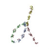 1q5cMC 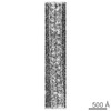 1051C 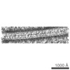 1053C M: atomic model generated by this map C: citing same article ( |
|---|
-Links
| EMDB pages | EMDB (EBI/PDBe) / EMDataResource |
|---|
-Map
| File | Download / File: emd_1052.map.gz / Format: CCP4 / Size: 41.5 MB / Type: IMAGE STORED AS SIGNED INTEGER (2 BYTES) | ||||||||||||||||||||||||||||||||||||||||||||||||||||||||||||||||||||
|---|---|---|---|---|---|---|---|---|---|---|---|---|---|---|---|---|---|---|---|---|---|---|---|---|---|---|---|---|---|---|---|---|---|---|---|---|---|---|---|---|---|---|---|---|---|---|---|---|---|---|---|---|---|---|---|---|---|---|---|---|---|---|---|---|---|---|---|---|---|
| Annotation | This is a 3D reconstruction map of an desmosome based on high pressure freezing/freeze-subsitution electron tomography | ||||||||||||||||||||||||||||||||||||||||||||||||||||||||||||||||||||
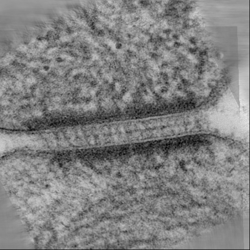
| Projections & slices | Image control
Images are generated by Spider. generated in cubic-lattice coordinate | ||||||||||||||||||||||||||||||||||||||||||||||||||||||||||||||||||||
| Voxel size | X=Y=Z: 7.266 Å | ||||||||||||||||||||||||||||||||||||||||||||||||||||||||||||||||||||
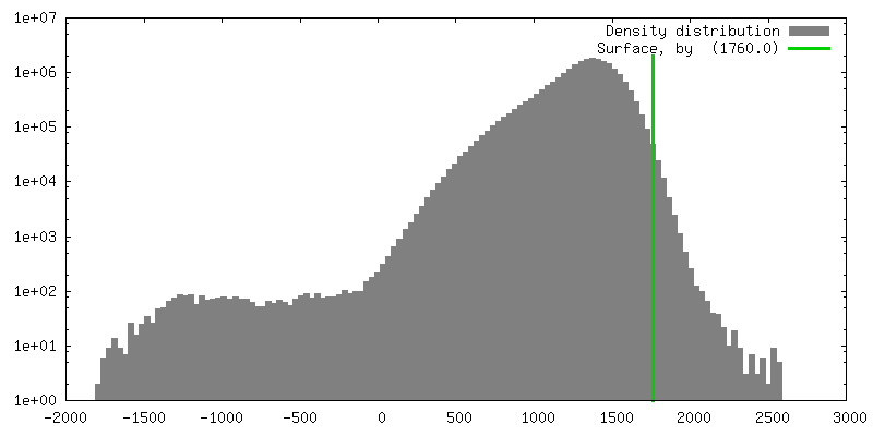
| Density |
| ||||||||||||||||||||||||||||||||||||||||||||||||||||||||||||||||||||
| Symmetry | Space group: 1 | ||||||||||||||||||||||||||||||||||||||||||||||||||||||||||||||||||||
| Details | EMDB XML:
CCP4 map header:
| ||||||||||||||||||||||||||||||||||||||||||||||||||||||||||||||||||||
 Z (Sec.)
 Y (Row.)
 X (Col.)


-Supplemental data
- Sample components
-Entire : mouse skin
| Entire | Name: mouse skin |
|---|---|
| Components |
|
-Supramolecule #1000: mouse skin
| Supramolecule | Name: mouse skin / type: sample / ID: 1000 Details: Skin from newborn mice frozen by high-pressure freezer followed by freeze-substitution, embedding in Epon resin and thin sectioning Oligomeric state: desmosome / Number unique components: 1 |
|---|
-Supramolecule #1: desmosome
| Supramolecule | Name: desmosome / type: organelle_or_cellular_component / ID: 1 / Name.synonym: desmosome Details: in-situ desmosome from frozen skin; fresh skin from newborn mice frozen immediately with high pressure freezer. Number of copies: 1 / Oligomeric state: unique / Recombinant expression: No / Database: NCBI |
|---|---|
| Source (natural) | Organism: |
-Experimental details
-Structure determination
| Method | negative staining, cryo EM |
|---|---|
 Processing Processing | electron tomography |
-Sample preparation
| Buffer | pH: 7.4 / Details: 0.5mM MgCl2 and 1-2mM CaCl2 in PBS |
|---|---|
| Staining | Type: NEGATIVE Details: staining en bloc freeze-substitution with 1% OsO4/0.1% uranyl acetate in acetone, thin section stained with 3% uranyl acetate/SATO Pb post-stain |
| Grid | Details: 200 mesh thin bar hexgonal cooper grid with formvar |
| Vitrification | Cryogen name: NITROGEN / Chamber humidity: 100 % / Chamber temperature: 90 K / Instrument: OTHER Details: Vitrification instrument: BalTec HPM 010. high pressure freezing at 2050 bar with liquid nitrigen Timed resolved state: 50msec / Method: High pressure freezing |
- Electron microscopy
| Microscope | FEI/PHILIPS CM200FEG |
|---|---|
| Temperature | Min: 293 K / Max: 293 K / Average: 293 K |
| Alignment procedure | Legacy - Astigmatism: objective lens astigmatism was corrected with thin carbon at 200-390kx Legacy - Electron beam tilt params: 0 |
| Details | dose corresponds to the cumulative dose for the entire the dataset. |
| Date | Nov 23, 2002 |
| Image recording | Category: CCD / Film or detector model: GATAN MULTISCAN / Number real images: 300 / Details: imaging directly on the CCD |
| Electron beam | Acceleration voltage: 200 kV / Electron source:  FIELD EMISSION GUN FIELD EMISSION GUN |
| Electron optics | Illumination mode: FLOOD BEAM / Imaging mode: BRIGHT FIELD / Cs: 2.0 mm / Nominal defocus max: 0.5 µm / Nominal defocus min: 0.3 µm / Nominal magnification: 50000 |
| Sample stage | Specimen holder: high tilt / Specimen holder model: OTHER / Tilt series - Axis1 - Angle increment: 1 ° |
-Image processing
| Details | details 50nm thin section stained with 3% uranyl acetate and followed by SATO Lead stain, picked on formvar coated grids and both sides coated 5-10nm amorphous carbon. |
|---|---|
| Final reconstruction | Algorithm: OTHER / Resolution.type: BY AUTHOR / Resolution: 30.0 Å / Resolution method: OTHER / Software - Name: IMOD Details: tomographic reconstruction from dual axis tilt series (tilt range:-78/+73; -68/+75,interval:1 degree). Number images used: 150 |
| CTF correction | Details: no CTF correction |
-Atomic model buiding 1
| Initial model | PDB ID: |
|---|---|
| Software | Name: AmiraMol 3.0 |
| Details | Protocol: Rigid Body. tomographic map used for fitting C-cadherin structure. #Results deposited in PDB database under codes 1Q55, 1Q5A, 1Q5B and 1Q5C. |
| Refinement | Space: REAL / Protocol: RIGID BODY FIT |
| Output model | PDB-1q55: PDB-1q5a: PDB-1q5b: PDB-1q5c: |

