 Movie
Movie Controller

[English] 日本語
 Yorodumi
- PDB-1q5a: S-shaped trans interactions of cadherins model based on fitting C... -
+ Open data

- Basic information
| Entry | Database: PDB / ID: 1q5a | |||||||||
|---|---|---|---|---|---|---|---|---|---|---|
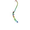
| Title | S-shaped trans interactions of cadherins model based on fitting C-cadherin (1L3W) to 3D map of desmosomes obtained by electron tomography | |||||||||
 Components Components | EP-cadherin | |||||||||
 Keywords Keywords | STRUCTURAL PROTEIN / CADHERIN / TRANS INTERACTION / DESMOSOME / JUNCTION / ADHESION | |||||||||
| Function / homology | 2-acetamido-2-deoxy-alpha-D-glucopyranose Function and homology information Function and homology information | |||||||||
| Biological species |  | |||||||||
| Method | ELECTRON MICROSCOPY / electron tomography / negative staining / cryo EM / Resolution: 30 Å | |||||||||
 Authors Authors | He, W. / Cowin, P. / Stokes, D.L. | |||||||||
 Citation Citation | Journal: Science / Year: 2003 Title: Untangling desmosomal knots with electron tomography. Authors: Wanzhong He / Pamela Cowin / David L Stokes /  Abstract: Cell adhesion by adherens junctions and desmosomes relies on interactions between cadherin molecules. However, the molecular interfaces that define molecular specificity and that mediate adhesion ...Cell adhesion by adherens junctions and desmosomes relies on interactions between cadherin molecules. However, the molecular interfaces that define molecular specificity and that mediate adhesion remain controversial. We used electron tomography of plastic sections from neonatal mouse skin to visualize the organization of desmosomes in situ. The resulting three-dimensional maps reveal individual cadherin molecules forming discrete groups and interacting through their tips. Fitting of an x-ray crystal structure for C-cadherin to these maps is consistent with a flexible intermolecular interface mediated by an exchange of amino-terminal tryptophans. This flexibility suggests a novel mechanism for generating both cis and trans interactions and for propagating these adhesive interactions along the junction. | |||||||||
| History |
|
- Structure visualization
| Movie |
Movie viewer |
|---|---|
| Structure viewer | Molecule: MolmilJmol/JSmol |
- Downloads & links
-Download
| PDBx/mmCIF format | 1q5a.cif.gz | 240.5 KB | Display | PDBx/mmCIF format |
|---|---|---|---|---|
| PDB format | pdb1q5a.ent.gz | 179.2 KB | Display | PDB format |
| PDBx/mmJSON format | 1q5a.json.gz | Tree view | PDBx/mmJSON format | |
| Others |  Other downloads Other downloads |
-Validation report
| Arichive directory | https://data.pdbj.org/pub/pdb/validation_reports/q5/1q5aftp://data.pdbj.org/pub/pdb/validation_reports/q5/1q5a | HTTPS FTP |
|---|
-Related structure data
| Related structure data |  1051MC  1052MC  1053C  1q55C  1q5bC 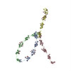 1q5cC C: citing same article ( M: map data used to model this data |
|---|---|
| Similar structure data |


-Links
 PDBj
PDBj


- Assembly
| Deposited unit | 
|
|---|---|
| 1 |
|
-Components
| #1: Protein | Mass: 97753.352 Da / Num. of mol.: 2 / Fragment: residues 1-546 of PDB entry 1L3W / Source method: isolated from a natural source / Details: Desmosome preparation from newborn mouse skin / Source: (natural) #2: Sugar | ChemComp-NAG /   Type: D-saccharide, beta linking / Mass: 221.208 Da / Num. of mol.: 26 Type: D-saccharide, beta linking / Mass: 221.208 Da / Num. of mol.: 26Source method: isolated from a genetically manipulated source Formula: C8H15NO6 #3: Sugar | ChemComp-NDG /   Type: D-saccharide, alpha linking / Mass: 221.208 Da / Num. of mol.: 4 Type: D-saccharide, alpha linking / Mass: 221.208 Da / Num. of mol.: 4Source method: isolated from a genetically manipulated source Formula: C8H15NO6 #4: Chemical | ChemComp-CA /   Mass: 40.078 Da / Num. of mol.: 24 / Source method: obtained synthetically / Formula: Ca Mass: 40.078 Da / Num. of mol.: 24 / Source method: obtained synthetically / Formula: CaCompound details | COMPOUND TRANS INTERACTION DEFINED BY N-TERMINAL TRYPTOPHAN SIDE CHAIN INSERTED INTO ANOTHER ...COMPOUND TRANS INTERACTIO | Has protein modification | Y | Sequence details | SEQUENCE SEQUENCE IS TAKEN FROM XENOPUS LAEVIS, AFRICAN CLAWED FROG, SWISSPROT ENTRY P33148, CADF_ ...SEQUENCE SEQUENCE IS TAKEN FROM XENOPUS LAEVIS, AFRICAN CLAWED FROG, SWISSPROT ENTRY P33148, CADF_XENLA BUT THE SOURCE ORGANISM IS MUS MUSCULUS. | |
|---|
-Experimental details
-Experiment
| Experiment | Method: ELECTRON MICROSCOPY |
|---|---|
| EM experiment | Aggregation state: TISSUE / 3D reconstruction method: electron tomography |
- Sample preparation
| Component |
| |||||||||||||||
|---|---|---|---|---|---|---|---|---|---|---|---|---|---|---|---|---|
| Buffer solution | Name: PBS with 1mM CaCl2 / pH: 7.4 / Details: PBS with 1mM CaCl2 | |||||||||||||||
| Specimen | Embedding applied: YES / Shadowing applied: NO / Staining applied: YES / Vitrification applied: YES | |||||||||||||||
| EM staining | Type: NEGATIVE / Material: Osmium tetroxide and Uranyl Acetate | |||||||||||||||
| Specimen support | Details: 200 mesh copper grid coated with formvar before picking 50nm thin section, then both side coated 5-10 nm thin carbon, operating at room temperature | |||||||||||||||
| EM embedding | Material: LX-112 resin | |||||||||||||||
| Vitrification | Cryogen name: NITROGEN Details: high pressure frozen and freezing substituted with 1% OsO4/0.1% Ur-Ac. in acetone, LX-112 resin embeded sample. 50nm thin section | |||||||||||||||
| Crystal grow | *PLUS Method: electron microscopy |
- Electron microscopy imaging
| Microscopy | Model: FEI/PHILIPS CM200FEG / Date: Nov 23, 2002 / Details: Dose is for whole dataset, not individual images |
|---|---|
| Electron gun | Electron source:  FIELD EMISSION GUN / Accelerating voltage: 200 kV / Illumination mode: FLOOD BEAM FIELD EMISSION GUN / Accelerating voltage: 200 kV / Illumination mode: FLOOD BEAM |
| Electron lens | Mode: BRIGHT FIELD / Nominal magnification: 50000 X / Calibrated magnification: 68276 X / Nominal defocus max: 500 nm / Nominal defocus min: 300 nm / Cs: 2 mm |
| Specimen holder | Temperature: 295 K / Tilt angle max: 79 ° / Tilt angle min: -78 ° |
| Image recording | Electron dose: 1200 e/Å2 / Film or detector model: GENERIC GATAN / Details: 1024x1024 pixels CCD. |
- Processing
| EM software |
| |||||||||||||||
|---|---|---|---|---|---|---|---|---|---|---|---|---|---|---|---|---|
| CTF correction | Details: no CTF correction. Imaging at underfocus 0.4 micron with CM200FEG microscope at 50,000 magnification | |||||||||||||||
| Symmetry | Point symmetry: C1 (asymmetric) | |||||||||||||||
| 3D reconstruction | Method: +/- 75 degree dual-axis electron tomography with IMOD Resolution: 30 Å / Actual pixel size: 7.266 Å Magnification calibration: CCD pixel size calibrated with gold crystal and silicon crystal Details: sectioned sample thickness 47.2 nm, +/- 75 degree dual-axis tilt-series with increment 1 degree dual-axis electron tomography with IMOD. 1.169nm alignment error Symmetry type: POINT | |||||||||||||||
| Atomic model building | Protocol: RIGID BODY FIT / Space: REAL Target criteria: best visual fit using the program AmiraMOL 3.0 Details: METHOD--tracking 3D density REFINEMENT PROTOCOL--rigid body | |||||||||||||||
| Atomic model building | PDB-ID: 1L3W Accession code: 1L3W / Source name: PDB / Type: experimental model | |||||||||||||||
| Refinement step | Cycle: LAST
|
