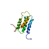
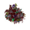
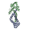
Journal: Elife / Year: 2015 Title: Structures of the scanning and engaged states of the mammalian SRP-ribosome complex. Authors: Rebecca M Voorhees / Ramanujan S Hegde / Abstract: The universally conserved signal recognition particle (SRP) is essential for the biogenesis of most integral membrane proteins. SRP scans the nascent chains of translating ribosomes, preferentially ...The universally conserved signal recognition particle (SRP) is essential for the biogenesis of most integral membrane proteins. SRP scans the nascent chains of translating ribosomes, preferentially engaging those with hydrophobic targeting signals, and delivers these ribosome-nascent chain complexes to the membrane. Here, we present structures of native mammalian SRP-ribosome complexes in the scanning and engaged states. These structures reveal the near-identical SRP architecture of these two states, show many of the SRP-ribosome interactions at atomic resolution, and suggest how the polypeptide-binding M domain selectively engages hydrophobic signals. The scanning M domain, pre-positioned at the ribosomal exit tunnel, is auto-inhibited by a C-terminal amphipathic helix occluding its hydrophobic binding groove. Upon engagement, the hydrophobic targeting signal displaces this amphipathic helix, which then acts as a protective lid over the signal. Biochemical experiments suggest how scanning and engagement are coordinated with translation elongation to minimize exposure of hydrophobic signals during membrane targeting.
A: Ribosomal protein uL2 D: Ribosomal protein uL18 G: Ribosomal protein eL8 H: Ribosomal protein uL6 J: Ribosomal protein uL5 L: Ribosomal protein eL13 M: Ribosomal protein eL14 N: Ribosomal protein eL15 O: Ribosomal protein uL13 Q: Ribosomal protein eL18 R: Ribosomal protein eL19 S: Ribosomal protein eL20 T: Ribosomal protein eL21 U: Ribosomal protein eL22 V: Ribosomal protein uL14 X: Ribosomal protein uL23 Y: Ribosomal protein uL24 Z: Ribosomal protein eL27 a: Ribosomal protein uL15 b: Ribosomal protein eL29 c: Ribosomal protein eL30 d: Ribosomal protein eL31 e: Ribosomal protein eL32 f: Ribosomal protein eL33 g: Ribosomal protein eL34 h: Ribosomal protein uL29 i: Ribosomal protein eL36 k: Ribosomal protein eL38 l: Ribosomal protein eL39 m: Ribosomal protein eL40 o: Ribosomal protein eL42 5: 28S ribosomal RNA 7: 5S ribosomal RNA 8: 5.8S ribosomal RNA B: Ribosomal protein uL3 C: Ribosomal protein uL4 E: Ribosomal protein eL6 F: Ribosomal protein uL30 I: Ribosomal protein uL16 P: Ribosomal protein uL22 W: Ribosomal protein eL24 j: Ribosomal protein eL37 n: Ribosomal protein eL41 p: Ribosomal protein eL43 r: Ribosomal protein eL28 K: Ribosomal protein uL11 q: Ribosomal protein uL10 z: SRP54 2: Nascent chain 3: Val tRNA 4: SRP 7S RNA 9: SRP19 6: SRP68 S2: 18S ribosomal RNA SA: Ribosomal protein uS2 SB: Ribosomal protein eS1 SC: Ribosomal protein uS5 SE: Ribosomal protein eS4 SG: Ribosomal protein eS6 SH: Ribosomal protein eS7 SI: Ribosomal protein eS8 SJ: Ribosomal protein uS4 SL: Ribosomal protein uS17 SN: Ribosomal protein uS15 SO: Ribosomal protein uS11 SV: Ribosomal protein eS21 SW: Ribosomal protein uS8 SX: Ribosomal protein uS12 SY: Ribosomal protein eS24 Sa: Ribosomal protein eS26 Sb: Ribosomal protein eS27 Se: Ribosomal protein eS30 SD: Ribosomal protein uS3 SF: Ribosomal protein uS7 SK: Ribosomal protein eS10 SM: Ribosomal protein eS12 SP: Ribosomal protein uS19 SQ: Ribosomal protein uS9 SR: Ribosomal protein eS17 SS: Ribosomal protein uS13 ST: Ribosomal protein eS19 SU: Ribosomal protein uS10 SZ: Ribosomal protein es25 Sc: Ribosomal protein eS28 Sd: Ribosomal protein uS14 Sf: Ribosomal protein eS31 Sg: Ribosomal protein RACK1 S1: SRP9 S4: SRP14 hetero molecules
Mass: 65.409 Da / Num. of mol.: 4 / Source method: obtained synthetically / Formula: Zn
-
Details
Sequence details
PROTEIN AND RNA IN THIS ENTRY ARE FROM ORYCTOLAGUS CUNICULUS, BUT THE MODELED SEQUENCES ARE FROM SUS SCROFA.
-
Experimental details
-
Experiment
Experiment
Method: ELECTRON MICROSCOPY
EM experiment
Aggregation state: PARTICLE / 3D reconstruction method: single particle reconstruction
-
Sample preparation
Component
ID
Name
Type
Parent-ID
Synonym
1
EngagedribosomeSRPcomplex
RIBOSOME
0
2
80Sribosome
RIBOSOME
1
mammalianribosome
3
Signalrecognitionparticle
1
SRP
Buffer solution
Name: 50 mM HEPES, 200 mM potassium acetate, 15 mM magnesium acetate, 1 mM DTT pH: 7.5 Details: 50 mM HEPES, 200 mM potassium acetate, 15 mM magnesium acetate, 1 mM DTT
Specimen
Embedding applied: NO / Shadowing applied: NO / Staining applied: NO / Vitrification applied: YES
Specimen support
Details: glow-discharged holey carbon grids (Quantifoil R2/2) coated with a ~70-Angstrom-thick layer of amorphous carbon
Vitrification
Instrument: FEI VITROBOT MARK IV / Cryogen name: ETHANE / Humidity: 100 % Details: 3 uL of sample was incubated on the grid for 30 seconds, blotted for 3 seconds, then flash-frozen in liquid ethane (FEI VITROBOT MARK IV). Method: 3 uL of sample was incubated on the grid for 30 seconds before blotting for 3 seconds
-
Electron microscopy imaging
Experimental equipment
Model: Titan Krios / Image courtesy: FEI Company
Microscopy
Model: FEI TITAN KRIOS / Date: Jun 16, 2014
Electron gun
Electron source: FIELD EMISSION GUN / Accelerating voltage: 300 kV / Illumination mode: FLOOD BEAM
Electron lens
Mode: BRIGHT FIELD / Nominal magnification: 59000 X / Nominal defocus max: 3500 nm / Nominal defocus min: 1000 nm / Cs: 2.7 mm
In the structure databanks used in Yorodumi, some data are registered as the other names, "COVID-19 virus" and "2019-nCoV". Here are the details of the virus and the list of structure data.
Jan 31, 2019. EMDB accession codes are about to change! (news from PDBe EMDB page)
EMDB accession codes are about to change! (news from PDBe EMDB page)
The allocation of 4 digits for EMDB accession codes will soon come to an end. Whilst these codes will remain in use, new EMDB accession codes will include an additional digit and will expand incrementally as the available range of codes is exhausted. The current 4-digit format prefixed with “EMD-” (i.e. EMD-XXXX) will advance to a 5-digit format (i.e. EMD-XXXXX), and so on. It is currently estimated that the 4-digit codes will be depleted around Spring 2019, at which point the 5-digit format will come into force.
The EM Navigator/Yorodumi systems omit the EMD- prefix.
Related info.:Q: What is EMD? / ID/Accession-code notation in Yorodumi/EM Navigator
Yorodumi is a browser for structure data from EMDB, PDB, SASBDB, etc.
This page is also the successor to EM Navigator detail page, and also detail information page/front-end page for Omokage search.
The word "yorodu" (or yorozu) is an old Japanese word meaning "ten thousand". "mi" (miru) is to see.
Related info.:EMDB / PDB / SASBDB / Comparison of 3 databanks / Yorodumi Search / Aug 31, 2016. New EM Navigator & Yorodumi / Yorodumi Papers / Jmol/JSmol / Function and homology information / Changes in new EM Navigator and Yorodumi
 Movie
Movie Controller

 Yorodumi
 Open data
 Basic information
 Components
 Keywords
 Function and homology information
 Authors
 Citation
 Structure visualization
 Downloads & links
 Other downloads
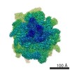


 PDBj
PDBj


 Assembly


 Mass: 24.305 Da / Num. of mol.: 166 / Source method: obtained synthetically / Formula: Mg
 Mass: 65.409 Da / Num. of mol.: 4 / Source method: obtained synthetically / Formula: Zn
 Sample preparation
 Electron microscopy imaging
 FIELD EMISSION GUN / Accelerating voltage: 300 kV / Illumination mode: FLOOD BEAM
 Processing