 Movie
Movie Controller


+ Open data

- Basic information
| Entry | Database: PDB / ID: 4p3e | ||||||
|---|---|---|---|---|---|---|---|
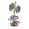
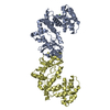
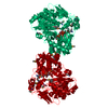
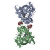
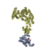
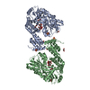
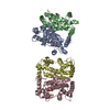
| Title | Structure of the human SRP S domain | ||||||
 Components Components |
| ||||||
 Keywords Keywords | RNA BINDING PROTEIN/RNA / SRP /  SRP RNA / SRP19 / SRP68 / ribonucleoprotein particle (RNP) / Arginine-rich motif (ARM) / RNA BINDING PROTEIN-RNA complex SRP RNA / SRP19 / SRP68 / ribonucleoprotein particle (RNP) / Arginine-rich motif (ARM) / RNA BINDING PROTEIN-RNA complex | ||||||
| Function / homology |  Function and homology information Function and homology informationSRP-dependent cotranslational protein targeting to membrane, signal sequence recognition / endoplasmic reticulum signal peptide binding / signal recognition particle, endoplasmic reticulum targeting / signal recognition particle binding / signal recognition particle / cotranslational protein targeting to membrane / 7S RNA binding / SRP-dependent cotranslational protein targeting to membrane / SRP-dependent cotranslational protein targeting to membrane / nuclear body ...SRP-dependent cotranslational protein targeting to membrane, signal sequence recognition / endoplasmic reticulum signal peptide binding / signal recognition particle, endoplasmic reticulum targeting / signal recognition particle binding / signal recognition particle / cotranslational protein targeting to membrane / 7S RNA binding / SRP-dependent cotranslational protein targeting to membrane / SRP-dependent cotranslational protein targeting to membrane / nuclear body / ribosome / response to xenobiotic stimulus / protein domain specific binding / focal adhesion / nucleolus / endoplasmic reticulum / RNA binding / cytosolSimilarity search - Function | ||||||
| Biological species |  Homo sapiens (human) Homo sapiens (human) | ||||||
| Method | X-RAY DIFFRACTION / SYNCHROTRON / MOLECULAR REPLACEMENT / Resolution: 3.5 Å | ||||||
 Authors Authors | Grotwinkel, J.T. / Wild, K. / Sinning, I. | ||||||
| Funding support |  Germany, 1items Germany, 1items
| ||||||
 Citation Citation | Journal: Science / Year: 2014 Title: SRP RNA remodeling by SRP68 explains its role in protein translocation. Authors: Grotwinkel, J.T. / Wild, K. / Segnitz, B. / Sinning, I. | ||||||
| History |
|
- Structure visualization
| Structure viewer | Molecule: MolmilJmol/JSmol |
|---|
- Downloads & links
-Download
| PDBx/mmCIF format | 4p3e.cif.gz | 148 KB | Display | PDBx/mmCIF format |
|---|---|---|---|---|
| PDB format | pdb4p3e.ent.gz | 108.2 KB | Display | PDB format |
| PDBx/mmJSON format | 4p3e.json.gz | Tree view | PDBx/mmJSON format | |
| Others |  Other downloads Other downloads |
-Validation report
| Arichive directory | https://data.pdbj.org/pub/pdb/validation_reports/p3/4p3eftp://data.pdbj.org/pub/pdb/validation_reports/p3/4p3e | HTTPS FTP |
|---|
-Related structure data
| Related structure data |  4p3fSC 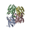 4p3gC 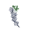 3ktvS S: Starting model for refinement C: citing same article ( |
|---|---|
| Similar structure data |


-Links
 PDBj
PDBj


- Assembly
| Deposited unit | 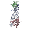
| ||||||||
|---|---|---|---|---|---|---|---|---|---|
| 1 |
| ||||||||
| Unit cell |
|
-Components
| #1: RNA chain | Mass: 40549.184 Da / Num. of mol.: 1 / Fragment: GB residues 234-358 / Mutation: C114G, G237U, G238A / Source method: obtained synthetically / Source: (synth.) Homo sapiens (human) / References: GenBank: 177793 |
|---|---|
| #2: Protein | Mass: 14791.062 Da / Num. of mol.: 1 / Fragment: UNP residues 1-120 Source method: isolated from a genetically manipulated source Source: (gene. exp.) Homo sapiens (human) / Gene: SRP19 / Production host:  Escherichia coli (E. coli) / References: UniProt: P09132 Escherichia coli (E. coli) / References: UniProt: P09132 |
| #3: Protein | / SRP68 / Signal recognition particle 68 kDa protein Mass: 25604.229 Da / Num. of mol.: 1 / Fragment: UNP residues 47-254 / Mutation: E108D Source method: isolated from a genetically manipulated source Source: (gene. exp.) Homo sapiens (human) / Gene: SRP68 / Production host: Escherichia coli (E. coli) / References: UniProt: Q9UHB9 |
| #4: Chemical | ChemComp-MG /   Mass: 24.305 Da / Num. of mol.: 4 / Source method: obtained synthetically / Formula: Mg Mass: 24.305 Da / Num. of mol.: 4 / Source method: obtained synthetically / Formula: Mg |
-Experimental details
-Experiment
| Experiment | Method: X-RAY DIFFRACTION / Number of used crystals: 1 |
|---|
- Sample preparation
| Crystal | Density Matthews: 2.87 Å3/Da / Density % sol: 57.09 % |
|---|---|
| Crystal grow | Temperature: 291 K / Method: vapor diffusion, sitting drop / pH: 5.2 / Details: Ammonium sulfate, Sodium malonate |
-Data collection
| Diffraction | Mean temperature: 100 K |
|---|---|
| Diffraction source | Source: SYNCHROTRON / Site: ESRF  / Beamline: ID29 / Wavelength: 0.9763 Å / Beamline: ID29 / Wavelength: 0.9763 Å |
| Detector | Type: DECTRIS PILATUS 6M / Detector: PIXEL / Date: May 8, 2013 |
| Radiation | Protocol: SINGLE WAVELENGTH / Scattering type: x-ray |
| Radiation wavelength | Wavelength: 0.9763 Å / Relative weight: 1 |
| Reflection | Resolution: 3.5→44.09 Å / Num. obs: 11703 / % possible obs: 99.5 % / Redundancy: 3.4 % / Biso Wilson estimate: 92.36 Å2 / Net I/σ(I): 9 |
| Reflection shell | Resolution: 3.5→3.85 Å / Redundancy: 5.7 % / Rmerge(I) obs: 0.36 / Rsym value: 0.021 / % possible all: 99.5 |
- Processing
| Software |
| |||||||||||||||||||||||||||||||||||
|---|---|---|---|---|---|---|---|---|---|---|---|---|---|---|---|---|---|---|---|---|---|---|---|---|---|---|---|---|---|---|---|---|---|---|---|---|
| Refinement | Method to determine structure: MOLECULAR REPLACEMENT Starting model: 4P3F and 3KTV Resolution: 3.5→44.089 Å / SU ML: 0.64 / Cross valid method: FREE R-VALUE / σ(F): 1.35 / Phase error: 37.42 / Stereochemistry target values: ML
| |||||||||||||||||||||||||||||||||||
| Solvent computation | Shrinkage radii: 0.9 Å / VDW probe radii: 1.11 Å / Solvent model: FLAT BULK SOLVENT MODEL | |||||||||||||||||||||||||||||||||||
| Displacement parameters | Biso max: 164.42 Å2 / Biso mean: 55.7406 Å2 / Biso min: 0 Å2 | |||||||||||||||||||||||||||||||||||
| Refinement step | Cycle: final / Resolution: 3.5→44.089 Å
| |||||||||||||||||||||||||||||||||||
| Refine LS restraints |
| |||||||||||||||||||||||||||||||||||
| LS refinement shell | Refine-ID: X-RAY DIFFRACTION / Total num. of bins used: 4
|
