 Movie
Movie Controller


+ Open data

- Basic information
| Entry | Database: PDB / ID: 2a96 | ||||||
|---|---|---|---|---|---|---|---|
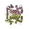
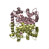
| Title | Crystal structure of phosphate tethered PhoN of S. typhimurium | ||||||
 Components Components | class A nonspecific acid phosphatase PhoN | ||||||
 Keywords Keywords |  HYDROLASE / Class-A bacterial non-specific acid phosphatase / PhoN protein HYDROLASE / Class-A bacterial non-specific acid phosphatase / PhoN protein | ||||||
| Function / homology |  Function and homology informationacid phosphatase / acid phosphatase activity / outer membrane-bounded periplasmic space Function and homology informationacid phosphatase / acid phosphatase activity / outer membrane-bounded periplasmic spaceSimilarity search - Function | ||||||
| Biological species |  Salmonella typhimurium (bacteria) Salmonella typhimurium (bacteria) | ||||||
| Method | X-RAY DIFFRACTION / Molecular replacement, Non-crystallographic symmetry averaging / Resolution: 2.5 Å | ||||||
 Authors Authors | Makde, R.D. / Mahajan, S.K. / Kumar, V. | ||||||
 Citation Citation | Journal: Biochemistry / Year: 2007 Title: Structure and Mutational Analysis of the PhoN Protein of Salmonella typhimurium Provide Insight into Mechanistic Details. Authors: Makde, R.D. / Mahajan, S.K. / Kumar, V. #1: Journal: Acta Crystallogr.,Sect.D / Year: 2003 Title: Purification, crystallization and preliminary x-ray diffraction studies of recombinant class A non-specific acid phosphatase of Salmonella typhimurium. Authors: Makde, R.D. / Kumar, V. / Rao, A.S. / Yadava, V.S. / Mahajan, S.K. | ||||||
| History |
|
- Structure visualization
| Structure viewer | Molecule: MolmilJmol/JSmol |
|---|
- Downloads & links
-Download
| PDBx/mmCIF format | 2a96.cif.gz | 184.7 KB | Display | PDBx/mmCIF format |
|---|---|---|---|---|
| PDB format | pdb2a96.ent.gz | 147.9 KB | Display | PDB format |
| PDBx/mmJSON format | 2a96.json.gz | Tree view | PDBx/mmJSON format | |
| Others |  Other downloads Other downloads |
-Validation report
| Arichive directory | https://data.pdbj.org/pub/pdb/validation_reports/a9/2a96ftp://data.pdbj.org/pub/pdb/validation_reports/a9/2a96 | HTTPS FTP |
|---|
-Related structure data
| Related structure data |  2akcC  2ipbC  1d2tS S: Starting model for refinement C: citing same article ( |
|---|---|
| Similar structure data |


-Links
 PDBj
PDBj
- Assembly
| Deposited unit | 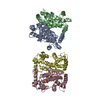
| |||||||||
|---|---|---|---|---|---|---|---|---|---|---|
| 1 | 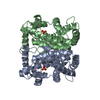
| |||||||||
| 2 | 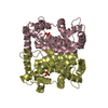
| |||||||||
| Unit cell |
| |||||||||
| Noncrystallographic symmetry (NCS) | NCS domain:
| |||||||||
| Details | The entry contains the crystallographic asymmetric unit consisting of four chains (two dimers). A dimer is the known biologically active state of the PHON protein. Chains A and B form one dimer while the chains C and D form the other dimer. |
-Components
| #1: Protein | Mass: 28437.137 Da / Num. of mol.: 4 Source method: isolated from a genetically manipulated source Source: (gene. exp.) Salmonella typhimurium (bacteria) / Gene: phoN / Plasmid: pSK- / Production host: Escherichia coli (E. coli) / Strain (production host): DH5alphaReferences: GenBank: 21913311, UniProt: Q8KRU6*PLUS, acid phosphatase#2: Chemical | ChemComp-PO4 / Phosphate  Mass: 94.971 Da / Num. of mol.: 4 / Source method: obtained synthetically / Formula: PO4 Mass: 94.971 Da / Num. of mol.: 4 / Source method: obtained synthetically / Formula: PO4#3: Water | ChemComp-HOH / | Water Mass: 18.015 Da / Num. of mol.: 275 / Source method: isolated from a natural source / Formula: H2O Mass: 18.015 Da / Num. of mol.: 275 / Source method: isolated from a natural source / Formula: H2O |
|---|
-Experimental details
-Experiment
| Experiment | Method: X-RAY DIFFRACTION / Number of used crystals: 1 |
|---|
- Sample preparation
| Crystal | Density Matthews: 2.15 Å3/Da / Density % sol: 42.8 % |
|---|---|
| Crystal grow | Temperature: 293 K / Method: vapor diffusion, hanging drop / pH: 7 Details: PEG-6000 12%, Glycerol 2%, 25mM phosphate, pH 7.0, VAPOR DIFFUSION, HANGING DROP, temperature 293K |
-Data collection
| Diffraction | Mean temperature: 293 K |
|---|---|
| Diffraction source | Source: ROTATING ANODE / Type: RIGAKU RU200 / Wavelength: 1.5418 Å |
| Detector | Type: RIGAKU RAXIS IIC / Detector: IMAGE PLATE / Date: Oct 18, 2001 / Details: OSMIC mirror |
| Radiation | Monochromator: Ni filter / Protocol: SINGLE WAVELENGTH / Monochromatic (M) / Laue (L): M / Scattering type: x-ray |
| Radiation wavelength | Wavelength: 1.5418 Å / Relative weight: 1 |
| Reflection | Resolution: 2.5→30 Å / Num. all: 27475 / Num. obs: 27475 / % possible obs: 90 % / Observed criterion σ(I): 0 / Redundancy: 1.4 % / Biso Wilson estimate: 34 Å2 / Rmerge(I) obs: 0.087 / Net I/σ(I): 8.1 |
| Reflection shell | Resolution: 2.5→2.64 Å / Redundancy: 1.3 % / Rmerge(I) obs: 0.243 / Mean I/σ(I) obs: 2.3 / Num. unique all: 4028 / % possible all: 89.9 |
- Processing
| Software |
| |||||||||||||||||||||||||||
|---|---|---|---|---|---|---|---|---|---|---|---|---|---|---|---|---|---|---|---|---|---|---|---|---|---|---|---|---|
| Refinement | Method to determine structure: Molecular replacement, Non-crystallographic symmetry averaging Starting model: PDB entry 1D2T Resolution: 2.5→20 Å / Isotropic thermal model: ISOTROPIC / Cross valid method: THROUGHOUT / σ(F): 0 / Stereochemistry target values: Engh & Huber Details: NCS restraints for equivalence of chain A and Chain C, and for chain B and Chain D.
| |||||||||||||||||||||||||||
| Solvent computation | Solvent model: Flat Model / Bsol: 50.4 Å2 | |||||||||||||||||||||||||||
| Displacement parameters | Biso mean: 28.7 Å2 | |||||||||||||||||||||||||||
| Refine analyze |
| |||||||||||||||||||||||||||
| Refinement step | Cycle: LAST / Resolution: 2.5→20 Å
| |||||||||||||||||||||||||||
| Refine LS restraints |
| |||||||||||||||||||||||||||
| Refine LS restraints NCS |
| |||||||||||||||||||||||||||
| LS refinement shell | Resolution: 2.5→2.59 Å
| |||||||||||||||||||||||||||
| Xplor file |
|
