 Movie
Movie Controller

[English] 日本語
 Yorodumi
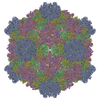
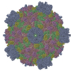
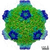
- PDB-3j17: Structure of a transcribing cypovirus by cryo-electron microscopy -
+ Open data

- Basic information
| Entry | Database: PDB / ID: 3j17 | ||||||
|---|---|---|---|---|---|---|---|
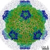
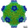
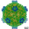
| Title | Structure of a transcribing cypovirus by cryo-electron microscopy | ||||||
 Components Components |
| ||||||
 Keywords Keywords | VIRUS / dsRNA virus Reoviridae transcribing cypovirus | ||||||
| Function / homology |  Function and homology information Function and homology information | ||||||
| Biological species |   Bombyx mori cypovirus 1 Bombyx mori cypovirus 1 | ||||||
| Method | ELECTRON MICROSCOPY / single particle reconstruction / cryo EM / Resolution: 4.1 Å | ||||||
 Authors Authors | Yang, C. / Ji, G. / Liu, H. / Zhang, K. / Liu, G. / Sun, F. / Zhu, P. / Cheng, L. | ||||||
 Citation Citation | Journal: Proc Natl Acad Sci U S A / Year: 2012 Title: Cryo-EM structure of a transcribing cypovirus. Authors: Chongwen Yang / Gang Ji / Hongrong Liu / Kai Zhang / Guangqiao Liu / Fei Sun / Ping Zhu / Lingpeng Cheng /  Abstract: Double-stranded RNA viruses in the family Reoviridae are capable of transcribing and capping nascent mRNA within an icosahedral viral capsid that remains intact throughout repeated transcription ...Double-stranded RNA viruses in the family Reoviridae are capable of transcribing and capping nascent mRNA within an icosahedral viral capsid that remains intact throughout repeated transcription cycles. However, how the highly coordinated mRNA transcription and capping process is facilitated by viral capsid proteins is still unknown. Cypovirus provides a good model system for studying the mRNA transcription and capping mechanism of viruses in the family Reoviridae. Here, we report a full backbone model of a transcribing cypovirus built from a near-atomic-resolution density map by cryoelectron microscopy. Compared with the structure of a nontranscribing cypovirus, the major capsid proteins of transcribing cypovirus undergo a series of conformational changes, giving rise to structural changes in the capsid shell: (i) an enlarged capsid chamber, which provides genomic RNA with more flexibility to move within the densely packed capsid, and (ii) a widened peripentonal channel in the capsid shell, which we confirmed to be a pathway for nascent mRNA. A rod-like structure attributable to a partially resolved nascent mRNA was observed in this channel. In addition, conformational change in the turret protein results in a relatively open turret at each fivefold axis. A GMP moiety, which is transferred to 5'-diphosphorylated mRNA during the mRNA capping reaction, was identified in the pocket-like guanylyltransferase domain of the turret protein. | ||||||
| History |
|
- Structure visualization
| Movie |
Movie viewer |
|---|---|
| Structure viewer | Molecule: MolmilJmol/JSmol |
- Downloads & links
-Download
| PDBx/mmCIF format | 3j17.cif.gz | 728.6 KB | Display | PDBx/mmCIF format |
|---|---|---|---|---|
| PDB format | pdb3j17.ent.gz | 606.8 KB | Display | PDB format |
| PDBx/mmJSON format | 3j17.json.gz | Tree view | PDBx/mmJSON format | |
| Others |  Other downloads Other downloads |
-Validation report
| Summary document | 3j17_validation.pdf.gz | 888.9 KB | Display | wwPDB validaton report |
|---|---|---|---|---|
| Full document | 3j17_full_validation.pdf.gz | 1 MB | Display | |
| Data in XML | 3j17_validation.xml.gz | 119.1 KB | Display | |
| Data in CIF | 3j17_validation.cif.gz | 174.2 KB | Display | |
| Arichive directory | https://data.pdbj.org/pub/pdb/validation_reports/j1/3j17ftp://data.pdbj.org/pub/pdb/validation_reports/j1/3j17 | HTTPS FTP |
-Related structure data
| Related structure data | 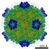 5376MC M: map data used to model this data C: citing same article ( |
|---|---|
| Similar structure data |


-Links
 PDBj
PDBj
- Assembly
| Deposited unit | 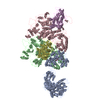
|
|---|---|
| 1 | x 60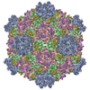
|
| 2 |
|
| 3 | x 5
|
| 4 | x 6
|
| 5 | 
|
| Symmetry | Point symmetry: (Schoenflies symbol: I (icosahedral)) |
-Components
| #1: Protein | Mass: 120489.984 Da / Num. of mol.: 1 / Source method: isolated from a natural source / Source: (natural) Bombyx mori cypovirus 1 / References: UniProt: Q914N6 | ||
|---|---|---|---|
| #2: Protein | Mass: 148696.062 Da / Num. of mol.: 2 / Source method: isolated from a natural source / Source: (natural) Bombyx mori cypovirus 1 / References: UniProt: D3JWE6, UniProt: Q6TS43*PLUS#3: Protein | Mass: 49906.176 Da / Num. of mol.: 2 / Source method: isolated from a natural source / Source: (natural) Bombyx mori cypovirus 1 / References: UniProt: C6K2M8 |
-Experimental details
-Experiment
| Experiment | Method: ELECTRON MICROSCOPY |
|---|---|
| EM experiment | Aggregation state: PARTICLE / 3D reconstruction method: single particle reconstruction |
- Sample preparation
| Component | Name: transcribing cypovirus / Type: VIRUS |
|---|---|
| Details of virus | Empty: NO / Enveloped: NO / Host category: INVERTEBRATES / Isolate: STRAIN / Type: VIRION |
| Natural host | Organism: Bombyx mori |
| Buffer solution | pH: 8 |
| Specimen | Embedding applied: NO / Shadowing applied: NO / Staining applied: NO / Vitrification applied: YES |
| Specimen support | Details: 300 mesh quantifoil |
| Vitrification | Instrument: FEI VITROBOT MARK IV / Cryogen name: ETHANE / Humidity: 100 % Details: Blotted for 4 seconds, plunged into ethane (FEI Vitrobot Mark IV) Method: blotted for 4s |
- Electron microscopy imaging
| Experimental equipment |  Model: Titan Krios / Image courtesy: FEI Company |
|---|---|
| Microscopy | Model: FEI TITAN KRIOS / Date: Jul 20, 2011 / Details: parallel beam |
| Electron gun | Electron source:  FIELD EMISSION GUN / Accelerating voltage: 300 kV / Illumination mode: OTHER FIELD EMISSION GUN / Accelerating voltage: 300 kV / Illumination mode: OTHER |
| Electron lens | Mode: BRIGHT FIELD / Nominal magnification: 75000 X / Calibrated magnification: 125390 X / Nominal defocus max: 3500 nm / Nominal defocus min: 800 nm / Cs: 2.7 mm / Camera length: 0 mm |
| Specimen holder | Specimen holder model: OTHER / Specimen holder type: Titan Krios / Temperature: 92 K / Tilt angle max: 0 ° / Tilt angle min: 0 ° |
| Image recording | Electron dose: 22 e/Å2 / Film or detector model: GATAN ULTRASCAN 4000 (4k x 4k) |
| Image scans | Num. digital images: 845 |
| Radiation | Protocol: SINGLE WAVELENGTH / Monochromatic (M) / Laue (L): M / Scattering type: x-ray |
| Radiation wavelength | Relative weight: 1 |
- Processing
| EM software |
| ||||||||||||
|---|---|---|---|---|---|---|---|---|---|---|---|---|---|
| CTF correction | Details: each image | ||||||||||||
| Symmetry | Point symmetry: I (icosahedral) | ||||||||||||
| 3D reconstruction | Method: Cross-common lines / Resolution: 4.1 Å / Resolution method: OTHER / Num. of particles: 8000 / Symmetry type: POINT | ||||||||||||
| Refinement step | Cycle: LAST
|
