Nonhomologous End-Joining (NHEJ) / double-strand break repair via nonhomologous end joining / DNA recombination / DNA-directed DNA polymerase / DNA-directed DNA polymerase activity / DNA binding / nucleoplasm / nucleus / metal ion binding Similarity search - Function
BRCT domain / DNA-directed DNA/RNA polymerase mu / DNA nucleotidylexotransferase (TdT) / DNA-directed DNA/RNA polymerase mu / DNA polymerase beta, palm domain / DNA polymerase beta palm / DNA polymerase lambda, fingers domain / Fingers domain of DNA polymerase lambda / DNA-directed DNA polymerase X / DNA polymerase beta-like, N-terminal domain / Helix-hairpin-helix domain ...BRCT domain / DNA-directed DNA/RNA polymerase mu / DNA nucleotidylexotransferase (TdT) / DNA-directed DNA/RNA polymerase mu / DNA polymerase beta, palm domain / DNA polymerase beta palm / DNA polymerase lambda, fingers domain / Fingers domain of DNA polymerase lambda / DNA-directed DNA polymerase X / DNA polymerase beta-like, N-terminal domain / Helix-hairpin-helix domain / DNA polymerase X family / DNA polymerase family X, binding site / DNA polymerase family X signature. / DNA polymerase lambda lyase domain superfamily / DNA polymerase family X / DNA polymerase beta, thumb domain / DNA polymerase beta thumb / DNA polymerase, thumb domain superfamily / BRCT domain profile. / BRCT domain / BRCT domain superfamily / Nucleotidyltransferase superfamily / Rossmann fold / 3-Layer(aba) Sandwich / Alpha Beta Similarity search - Domain/homology
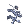
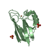
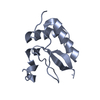
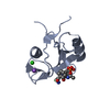
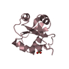
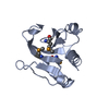
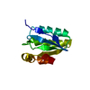
Mass: 11420.022 Da / Num. of mol.: 1 / Fragment: BRCT domain / Mutation: S20G Source method: isolated from a genetically manipulated source Source: (gene. exp.) Homo sapiens (human) / Gene: POLM / Production host: Escherichia coli (E. coli) / References: UniProt: Q9NP87, DNA-directed DNA polymerase
-
Experimental details
-
Experiment
Experiment
Method: SOLUTION NMR
NMR experiment
Conditions-ID
Experiment-ID
Solution-ID
Type
1
1
1
3D 13C-separated NOESY
1
2
1
3D 15N-separated NOESY
-
Sample preparation
Details
Contents: 1mM polymerase mu BRCT domain, 25 mM d-Tris, 50 mM KCl, 0.02% NaN3, 5 mM DTT, pH 7.9, 90% H2O, 10% D2O Solvent system: 90% H2O/10% D2O
Sample conditions
Ionic strength: 50 mM KCl / pH: 7.9 / Pressure: ambient / Temperature: 283 K
-
NMR measurement
Radiation
Protocol: SINGLE WAVELENGTH / Monochromatic (M) / Laue (L): M
Radiation wavelength
Relative weight: 1
NMR spectrometer
Type
Manufacturer
Model
Field strength (MHz)
Spectrometer-ID
Varian INOVA
Varian
INOVA
600
1
Varian INOVA
Varian
INOVA
800
2
-
Processing
NMR software
Name
Version
Developer
Classification
VNMR
1.1C, VNMRJ1.1C
Varian
collection
NMRPipe
2003, 2004
Delaglio, Grzesiek, Vuister, Zhu, Pfeifer, andBax
processing
NMRView
5.0.4
JohnsonandBlevins
processing
CYANA
2.1
Guentert, Mumenthaler, Herrmann, andWuthrich
structuresolution
ARIA
2.0a
Habeck, Rieping, Linge, andNilges
refinement
Refinement
Method: Torsion angle dynamics, molecular dynamics refinement in explicit water solvent Software ordinal: 1 Details: The initial structures were computed using the auto noeassign feature of CYANA 2.1; 1503 NOE distance restraints were assigned, and 147 dihedral angle restraints were used in the calculation. ...Details: The initial structures were computed using the auto noeassign feature of CYANA 2.1; 1503 NOE distance restraints were assigned, and 147 dihedral angle restraints were used in the calculation. The 20 best CYANA structures were refined in explicit solvent using ARIA 2.0a with the 1506 NOE distance restraints, 147 dihedral angle restraints, 42 H-bond restraints, and 48 residual dipolar coupling restraints.
NMR representative
Selection criteria: minimized average structure
NMR ensemble
Conformer selection criteria: structures with the lowest energy Conformers calculated total number: 100 / Conformers submitted total number: 11
+
About Yorodumi
-
News
-
Feb 9, 2022. New format data for meta-information of EMDB entries
New format data for meta-information of EMDB entries
Version 3 of the EMDB header file is now the official format.
The previous official version 1.9 will be removed from the archive.
In the structure databanks used in Yorodumi, some data are registered as the other names, "COVID-19 virus" and "2019-nCoV". Here are the details of the virus and the list of structure data.
Jan 31, 2019. EMDB accession codes are about to change! (news from PDBe EMDB page)
EMDB accession codes are about to change! (news from PDBe EMDB page)
The allocation of 4 digits for EMDB accession codes will soon come to an end. Whilst these codes will remain in use, new EMDB accession codes will include an additional digit and will expand incrementally as the available range of codes is exhausted. The current 4-digit format prefixed with “EMD-” (i.e. EMD-XXXX) will advance to a 5-digit format (i.e. EMD-XXXXX), and so on. It is currently estimated that the 4-digit codes will be depleted around Spring 2019, at which point the 5-digit format will come into force.
The EM Navigator/Yorodumi systems omit the EMD- prefix.
Related info.:Q: What is EMD? / ID/Accession-code notation in Yorodumi/EM Navigator
Yorodumi is a browser for structure data from EMDB, PDB, SASBDB, etc.
This page is also the successor to EM Navigator detail page, and also detail information page/front-end page for Omokage search.
The word "yorodu" (or yorozu) is an old Japanese word meaning "ten thousand". "mi" (miru) is to see.
Related info.:EMDB / PDB / SASBDB / Comparison of 3 databanks / Yorodumi Search / Aug 31, 2016. New EM Navigator & Yorodumi / Yorodumi Papers / Jmol/JSmol / Function and homology information / Changes in new EM Navigator and Yorodumi
 Movie
Movie Controller

 Yorodumi
 Open data
 Basic information
 Components
 Keywords
 Function and homology information
 Homo sapiens (human)
 Authors
 Citation
 Structure visualization
 Downloads & links
 Other downloads
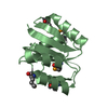


 PDBj
PDBj


 Assembly
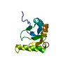

 Sample preparation
 Processing
 NMRPipe