 Movie
Movie Controller

[English] 日本語
 Yorodumi
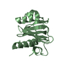
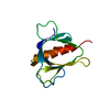
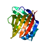
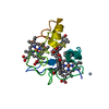
- PDB-3h9x: Crystal Structure of the PSPTO_3016 protein from Pseudomonas syri... -
+ Open data

- Basic information
| Entry | Database: PDB / ID: 3h9x | ||||||
|---|---|---|---|---|---|---|---|
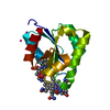
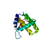
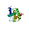
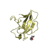
| Title | Crystal Structure of the PSPTO_3016 protein from Pseudomonas syringae, Northeast Structural Genomics Consortium Target PsR293 | ||||||
 Components Components | uncharacterized protein PSPTO_3016 | ||||||
 Keywords Keywords |  structural genomics / unknown function / alpha-beta protein. / PSI-2 / Protein Structure Initiative / Northeast Structural Genomics Consortium / NESG structural genomics / unknown function / alpha-beta protein. / PSI-2 / Protein Structure Initiative / Northeast Structural Genomics Consortium / NESG | ||||||
| Function / homology | Aspartate Aminotransferase, domain 1 - #30 / Uncharacterised protein YjbR / YjbR-like superfamily / YjbR / Aspartate Aminotransferase, domain 1 / Alpha-Beta Complex / Alpha Beta / MmcQ/YjbR family DNA-binding protein Function and homology information Function and homology information | ||||||
| Biological species |  Pseudomonas syringae pv. tomato (bacteria) Pseudomonas syringae pv. tomato (bacteria) | ||||||
| Method | X-RAY DIFFRACTION / SYNCHROTRON / SAD / Resolution: 2.51 Å | ||||||
 Authors Authors | Seetharaman, J. / Lew, S. / Forouhar, F. / Hamilton, H. / Xiao, R. / Ciccosanti, C. / Foote, E.L. / Zhao, L. / Everett, J.K. / Nair, R. ...Seetharaman, J. / Lew, S. / Forouhar, F. / Hamilton, H. / Xiao, R. / Ciccosanti, C. / Foote, E.L. / Zhao, L. / Everett, J.K. / Nair, R. / Acton, T.B. / Rost, B. / Montelione, G.T. / Hunt, J.F. / Tong, L. / Northeast Structural Genomics Consortium (NESG) | ||||||
 Citation Citation | Journal: J.Struct.Funct.Genom. / Year: 2012 Title: Solution NMR and X-ray crystal structures of Pseudomonas syringae Pspto_3016 from protein domain family PF04237 (DUF419) adopt a "double wing" DNA binding motif. Authors: Feldmann, E.A. / Seetharaman, J. / Ramelot, T.A. / Lew, S. / Zhao, L. / Hamilton, K. / Ciccosanti, C. / Xiao, R. / Acton, T.B. / Everett, J.K. / Tong, L. / Montelione, G.T. / Kennedy, M.A. | ||||||
| History |
|
- Structure visualization
| Structure viewer | Molecule: MolmilJmol/JSmol |
|---|
- Downloads & links
-Download
| PDBx/mmCIF format | 3h9x.cif.gz | 101.9 KB | Display | PDBx/mmCIF format |
|---|---|---|---|---|
| PDB format | pdb3h9x.ent.gz | 84.8 KB | Display | PDB format |
| PDBx/mmJSON format | 3h9x.json.gz | Tree view | PDBx/mmJSON format | |
| Others |  Other downloads Other downloads |
-Validation report
| Arichive directory | https://data.pdbj.org/pub/pdb/validation_reports/h9/3h9xftp://data.pdbj.org/pub/pdb/validation_reports/h9/3h9x | HTTPS FTP |
|---|
-Related structure data

-Links
 PDBj
PDBj- Assembly
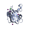
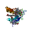
| Deposited unit | 
| ||||||||
|---|---|---|---|---|---|---|---|---|---|
| 1 | 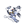
| ||||||||
| 2 | 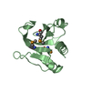
| ||||||||
| 3 | 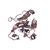
| ||||||||
| 4 | 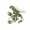
| ||||||||
| Unit cell |
|
-Components
| #1: Protein | Mass: 14970.576 Da / Num. of mol.: 4 Source method: isolated from a genetically manipulated source Source: (gene. exp.) Pseudomonas syringae pv. tomato (bacteria)Strain: DC3000 / Gene: PSPTO3016, PSPTO_3016 / Plasmid: pET 21-23C / Production host: Escherichia coli BL21(DE3) (bacteria) / Strain (production host): BL21(DE3)+ Magic / References: UniProt: Q880Y4#2: Water | ChemComp-HOH / | Water Mass: 18.015 Da / Num. of mol.: 37 / Source method: isolated from a natural source / Formula: H2O Mass: 18.015 Da / Num. of mol.: 37 / Source method: isolated from a natural source / Formula: H2O |
|---|
-Experimental details
-Experiment
| Experiment | Method: X-RAY DIFFRACTION / Number of used crystals: 1 |
|---|
- Sample preparation
| Crystal | Density Matthews: 2.43 Å3/Da / Density % sol: 49.47 % |
|---|---|
| Crystal grow | Temperature: 277 K / Method: vapor diffusion, hanging drop / pH: 4.2 Details: Protein solution: 100mM NaCl, 5mM DTT, 0.02% NaN3, 10mM Tris-HCl (pH 7.5) . Reservoir solution: 100mM NaCitrate (pH 4.2), 20% PEG8k, and 60mM NaNO3. , VAPOR DIFFUSION, HANGING DROP, temperature 277K |
-Data collection
| Diffraction | Mean temperature: 100 K |
|---|---|
| Diffraction source | Source: SYNCHROTRON / Site: NSLS  / Beamline: X4A / Wavelength: 0.97896 Å / Beamline: X4A / Wavelength: 0.97896 Å |
| Detector | Type: ADSC QUANTUM 4 / Detector: CCD / Date: Apr 12, 2009 / Details: mirrors |
| Radiation | Monochromator: Si 111 CHANNEL / Protocol: SINGLE WAVELENGTH / Monochromatic (M) / Laue (L): M / Scattering type: x-ray |
| Radiation wavelength | Wavelength: 0.97896 Å / Relative weight: 1 |
| Reflection | Resolution: 2.5→30 Å / Num. all: 38733 / Num. obs: 37487 / % possible obs: 96.9 % / Observed criterion σ(F): 0 / Observed criterion σ(I): 0 / Redundancy: 1.9 % / Biso Wilson estimate: 23 Å2 / Rmerge(I) obs: 0.071 / Rsym value: 0.06 / Net I/σ(I): 12.18 |
| Reflection shell | Resolution: 2.5→2.59 Å / Redundancy: 1.8 % / Rmerge(I) obs: 0.255 / Mean I/σ(I) obs: 2.37 / Num. unique all: 3919 / Rsym value: 0.249 / % possible all: 93.5 |
- Processing
| Software |
| ||||||||||||||||||||||||||||||||
|---|---|---|---|---|---|---|---|---|---|---|---|---|---|---|---|---|---|---|---|---|---|---|---|---|---|---|---|---|---|---|---|---|---|
| Refinement | Method to determine structure: SAD / Resolution: 2.51→19.94 Å / Rfactor Rfree error: 0.005 / Data cutoff high absF: 326319.906 / Data cutoff low absF: 0 / Isotropic thermal model: RESTRAINED / Cross valid method: THROUGHOUT / σ(F): 2 / σ(I): 2 / Stereochemistry target values: Engh & Huber
| ||||||||||||||||||||||||||||||||
| Solvent computation | Solvent model: FLAT MODEL / Bsol: 26.721 Å2 / ksol: 0.4 e/Å3 | ||||||||||||||||||||||||||||||||
| Displacement parameters | Biso mean: 49.7 Å2
| ||||||||||||||||||||||||||||||||
| Refine analyze |
| ||||||||||||||||||||||||||||||||
| Refinement step | Cycle: LAST / Resolution: 2.51→19.94 Å
| ||||||||||||||||||||||||||||||||
| Refine LS restraints |
| ||||||||||||||||||||||||||||||||
| LS refinement shell | Resolution: 2.5→2.59 Å / Rfactor Rfree error: 0.027 / Total num. of bins used: 10
|
