 Movie
Movie Controller

[English] 日本語
 Yorodumi
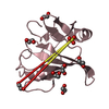
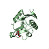
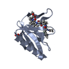
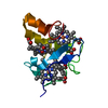
- PDB-4bh5: LytM domain of EnvC, an activator of cell wall amidases in Escher... -
+ Open data

- Basic information
| Entry | Database: PDB / ID: 4bh5 | ||||||
|---|---|---|---|---|---|---|---|
| Title | LytM domain of EnvC, an activator of cell wall amidases in Escherichia coli | ||||||
 Components Components | MUREIN HYDROLASE ACTIVATOR ENVC | ||||||
 Keywords Keywords |  CELL CYCLE / AMIDASE ACTIVATOR / AUTOLYSIN / CYTOKINESIS / MORPHOGENESIS / SACCULUS / PEPTIDOGLYCAN CELL CYCLE / AMIDASE ACTIVATOR / AUTOLYSIN / CYTOKINESIS / MORPHOGENESIS / SACCULUS / PEPTIDOGLYCAN | ||||||
| Function / homology |  Function and homology information Function and homology informationpeptidoglycan-based cell wall biogenesis / septum digestion after cytokinesis / cell division site / response to radiation / metalloendopeptidase activity / outer membrane-bounded periplasmic space / periplasmic space / hydrolase activity / response to xenobiotic stimulus / cell cycle / plasma membraneSimilarity search - Function | ||||||
| Biological species |  ESCHERICHIA COLI (E. coli) ESCHERICHIA COLI (E. coli) | ||||||
| Method | X-RAY DIFFRACTION / SYNCHROTRON / MOLECULAR REPLACEMENT / Resolution: 1.57 Å | ||||||
 Authors Authors | Morlot, C. / Peters, N.T. / Yang, D.C. / Uehara, T. / Vernet, T. / Bernhardt, T.G. | ||||||
 Citation Citation | Journal: Mol.Microbiol. / Year: 2013 Title: Structure-Function Analysis of the Lytm Domain of Envc, an Activator of Cell Wall Remodeling at the Escherichia Coli Division Site. Authors: Peters, N.T. / Morlot, C. / Yang, D.C. / Uehara, T. / Vernet, T. / Bernhardt, T.G. | ||||||
| History |
|
- Structure visualization
| Structure viewer | Molecule: MolmilJmol/JSmol |
|---|
- Downloads & links
-Download
| PDBx/mmCIF format | 4bh5.cif.gz | 121.6 KB | Display | PDBx/mmCIF format |
|---|---|---|---|---|
| PDB format | pdb4bh5.ent.gz | 93.3 KB | Display | PDB format |
| PDBx/mmJSON format | 4bh5.json.gz | Tree view | PDBx/mmJSON format | |
| Others |  Other downloads Other downloads |
-Validation report
| Arichive directory | https://data.pdbj.org/pub/pdb/validation_reports/bh/4bh5ftp://data.pdbj.org/pub/pdb/validation_reports/bh/4bh5 | HTTPS FTP |
|---|
-Related structure data
| Related structure data |  3ne8S S: Starting model for refinement |
|---|---|
| Similar structure data |


-Links
 PDBj
PDBj- Assembly
| Deposited unit | 
| ||||||||
|---|---|---|---|---|---|---|---|---|---|
| 1 | 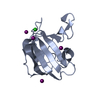
| ||||||||
| 2 | 
| ||||||||
| 3 | 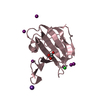
| ||||||||
| 4 | 
| ||||||||
| Unit cell |
|
-Components
-Protein , 1 types, 4 molecules ABCD
| #1: Protein | Mass: 15252.239 Da / Num. of mol.: 4 / Fragment: LYTM DOMAIN, RESIDUES 278-419 Source method: isolated from a genetically manipulated source Source: (gene. exp.) ESCHERICHIA COLI (E. coli) / Strain: K-12 / Plasmid: PTB104 / Production host: ESCHERICHIA COLI (E. coli) / Strain (production host): BL21(DE3) / References: UniProt: P37690 |
|---|
-Non-polymers , 5 types, 419 molecules 


| #2: Chemical | ChemComp-IOD / Iodide Mass: 126.904 Da / Num. of mol.: 23 / Source method: obtained synthetically / Formula: I Mass: 126.904 Da / Num. of mol.: 23 / Source method: obtained synthetically / Formula: I#3: Chemical | ChemComp-CL / Chloride Mass: 35.453 Da / Num. of mol.: 4 / Source method: obtained synthetically / Formula: Cl Mass: 35.453 Da / Num. of mol.: 4 / Source method: obtained synthetically / Formula: Cl#4: Chemical | Glycerol Mass: 92.094 Da / Num. of mol.: 2 / Source method: obtained synthetically / Formula: C3H8O3 Mass: 92.094 Da / Num. of mol.: 2 / Source method: obtained synthetically / Formula: C3H8O3#5: Chemical | ChemComp-K / |  Mass: 39.098 Da / Num. of mol.: 1 / Source method: obtained synthetically / Formula: K Mass: 39.098 Da / Num. of mol.: 1 / Source method: obtained synthetically / Formula: K#6: Water | ChemComp-HOH / | WaterMass: 18.015 Da / Num. of mol.: 389 / Source method: isolated from a natural source / Formula: H2O |
|---|
-Experimental details
-Experiment
| Experiment | Method: X-RAY DIFFRACTION / Number of used crystals: 1 |
|---|
- Sample preparation
| Crystal | Density Matthews: 2.02 Å3/Da / Density % sol: 39.2 % / Description: NONE |
|---|---|
| Crystal grow | Temperature: 293 K / Method: vapor diffusion / pH: 6.9 / Details: 0.2 M SODIUM IODIDE, 17% PEG 3350, PH 6.9 |
-Data collection
| Diffraction | Mean temperature: 100 K |
|---|---|
| Diffraction source | Source: SYNCHROTRON / Site: ESRF  / Beamline: ID14-1 / Wavelength: 0.9334 / Beamline: ID14-1 / Wavelength: 0.9334 |
| Detector | Type: ADSC QUANTUM 210 / Detector: CCD / Date: Nov 6, 2010 |
| Radiation | Protocol: SINGLE WAVELENGTH / Monochromatic (M) / Laue (L): M / Scattering type: x-ray |
| Radiation wavelength | Wavelength: 0.9334 Å / Relative weight: 1 |
| Reflection | Resolution: 1.56→46.4 Å / Num. obs: 64864 / % possible obs: 96.4 % / Observed criterion σ(I): -3 / Redundancy: 2.89 % / Rmerge(I) obs: 0.076 / Net I/σ(I): 15.9 |
| Reflection shell | Resolution: 1.56→1.66 Å / Redundancy: 2.19 % / Rmerge(I) obs: 0.493 / Mean I/σ(I) obs: 2.8 / % possible all: 73 |
- Processing
| Software |
| ||||||||||||||||||||||||||||||||||||||||||||||||||||||||||||||||||||||||||||||||||||||||||||||||||||||||||||||||||||||||||||||||||||||||||||||||||||||||||||||||||||||||||||||||||||||
|---|---|---|---|---|---|---|---|---|---|---|---|---|---|---|---|---|---|---|---|---|---|---|---|---|---|---|---|---|---|---|---|---|---|---|---|---|---|---|---|---|---|---|---|---|---|---|---|---|---|---|---|---|---|---|---|---|---|---|---|---|---|---|---|---|---|---|---|---|---|---|---|---|---|---|---|---|---|---|---|---|---|---|---|---|---|---|---|---|---|---|---|---|---|---|---|---|---|---|---|---|---|---|---|---|---|---|---|---|---|---|---|---|---|---|---|---|---|---|---|---|---|---|---|---|---|---|---|---|---|---|---|---|---|---|---|---|---|---|---|---|---|---|---|---|---|---|---|---|---|---|---|---|---|---|---|---|---|---|---|---|---|---|---|---|---|---|---|---|---|---|---|---|---|---|---|---|---|---|---|---|---|---|---|
| Refinement | Method to determine structure: MOLECULAR REPLACEMENT Starting model: PDB ENTRY 3NE8 Resolution: 1.57→46.4 Å / Cor.coef. Fo:Fc: 0.961 / Cor.coef. Fo:Fc free: 0.941 / SU B: 1.721 / SU ML: 0.06 / Cross valid method: THROUGHOUT / ESU R: 0.091 / ESU R Free: 0.095 / Stereochemistry target values: MAXIMUM LIKELIHOOD / Details: HYDROGENS HAVE BEEN ADDED IN THE RIDING POSITIONS.
| ||||||||||||||||||||||||||||||||||||||||||||||||||||||||||||||||||||||||||||||||||||||||||||||||||||||||||||||||||||||||||||||||||||||||||||||||||||||||||||||||||||||||||||||||||||||
| Solvent computation | Ion probe radii: 0.8 Å / Shrinkage radii: 0.8 Å / VDW probe radii: 1.4 Å / Solvent model: BABINET MODEL WITH MASK | ||||||||||||||||||||||||||||||||||||||||||||||||||||||||||||||||||||||||||||||||||||||||||||||||||||||||||||||||||||||||||||||||||||||||||||||||||||||||||||||||||||||||||||||||||||||
| Displacement parameters | Biso mean: 14.897 Å2
| ||||||||||||||||||||||||||||||||||||||||||||||||||||||||||||||||||||||||||||||||||||||||||||||||||||||||||||||||||||||||||||||||||||||||||||||||||||||||||||||||||||||||||||||||||||||
| Refinement step | Cycle: LAST / Resolution: 1.57→46.4 Å
| ||||||||||||||||||||||||||||||||||||||||||||||||||||||||||||||||||||||||||||||||||||||||||||||||||||||||||||||||||||||||||||||||||||||||||||||||||||||||||||||||||||||||||||||||||||||
| Refine LS restraints |
|