 ムービー
ムービー コントローラー


+ データを開く

- 基本情報
基本情報
| 登録情報 | データベース: PDB / ID: 3j41 | ||||||
|---|---|---|---|---|---|---|---|
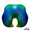
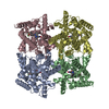
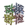
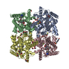
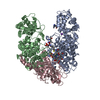
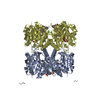
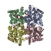
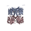
| タイトル | Pseudo-atomic model of the Aquaporin-0/Calmodulin complex derived from electron microscopy | ||||||
 要素 要素 |
| ||||||
 キーワード キーワード | TRANSPORT PROTEIN/CALCIUM BINDING /  calcium regulation / water channel (アクアポリン) / membrane protein complex (生体膜) / TRANSPORT PROTEIN-CALCIUM BINDING complex calcium regulation / water channel (アクアポリン) / membrane protein complex (生体膜) / TRANSPORT PROTEIN-CALCIUM BINDING complex | ||||||
| 機能・相同性 |  機能・相同性情報 機能・相同性情報gap junction-mediated intercellular transport / water channel activity / water transport / : / structural constituent of eye lens / establishment of protein localization to mitochondrial membrane / ギャップ結合 / type 3 metabotropic glutamate receptor binding / CAM型光合成 / Cam-PDE 1 activation ...gap junction-mediated intercellular transport / water channel activity / water transport / : / structural constituent of eye lens / establishment of protein localization to mitochondrial membrane / ギャップ結合 / type 3 metabotropic glutamate receptor binding / CAM型光合成 / Cam-PDE 1 activation / Sodium/Calcium exchangers / lens development in camera-type eye / response to stimulus / Calmodulin induced events / Reduction of cytosolic Ca++ levels / Activation of Ca-permeable Kainate Receptor / CREB1 phosphorylation through the activation of CaMKII/CaMKK/CaMKIV cascasde / regulation of synaptic vesicle endocytosis / Loss of phosphorylation of MECP2 at T308 / CREB1 phosphorylation through the activation of Adenylate Cyclase / PKA activation / negative regulation of high voltage-gated calcium channel activity / regulation of synaptic vesicle exocytosis / CaMK IV-mediated phosphorylation of CREB / Glycogen breakdown (glycogenolysis) / negative regulation of calcium ion export across plasma membrane / organelle localization by membrane tethering / Activation of RAC1 downstream of NMDARs / regulation of cardiac muscle cell action potential / mitochondrion-endoplasmic reticulum membrane tethering / CLEC7A (Dectin-1) induces NFAT activation / autophagosome membrane docking / response to corticosterone / positive regulation of ryanodine-sensitive calcium-release channel activity / Negative regulation of NMDA receptor-mediated neuronal transmission / nitric-oxide synthase binding / regulation of cell communication by electrical coupling involved in cardiac conduction / Unblocking of NMDA receptors, glutamate binding and activation / negative regulation of peptidyl-threonine phosphorylation / Synthesis of IP3 and IP4 in the cytosol / Phase 0 - rapid depolarisation / protein phosphatase activator activity / RHO GTPases activate PAKs / positive regulation of cyclic-nucleotide phosphodiesterase activity / positive regulation of phosphoprotein phosphatase activity / 長期増強 / Ion transport by P-type ATPases / Uptake and function of anthrax toxins / adenylate cyclase binding / Calcineurin activates NFAT / Regulation of MECP2 expression and activity / catalytic complex / DARPP-32 events / detection of calcium ion / positive regulation of cell adhesion / negative regulation of ryanodine-sensitive calcium-release channel activity / Smooth Muscle Contraction / RHO GTPases activate IQGAPs / regulation of cardiac muscle contraction / calcium channel inhibitor activity / cellular response to interferon-beta / positive regulation of DNA binding / regulation of cardiac muscle contraction by regulation of the release of sequestered calcium ion / Protein methylation / enzyme regulator activity / voltage-gated potassium channel complex / phosphatidylinositol 3-kinase binding / Activation of AMPK downstream of NMDARs / eNOS activation / regulation of release of sequestered calcium ion into cytosol by sarcoplasmic reticulum / regulation of calcium-mediated signaling / positive regulation of protein dephosphorylation / titin binding / regulation of ryanodine-sensitive calcium-release channel activity / Tetrahydrobiopterin (BH4) synthesis, recycling, salvage and regulation / Ion homeostasis / positive regulation of protein autophosphorylation / sperm midpiece / response to amphetamine / calcium channel complex / activation of adenylate cyclase activity / substantia nigra development / adenylate cyclase activator activity / 視覚 / Ras activation upon Ca2+ influx through NMDA receptor / regulation of heart rate / nitric-oxide synthase regulator activity / protein serine/threonine kinase activator activity / sarcomere / FCERI mediated Ca+2 mobilization / FCGR3A-mediated IL10 synthesis / VEGFR2 mediated vascular permeability / positive regulation of peptidyl-threonine phosphorylation / Antigen activates B Cell Receptor (BCR) leading to generation of second messengers / VEGFR2 mediated cell proliferation / regulation of cytokinesis / positive regulation of nitric-oxide synthase activity / Translocation of SLC2A4 (GLUT4) to the plasma membrane / spindle microtubule / ミトコンドリア類似検索 - 分子機能 | ||||||
| 生物種 |  Homo sapiens (ヒト) Homo sapiens (ヒト) Ovis aries (ヒツジ) Ovis aries (ヒツジ) | ||||||
| 手法 | 電子顕微鏡法 / 単粒子再構成法 / ネガティブ染色法 / 解像度: 25 Å | ||||||
 データ登録者 データ登録者 | Reichow, S.L. / Clemens, D.M. / Freites, J.A. / Nemeth-Cahalan, K.L. / Heyden, M. / Tobias, D.J. / Hall, J.E. / Gonen, T. | ||||||
 引用 引用 | ジャーナル: Nat Struct Mol Biol / 年: 2013 タイトル: Allosteric mechanism of water-channel gating by Ca2+-calmodulin. 著者: Steve L Reichow / Daniel M Clemens / J Alfredo Freites / Karin L Németh-Cahalan / Matthias Heyden / Douglas J Tobias / James E Hall / Tamir Gonen /  要旨: Calmodulin (CaM) is a universal regulatory protein that communicates the presence of calcium to its molecular targets and correspondingly modulates their function. This key signaling protein is ...Calmodulin (CaM) is a universal regulatory protein that communicates the presence of calcium to its molecular targets and correspondingly modulates their function. This key signaling protein is important for controlling the activity of hundreds of membrane channels and transporters. However, understanding of the structural mechanisms driving CaM regulation of full-length membrane proteins has remained elusive. In this study, we determined the pseudoatomic structure of full-length mammalian aquaporin-0 (AQP0, Bos taurus) in complex with CaM, using EM to elucidate how this signaling protein modulates water-channel function. Molecular dynamics and functional mutation studies reveal how CaM binding inhibits AQP0 water permeability by allosterically closing the cytoplasmic gate of AQP0. Our mechanistic model provides new insight, only possible in the context of the fully assembled channel, into how CaM regulates multimeric channels by facilitating cooperativity between adjacent subunits. | ||||||
| 履歴 |
|
- 構造の表示
構造の表示
| ムービー |
ムービービューア |
|---|---|
| 構造ビューア | 分子: MolmilJmol/JSmol |
- ダウンロードとリンク
-ダウンロード
| PDBx/mmCIF形式 | 3j41.cif.gz | 231.7 KB | 表示 | PDBx/mmCIF形式 |
|---|---|---|---|---|
| PDB形式 | pdb3j41.ent.gz | 182.8 KB | 表示 | PDB形式 |
| PDBx/mmJSON形式 | 3j41.json.gz | ツリー表示 | PDBx/mmJSON形式 | |
| その他 |  その他のダウンロード その他のダウンロード |
-検証レポート
| アーカイブディレクトリ | https://data.pdbj.org/pub/pdb/validation_reports/j4/3j41ftp://data.pdbj.org/pub/pdb/validation_reports/j4/3j41 | HTTPS FTP |
|---|
-関連構造データ
-リンク
 PDBj
PDBj


- 集合体
集合体
| 登録構造単位 | 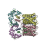
|
|---|---|
| 1 |
|
-要素
| #1: タンパク質 | / Aquaporin-0 分子量: 28244.865 Da / 分子数: 4 / 断片: SEE REMARK 999 / 由来タイプ: 天然 / 由来: (天然) Ovis aries (ヒツジ) / 組織: eye lensLens (anatomy) / 参照: UniProt: Q6J8I9*PLUS#2: タンパク質 | カルモジュリン / CaM分子量: 16852.545 Da / 分子数: 2 / 由来タイプ: 組換発現 / 由来: (組換発現) Homo sapiens (ヒト)遺伝子: CALM1, CALM, CAM, CAM1, CALM2, CAM2, CAMB, CALM3, CALML2, CAM3, CAMC, CAMIII プラスミド: pET / 発現宿主:  Escherichia coli (大腸菌) / 株 (発現宿主): BL21 / 参照: UniProt: P62158, UniProt: P0DP23*PLUS Escherichia coli (大腸菌) / 株 (発現宿主): BL21 / 参照: UniProt: P62158, UniProt: P0DP23*PLUS#3: 化合物 | ChemComp-CA /   分子量: 40.078 Da / 分子数: 8 / 由来タイプ: 合成 / 式: Ca 分子量: 40.078 Da / 分子数: 8 / 由来タイプ: 合成 / 式: Ca配列の詳細 | CHAINS A, B, C, AND D (AQUAPORIN) ARE FROM OVIS ARIES, BUT THE MODELED SEQUENCE IS FROM BOS TAURUS ...CHAINS A, B, C, AND D (AQUAPORIN) ARE FROM OVIS ARIES, BUT THE MODELED SEQUENCE IS FROM BOS TAURUS (UNP P06624). | |
|---|
-実験情報
-実験
| 実験 | 手法: 電子顕微鏡法 |
|---|---|
| EM実験 | 試料の集合状態: PARTICLE / 3次元再構成法: 単粒子再構成法 |
- 試料調製
試料調製
| 構成要素 |
| ||||||||||||||||||||
|---|---|---|---|---|---|---|---|---|---|---|---|---|---|---|---|---|---|---|---|---|---|
| 分子量 | 値: 0.13 MDa / 実験値: YES | ||||||||||||||||||||
| 緩衝液 | 名称: 25mM HEPES, pH 7.4, 5mM CaCl2, 0.3% decylmaltoside / pH: 7.4 / 詳細: 25mM HEPES, pH 7.4, 5mM CaCl2, 0.3% decylmaltoside | ||||||||||||||||||||
| 試料 | 濃度: 0.02 mg/ml / 包埋: NO / シャドウイング: NO / 染色: YES / 凍結: NO 詳細: 25mM HEPES, 5mM CaCl2, 0.3% decylmaltoside (Stain Details 0.75% Uranyl Formate) | ||||||||||||||||||||
| 染色 | タイプ: NEGATIVE / 染色剤: Uranyl Formate | ||||||||||||||||||||
| 試料支持 | 詳細: 400 mesh carbon coated grid (Ted Pella) |
- 電子顕微鏡撮影
| 顕微鏡 | モデル: FEI TECNAI 12 / 日付: 2010年2月25日 |
|---|---|
| 電子銃 | 電子線源: LAB6 / 加速電圧: 120 kV / 照射モード: FLOOD BEAM / Electron beam tilt params: 0 |
| 電子レンズ | モード: BRIGHT FIELDBright-field microscopy / 倍率(公称値): 52000 X / 倍率(補正後): 52000 X / 最大 デフォーカス(公称値): 2000 nm / 最小 デフォーカス(公称値): 1000 nm / Cs: 2 mm 非点収差 : Objective lens astigmatism was corrected at 100,000 times magnificationカメラ長: 0 mm |
| 試料ホルダ | 試料ホルダーモデル: OTHER / 資料ホルダタイプ: FEI Single-Tilt / 傾斜角・最大: 50 ° / 傾斜角・最小: 0 ° |
| 撮影 | 電子線照射量: 15 e/Å2 / フィルム・検出器のモデル: KODAK SO-163 FILM |
| 画像スキャン | デジタル画像の数: 200 |
| 放射 | プロトコル: SINGLE WAVELENGTH / 単色(M)・ラウエ(L): M / 散乱光タイプ: x-ray |
| 放射波長 | 相対比: 1 |
- 解析
解析
| EMソフトウェア |
| |||||||||||||||
|---|---|---|---|---|---|---|---|---|---|---|---|---|---|---|---|---|
| CTF補正 | 詳細: CTF-TILT, each micrograph | |||||||||||||||
| 対称性 | 点対称性: C2 (2回回転対称) | |||||||||||||||
| 3次元再構成 | 手法: Random Conical Tilt / 解像度: 25 Å / 解像度の算出法: FSC 0.5 CUT-OFF / 粒子像の数: 11720 / ピクセルサイズ(公称値): 3.98 Å / ピクセルサイズ(実測値): 3.98 Å / 倍率補正: AQP0 cyrstal 詳細: Final Map with C2 Symmetry and Filtered to 25 Angstrom (Single particle details: Particles were selected from a tilted pair dataset at 0 and 50 degree tilt using SPIDER. An initial ...詳細: Final Map with C2 Symmetry and Filtered to 25 Angstrom (Single particle details: Particles were selected from a tilted pair dataset at 0 and 50 degree tilt using SPIDER. An initial reconstruction was generated using random conical tilt methods in SPIDER and refined in FREALIGN.) (Single particle--Applied symmetry: C2) 対称性のタイプ: POINT | |||||||||||||||
| 原子モデル構築 | プロトコル: RIGID BODY FIT / 空間: REAL / Target criteria: cross-correlation 詳細: REFINEMENT PROTOCOL--rigid body DETAILS--A complete model of AQP0-CaM was built by fitting 2B6P and 1NWD into the EM map in Chimera. Loops connecting the two structures were built using COOT ...詳細: REFINEMENT PROTOCOL--rigid body DETAILS--A complete model of AQP0-CaM was built by fitting 2B6P and 1NWD into the EM map in Chimera. Loops connecting the two structures were built using COOT and the final model was energy minimized to remove steric clashes. The geometries of the modeled loops (AQP0 residues 222-227) were not refined due to lack of resolution in the experimental map. | |||||||||||||||
| 原子モデル構築 | 3D fitting-ID: 1 / Source name: PDB / タイプ: experimental model
| |||||||||||||||
| 精密化ステップ | サイクル: LAST
|
