 Movie
Movie Controller

[English] 日本語
 Yorodumi
- PDB-3v9b: Crystal structure of the catalytic domain of PDE4D2 with (S)-N-(3... -
+ Open data

- Basic information
| Entry | Database: PDB / ID: 3v9b | ||||||
|---|---|---|---|---|---|---|---|
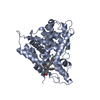
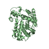
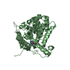
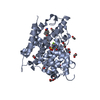
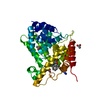
| Title | Crystal structure of the catalytic domain of PDE4D2 with (S)-N-(3-{1-[1-(3-Cyclopropylmethoxy-4-difluoromethoxyphenyl)-2-(1-oxypyridin-4-yl)-ethyl]-1H-pyrazl-3-yl}phenyl)acetamide | ||||||
 Components Components | cAMP-specific 3',5'-cyclic phosphodiesterase 4D | ||||||
 Keywords Keywords | HYDROLASE/HYDROLASE INHIBITOR /  hydrolase / 3' / 5'-Cyclic-AMP Phosphodiesterases / Cyclic nucleotide / phosphodiesterases / HYDROLASE-HYDROLASE INHIBITOR complex hydrolase / 3' / 5'-Cyclic-AMP Phosphodiesterases / Cyclic nucleotide / phosphodiesterases / HYDROLASE-HYDROLASE INHIBITOR complex | ||||||
| Function / homology |  Function and homology information Function and homology informationsignaling receptor regulator activity / negative regulation of relaxation of cardiac muscle / negative regulation of heart contraction / 3',5'-cyclic-AMP phosphodiesterase / positive regulation of interleukin-5 production / regulation of cardiac muscle cell contraction / establishment of endothelial barrier / negative regulation of cAMP-mediated signaling / beta-2 adrenergic receptor binding / regulation of calcium ion transmembrane transport via high voltage-gated calcium channel ...signaling receptor regulator activity / negative regulation of relaxation of cardiac muscle / negative regulation of heart contraction / 3',5'-cyclic-AMP phosphodiesterase / positive regulation of interleukin-5 production / regulation of cardiac muscle cell contraction / establishment of endothelial barrier / negative regulation of cAMP-mediated signaling / beta-2 adrenergic receptor binding / regulation of calcium ion transmembrane transport via high voltage-gated calcium channel / positive regulation of heart rate / heterocyclic compound binding / adrenergic receptor signaling pathway / voltage-gated calcium channel complex / regulation of cell communication by electrical coupling involved in cardiac conduction / cAMP catabolic process / calcium channel regulator activity / cAMP-mediated signaling / 3',5'-cyclic-nucleotide phosphodiesterase activity / 3',5'-cyclic-AMP phosphodiesterase activity / DARPP-32 events / negative regulation of peptidyl-serine phosphorylation / regulation of release of sequestered calcium ion into cytosol by sarcoplasmic reticulum / cAMP binding / cellular response to cAMP / cellular response to epinephrine stimulus / calcium channel complex / positive regulation of interleukin-2 production / regulation of heart rate / positive regulation of type II interferon production / ATPase binding / T cell receptor signaling pathway / G alpha (s) signalling events / scaffold protein binding / transmembrane transporter binding / apical plasma membrane / centrosome / perinuclear region of cytoplasm / enzyme binding / signal transduction / membrane / metal ion binding / nucleus / plasma membrane / cytosolSimilarity search - Function | ||||||
| Biological species |  Homo sapiens (human) Homo sapiens (human) | ||||||
| Method | X-RAY DIFFRACTION / SYNCHROTRON / MOLECULAR REPLACEMENT / Resolution: 2.1 Å | ||||||
 Authors Authors | Kim, H.T. / Chang, H.J. | ||||||
 Citation Citation | Journal: Org.Biomol.Chem. / Year: 2012 Title: Stereoselective synthesis of novel pyrazole derivatives using tert-butansulfonamide as a chiral auxiliary Authors: Park, C.M. / Jeon, D.J. | ||||||
| History |
|
- Structure visualization
| Structure viewer | Molecule: MolmilJmol/JSmol |
|---|
- Downloads & links
-Download
| PDBx/mmCIF format | 3v9b.cif.gz | 281.5 KB | Display | PDBx/mmCIF format |
|---|---|---|---|---|
| PDB format | pdb3v9b.ent.gz | 226.3 KB | Display | PDB format |
| PDBx/mmJSON format | 3v9b.json.gz | Tree view | PDBx/mmJSON format | |
| Others |  Other downloads Other downloads |
-Validation report
| Arichive directory | https://data.pdbj.org/pub/pdb/validation_reports/v9/3v9bftp://data.pdbj.org/pub/pdb/validation_reports/v9/3v9b | HTTPS FTP |
|---|
-Related structure data
| Related structure data | 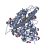 3sl8S S: Starting model for refinement |
|---|---|
| Similar structure data |


-Links
 PDBj
PDBj


- Assembly
| Deposited unit | 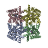
| ||||||||
|---|---|---|---|---|---|---|---|---|---|
| 1 | 
| ||||||||
| 2 | 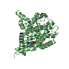
| ||||||||
| 3 | 
| ||||||||
| 4 | 
| ||||||||
| Unit cell |
|
-Components
| #1: Protein | Mass: 41394.773 Da / Num. of mol.: 4 / Fragment: catalytic domain, UNP residues 381-740 Source method: isolated from a genetically manipulated source Source: (gene. exp.) Homo sapiens (human) / Gene: PDE4D / Plasmid: pET15b / Production host:  Escherichia coli (E. coli) / Strain (production host): BL21(DE3) Escherichia coli (E. coli) / Strain (production host): BL21(DE3)References: UniProt: Q08499, 3',5'-cyclic-nucleotide phosphodiesterase#2: Chemical | ChemComp-IHM /   Mass: 534.554 Da / Num. of mol.: 4 / Source method: obtained synthetically / Formula: C29H28F2N4O4 Mass: 534.554 Da / Num. of mol.: 4 / Source method: obtained synthetically / Formula: C29H28F2N4O4#3: Chemical | ChemComp-ZN /   Mass: 65.409 Da / Num. of mol.: 8 / Source method: obtained synthetically / Formula: Zn Mass: 65.409 Da / Num. of mol.: 8 / Source method: obtained synthetically / Formula: Zn#4: Water | ChemComp-HOH / | Water Mass: 18.015 Da / Num. of mol.: 414 / Source method: isolated from a natural source / Formula: H2O Mass: 18.015 Da / Num. of mol.: 414 / Source method: isolated from a natural source / Formula: H2O |
|---|
-Experimental details
-Experiment
| Experiment | Method: X-RAY DIFFRACTION / Number of used crystals: 1 |
|---|
- Sample preparation
| Crystal | Density Matthews: 2.65 Å3/Da / Density % sol: 53.64 % |
|---|---|
| Crystal grow | Temperature: 277 K / Method: vapor diffusion, hanging drop / pH: 7.5 Details: 20% PEG 3350, 25%(v/v) ethylene glycol, 10%(v/v) isopropanol, 100mM HEPES, pH 7.5, VAPOR DIFFUSION, HANGING DROP, temperature 277K |
-Data collection
| Diffraction | Mean temperature: 100 K |
|---|---|
| Diffraction source | Source: SYNCHROTRON / Site: PAL/PLS  / Beamline: 6B / Wavelength: 1.1 Å / Beamline: 6B / Wavelength: 1.1 Å |
| Detector | Type: ADSC QUANTUM 210r / Detector: CCD / Date: Nov 5, 2005 |
| Radiation | Monochromator: double mirrors / Protocol: SINGLE WAVELENGTH / Monochromatic (M) / Laue (L): M / Scattering type: x-ray |
| Radiation wavelength | Wavelength: 1.1 Å / Relative weight: 1 |
| Reflection | Resolution: 2.1→50 Å / Num. all: 288261 / Num. obs: 97786 / % possible obs: 94.2 % / Observed criterion σ(F): 1 / Observed criterion σ(I): 1 / Redundancy: 2.9 % / Biso Wilson estimate: 14.1 Å2 / Rmerge(I) obs: 0.093 / Rsym value: 0.07 / Net I/σ(I): 14.58 |
| Reflection shell | Resolution: 2.1→2.18 Å / Redundancy: 2.6 % / Rmerge(I) obs: 0.445 / Mean I/σ(I) obs: 2.41 / Num. unique all: 8984 / Rsym value: 0.408 / % possible all: 87.5 |
- Processing
| Software |
| ||||||||||||||||||||||||||||||||||||
|---|---|---|---|---|---|---|---|---|---|---|---|---|---|---|---|---|---|---|---|---|---|---|---|---|---|---|---|---|---|---|---|---|---|---|---|---|---|
| Refinement | Method to determine structure: MOLECULAR REPLACEMENT Starting model: PDB ENTRY 3SL8 Resolution: 2.1→35.41 Å / Rfactor Rfree error: 0.003 / Data cutoff high absF: 239640.86 / Data cutoff low absF: 0 / Isotropic thermal model: RESTRAINED / Cross valid method: THROUGHOUT / σ(F): 0 / Details: BULK SOLVENT MODEL USED
| ||||||||||||||||||||||||||||||||||||
| Solvent computation | Solvent model: FLAT MODEL / Bsol: 55.546 Å2 / ksol: 0.42 e/Å3 | ||||||||||||||||||||||||||||||||||||
| Displacement parameters | Biso mean: 28.4 Å2
| ||||||||||||||||||||||||||||||||||||
| Refine analyze |
| ||||||||||||||||||||||||||||||||||||
| Refinement step | Cycle: LAST / Resolution: 2.1→35.41 Å
| ||||||||||||||||||||||||||||||||||||
| Refine LS restraints |
| ||||||||||||||||||||||||||||||||||||
| LS refinement shell | Resolution: 2.1→2.18 Å / Total num. of bins used: 6
| ||||||||||||||||||||||||||||||||||||
| Xplor file |
|
