 Movie
Movie Controller

[English] 日本語
 Yorodumi
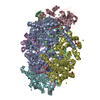
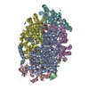
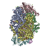
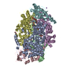
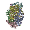
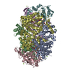
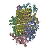
- PDB-5n2a: METHYL-COENZYME M REDUCTASE III FROM METHANOTORRIS FORMICICUS TRI... -
+ Open data

- Basic information
| Entry | Database: PDB / ID: 5n2a | |||||||||
|---|---|---|---|---|---|---|---|---|---|---|
| Title | METHYL-COENZYME M REDUCTASE III FROM METHANOTORRIS FORMICICUS TRIGONAL FORM | |||||||||
 Components Components |
| |||||||||
 Keywords Keywords |  TRANSFERASE / POST-TRANSLATIONAL MODIFICATION / BINDING SITES / CATALYSIS / COENZYMES / DISULFIDES / HYDROGEN / HYDROGEN BONDING / LIGANDS / MESNA / METALLOPORPHYRINS / METHANE / METHANOCOCCALES / NICKEL / OXIDATION-REDUCTION / OXIDOREDUCTASES / PHOSPHOTHREONINE / PROTEIN CONFORMATION / PROTEIN FOLDING / PROTEIN STRUCTURE / THERMOPHILE / AUTOTROPH / HYDROXY-TRYPTOPHANE TRANSFERASE / POST-TRANSLATIONAL MODIFICATION / BINDING SITES / CATALYSIS / COENZYMES / DISULFIDES / HYDROGEN / HYDROGEN BONDING / LIGANDS / MESNA / METALLOPORPHYRINS / METHANE / METHANOCOCCALES / NICKEL / OXIDATION-REDUCTION / OXIDOREDUCTASES / PHOSPHOTHREONINE / PROTEIN CONFORMATION / PROTEIN FOLDING / PROTEIN STRUCTURE / THERMOPHILE / AUTOTROPH / HYDROXY-TRYPTOPHANE | |||||||||
| Function / homology |  Function and homology informationcoenzyme-B sulfoethylthiotransferase / coenzyme-B sulfoethylthiotransferase activity / methanogenesis / metal ion binding Function and homology informationcoenzyme-B sulfoethylthiotransferase / coenzyme-B sulfoethylthiotransferase activity / methanogenesis / metal ion bindingSimilarity search - Function | |||||||||
| Biological species |  Methanotorris formicicus Mc-S-70 (archaea) Methanotorris formicicus Mc-S-70 (archaea) | |||||||||
| Method | X-RAY DIFFRACTION / SYNCHROTRON / MOLECULAR REPLACEMENT / Resolution: 2.8 Å | |||||||||
 Authors Authors | Wagner, T. / Wegner, C.E. / Ermler, U. / Shima, S. | |||||||||
 Citation Citation | Journal: J.Bacteriol. / Year: 2017 Title: Phylogenetic and Structural Comparisons of the Three Types of Methyl Coenzyme M Reductase from Methanococcales and Methanobacteriales. Authors: Wagner, T. / Wegner, C.E. / Kahnt, J. / Ermler, U. / Shima, S. | |||||||||
| History |
|
- Structure visualization
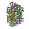
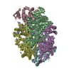
| Structure viewer | Molecule: MolmilJmol/JSmol |
|---|
- Downloads & links
-Download
| PDBx/mmCIF format | 5n2a.cif.gz | 495.4 KB | Display | PDBx/mmCIF format |
|---|---|---|---|---|
| PDB format | pdb5n2a.ent.gz | 404.8 KB | Display | PDB format |
| PDBx/mmJSON format | 5n2a.json.gz | Tree view | PDBx/mmJSON format | |
| Others |  Other downloads Other downloads |
-Validation report
| Arichive directory | https://data.pdbj.org/pub/pdb/validation_reports/n2/5n2aftp://data.pdbj.org/pub/pdb/validation_reports/n2/5n2a | HTTPS FTP |
|---|
-Related structure data
| Related structure data |  5n1qC 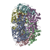 5n28C  5a8wS S: Starting model for refinement C: citing same article ( |
|---|---|
| Similar structure data |

-Links
 PDBj
PDBj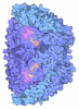


- Assembly
| Deposited unit | 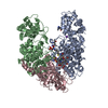
| ||||||||
|---|---|---|---|---|---|---|---|---|---|
| 1 | 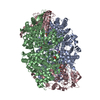
| ||||||||
| Unit cell |
| ||||||||
| Components on special symmetry positions |
|
-Components
-Protein , 1 types, 1 molecules A
| #1: Protein | Coenzyme-B sulfoethylthiotransferase Mass: 61180.930 Da / Num. of mol.: 1 / Source method: isolated from a natural source Details: IN CHAIN A, RESIDUE 260 IS A N1-METHYLHISTIDINE. RESIDUE 274 IS A C5-(S)-METHYLARGININE. RESIDUE 402 IS A C2-(S)-METHYLGLUTAMINE. RESIDUE 429 IS A POSSIBLE 6-HYDROXY-TRYPTOPHANE. RESIDUE 447 IS A THIOGLYCINE. Source: (natural) Methanotorris formicicus Mc-S-70 (archaea)Cell line: / / Organ: / / Plasmid details: DSMZ / Variant: Wild-type / Tissue: / References: UniProt: H1KXL5, coenzyme-B sulfoethylthiotransferase |
|---|
-Methyl-coenzyme M reductase, ... , 2 types, 2 molecules BC
| #2: Protein | Coenzyme-B sulfoethylthiotransferase Mass: 47541.703 Da / Num. of mol.: 1 / Source method: isolated from a natural source Source: (natural) Methanotorris formicicus Mc-S-70 (archaea)Cell line: / / Organ: / / Plasmid details: DSMZ / Variant: Wild-type / Tissue: / References: UniProt: H1KXL9, coenzyme-B sulfoethylthiotransferase |
|---|---|
| #3: Protein | Coenzyme-B sulfoethylthiotransferase Mass: 30274.383 Da / Num. of mol.: 1 / Source method: isolated from a natural source Source: (natural) Methanotorris formicicus Mc-S-70 (archaea)Cell line: / / Organ: / / Plasmid details: DSMZ / Variant: Wild-type / Tissue: / References: UniProt: H1KXL6, coenzyme-B sulfoethylthiotransferase |
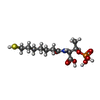
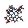
-Non-polymers , 5 types, 5 molecules 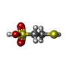


| #4: Chemical | ChemComp-COM /  Mass: 142.197 Da / Num. of mol.: 1 / Source method: obtained synthetically / Formula: C2H6O3S2 Mass: 142.197 Da / Num. of mol.: 1 / Source method: obtained synthetically / Formula: C2H6O3S2 |
|---|---|
| #5: Chemical | ChemComp-TP7 / Coenzyme B Mass: 343.334 Da / Num. of mol.: 1 / Source method: obtained synthetically / Formula: C11H22NO7PS Mass: 343.334 Da / Num. of mol.: 1 / Source method: obtained synthetically / Formula: C11H22NO7PS |
| #6: Chemical | ChemComp-F43 / Cofactor F430 Mass: 906.580 Da / Num. of mol.: 1 / Source method: obtained synthetically / Formula: C42H51N6NiO13 Mass: 906.580 Da / Num. of mol.: 1 / Source method: obtained synthetically / Formula: C42H51N6NiO13 |
| #7: Chemical | ChemComp-K /  Mass: 39.098 Da / Num. of mol.: 1 / Source method: obtained synthetically / Formula: K Mass: 39.098 Da / Num. of mol.: 1 / Source method: obtained synthetically / Formula: K |
| #8: Chemical | ChemComp-BR / Bromide Mass: 79.904 Da / Num. of mol.: 1 / Source method: obtained synthetically / Formula: Br Mass: 79.904 Da / Num. of mol.: 1 / Source method: obtained synthetically / Formula: Br |
-Experimental details
-Experiment
| Experiment | Method: X-RAY DIFFRACTION / Number of used crystals: 1 |
|---|
- Sample preparation
| Crystal | Density Matthews: 2.75 Å3/Da / Density % sol: 55.34 % / Description: Yellow cubic crystals |
|---|---|
| Crystal grow | Temperature: 291 K / Method: vapor diffusion, sitting drop / pH: 6.5 Details: Three single crystals appeared after one year and could be immediately fished. The drop contained a mixture of 0.8 ul of 35 mg/ml of MCR III Methanotorris formicicus and 0.8 ul of the ...Details: Three single crystals appeared after one year and could be immediately fished. The drop contained a mixture of 0.8 ul of 35 mg/ml of MCR III Methanotorris formicicus and 0.8 ul of the reservoir solution containing 200 mM potassium bromide, 200 mM potassium thiocyanate, 100 mM Na cacodylate pH 6.5, 3% (w/v) PGA-LM, and 30% PEG 400 (v/v). |
-Data collection
| Diffraction | Mean temperature: 100 K |
|---|---|
| Diffraction source | Source: SYNCHROTRON / Site: SLS  / Beamline: X10SA / Wavelength: 0.99998 Å / Beamline: X10SA / Wavelength: 0.99998 Å |
| Detector | Type: DECTRIS PILATUS3 6M / Detector: PIXEL / Date: May 22, 2014 |
| Radiation | Protocol: SINGLE WAVELENGTH / Monochromatic (M) / Laue (L): M / Scattering type: x-ray |
| Radiation wavelength | Wavelength: 0.99998 Å / Relative weight: 1 |
| Reflection | Resolution: 2.8→48.13 Å / Num. obs: 37773 / % possible obs: 99.9 % / Redundancy: 5 % / Biso Wilson estimate: 62.56 Å2 / CC1/2: 0.987 / Rmerge(I) obs: 0.188 / Rpim(I) all: 0.094 / Net I/σ(I): 7.4 |
| Reflection shell | Resolution: 2.8→2.95 Å / Redundancy: 4.9 % / Rmerge(I) obs: 0.739 / Mean I/σ(I) obs: 2.3 / Num. unique obs: 5454 / CC1/2: 0.334 / Rpim(I) all: 0.371 / % possible all: 100 |
- Processing
| Software |
| ||||||||||||||||||||||||||||||||||||||||||||||||||||||||||||||||||||||||||||||||||||||||||||||||||||||||||||||||||
|---|---|---|---|---|---|---|---|---|---|---|---|---|---|---|---|---|---|---|---|---|---|---|---|---|---|---|---|---|---|---|---|---|---|---|---|---|---|---|---|---|---|---|---|---|---|---|---|---|---|---|---|---|---|---|---|---|---|---|---|---|---|---|---|---|---|---|---|---|---|---|---|---|---|---|---|---|---|---|---|---|---|---|---|---|---|---|---|---|---|---|---|---|---|---|---|---|---|---|---|---|---|---|---|---|---|---|---|---|---|---|---|---|---|---|---|
| Refinement | Method to determine structure: MOLECULAR REPLACEMENT Starting model: 5A8W Resolution: 2.8→39.85 Å / Cor.coef. Fo:Fc: 0.918 / Cor.coef. Fo:Fc free: 0.8968 / Cross valid method: THROUGHOUT / σ(F): 0 / SU Rfree Blow DPI: 0.306
| ||||||||||||||||||||||||||||||||||||||||||||||||||||||||||||||||||||||||||||||||||||||||||||||||||||||||||||||||||
| Displacement parameters | Biso mean: 61.19 Å2
| ||||||||||||||||||||||||||||||||||||||||||||||||||||||||||||||||||||||||||||||||||||||||||||||||||||||||||||||||||
| Refine analyze | Luzzati coordinate error obs: 0.369 Å | ||||||||||||||||||||||||||||||||||||||||||||||||||||||||||||||||||||||||||||||||||||||||||||||||||||||||||||||||||
| Refinement step | Cycle: 1 / Resolution: 2.8→39.85 Å
| ||||||||||||||||||||||||||||||||||||||||||||||||||||||||||||||||||||||||||||||||||||||||||||||||||||||||||||||||||
| Refine LS restraints |
| ||||||||||||||||||||||||||||||||||||||||||||||||||||||||||||||||||||||||||||||||||||||||||||||||||||||||||||||||||
| LS refinement shell | Resolution: 2.8→2.88 Å / Total num. of bins used: 19
| ||||||||||||||||||||||||||||||||||||||||||||||||||||||||||||||||||||||||||||||||||||||||||||||||||||||||||||||||||
| Refinement TLS params. | Method: refined / Refine-ID: X-RAY DIFFRACTION
| ||||||||||||||||||||||||||||||||||||||||||||||||||||||||||||||||||||||||||||||||||||||||||||||||||||||||||||||||||
| Refinement TLS group |
|
